. 2023 Mar 4;26(4):106339.
doi: 10.1016/j.isci.2023.106339. eCollection 2023 Apr 21.

MicroRNAs emerging coordinate with placental mammals alter pathways in endometrial epithelia important for endometrial function

Affiliations

MicroRNAs emerging coordinate with placental mammals alter pathways in endometrial epithelia important for endometrial function

Laura Hume et al. iScience. .

Abstract

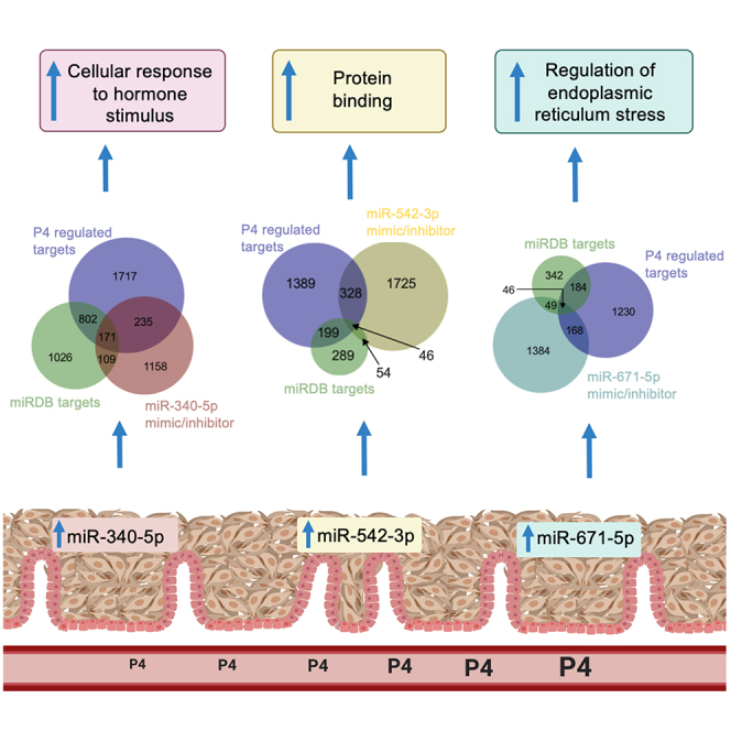
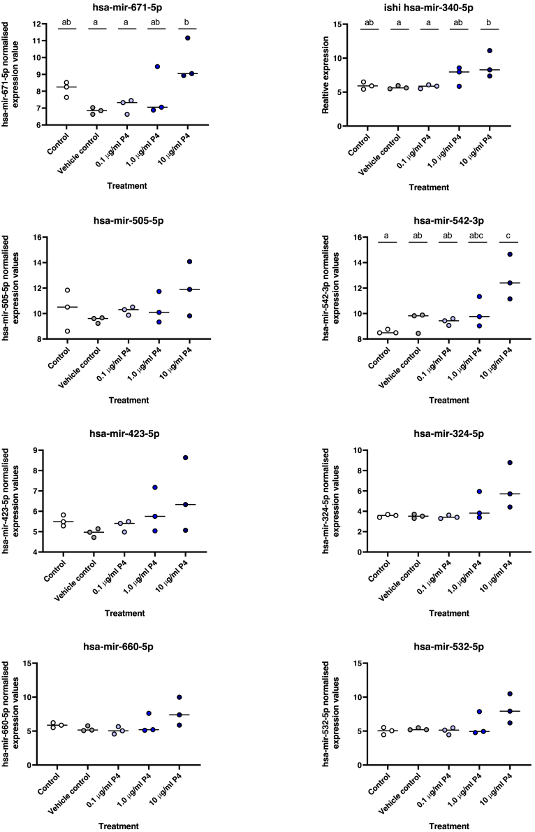
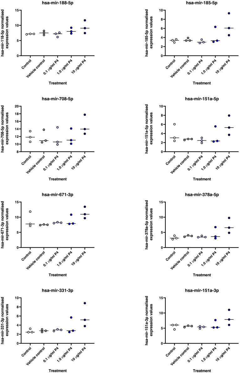
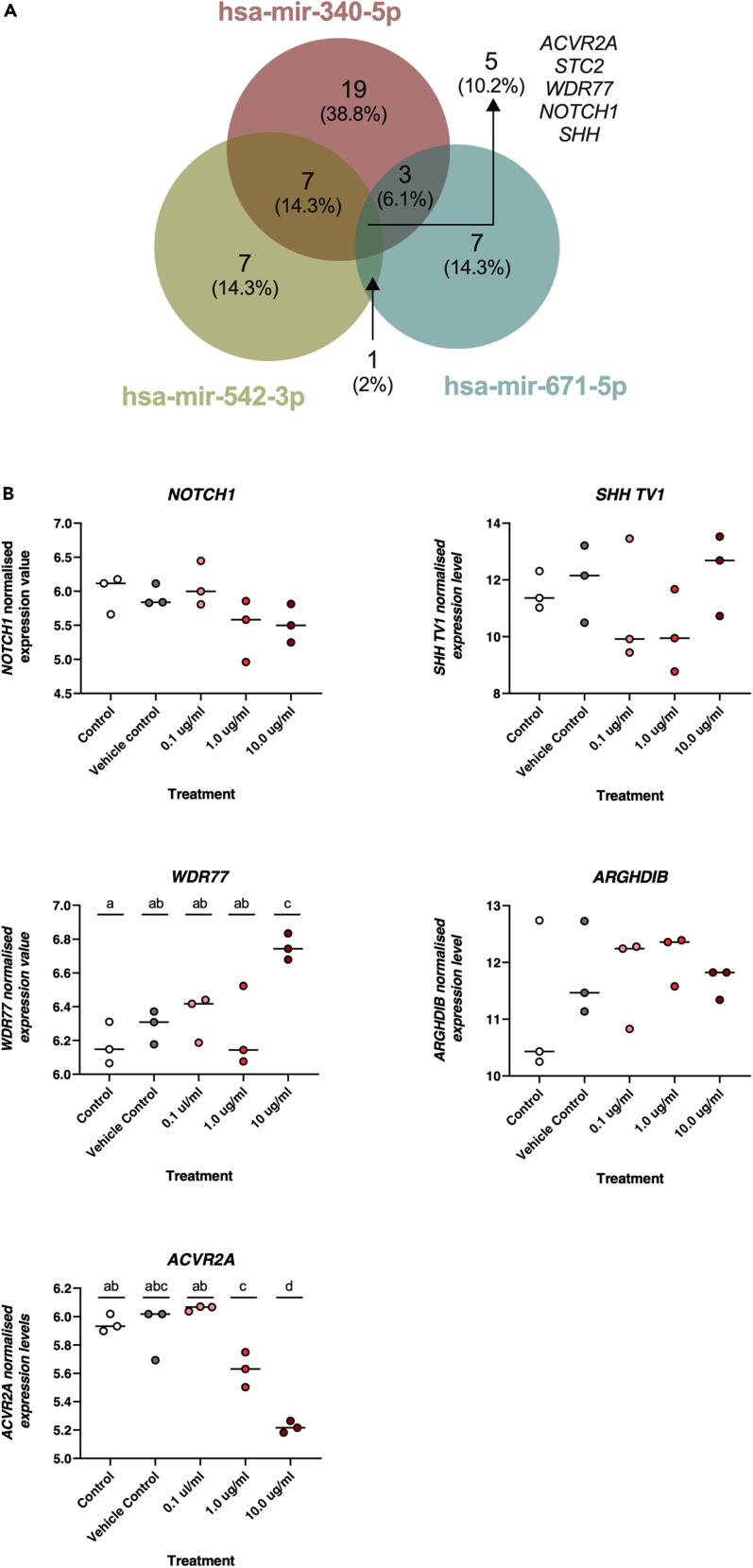
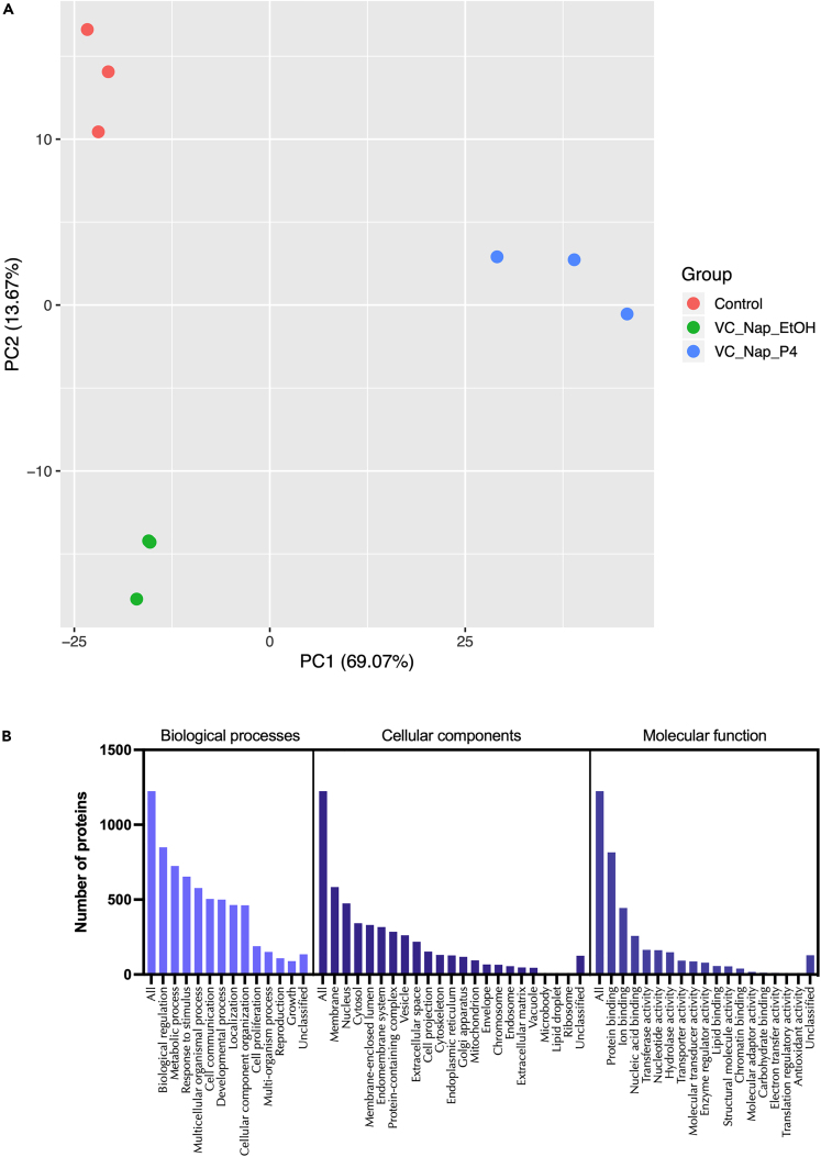
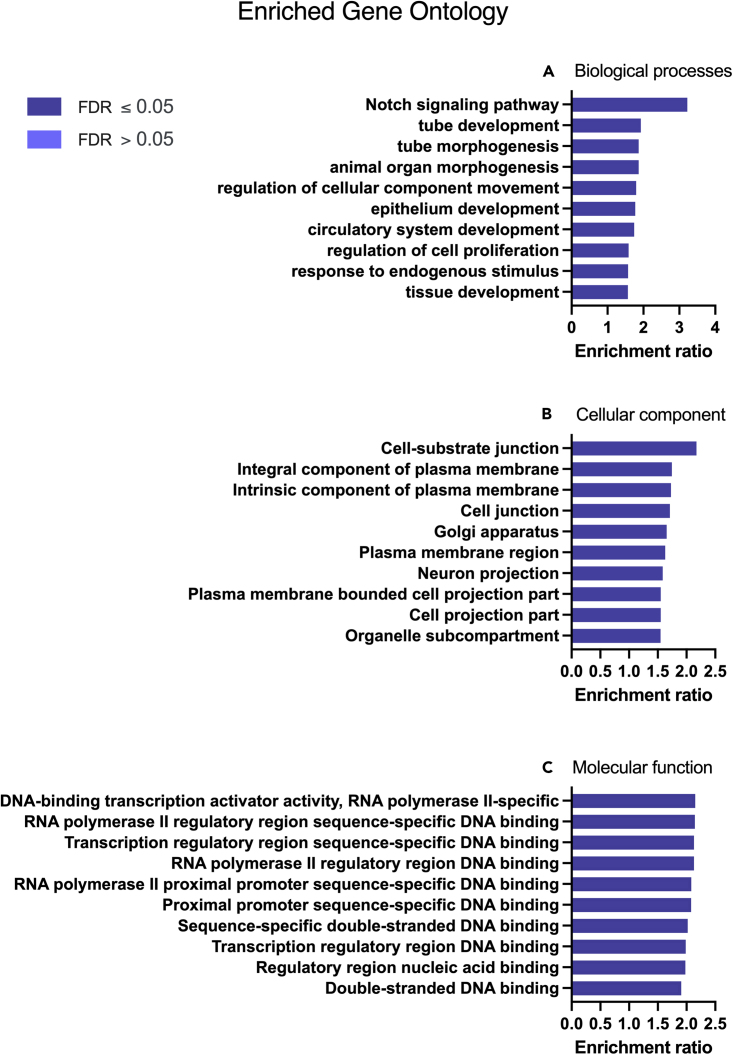
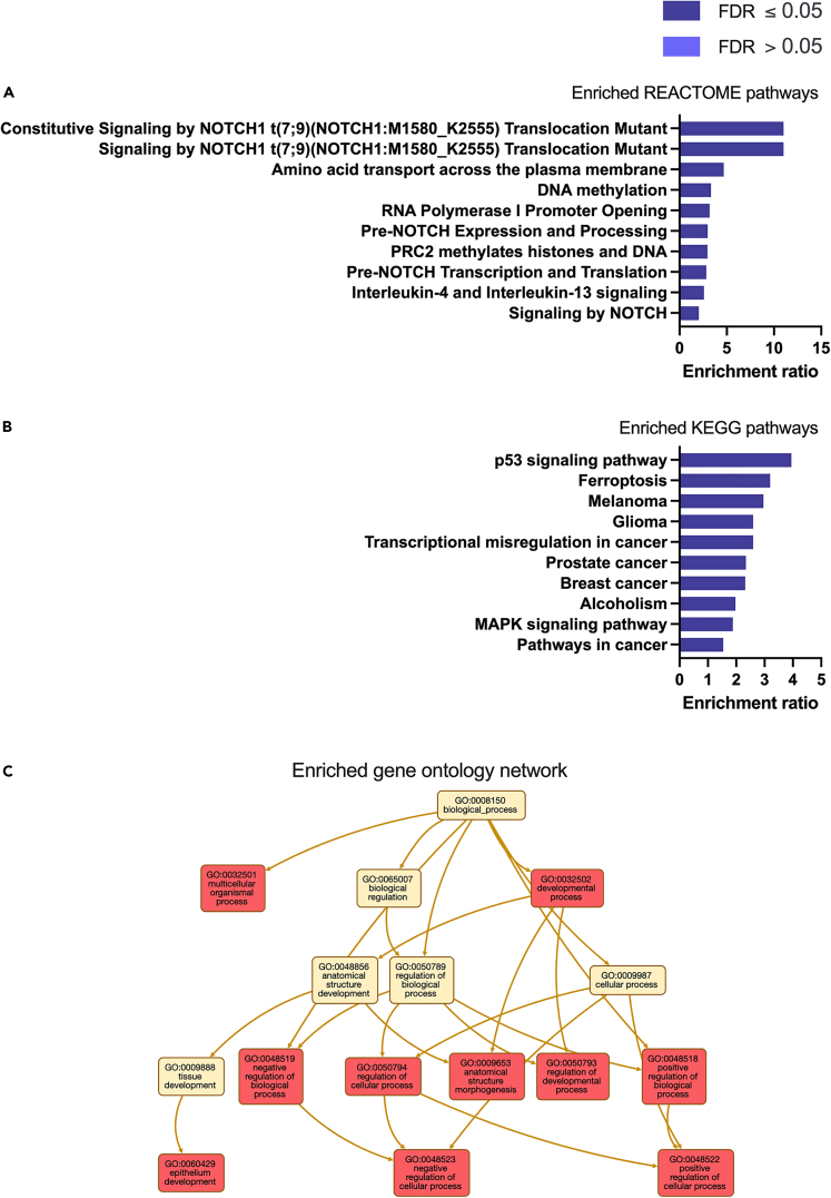
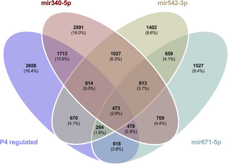
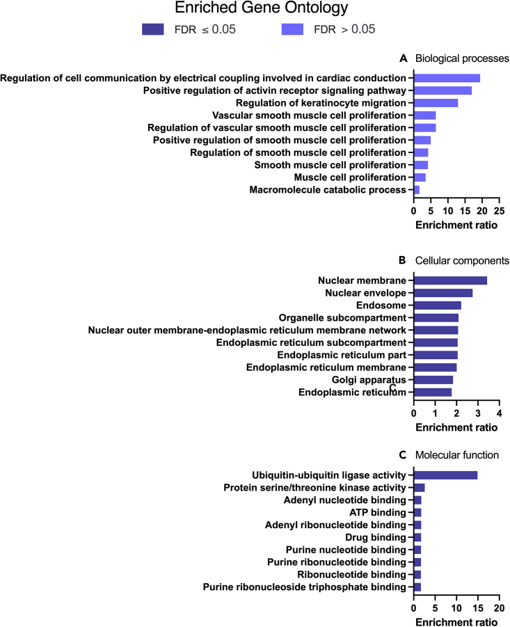
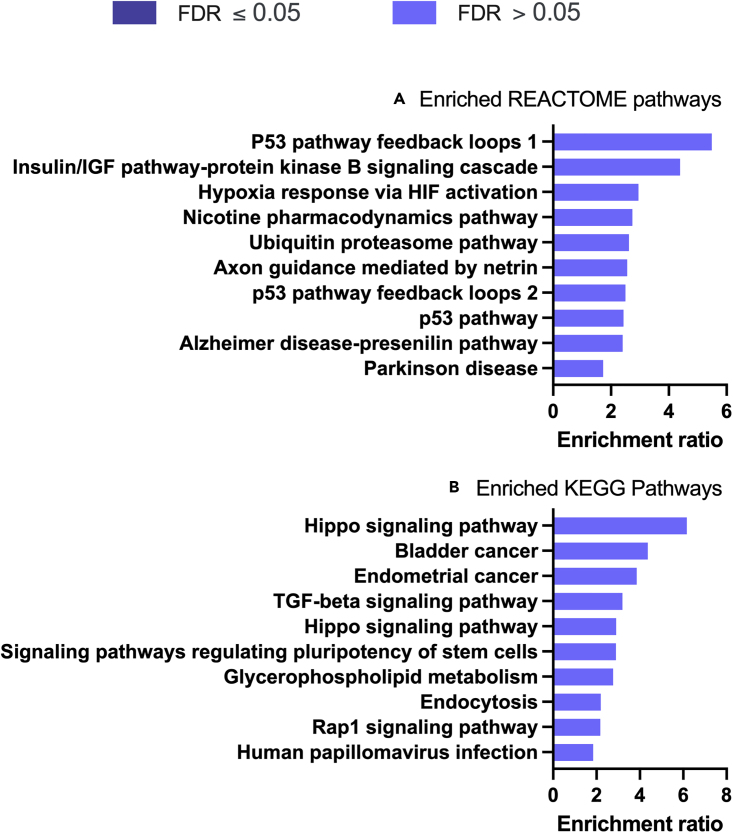
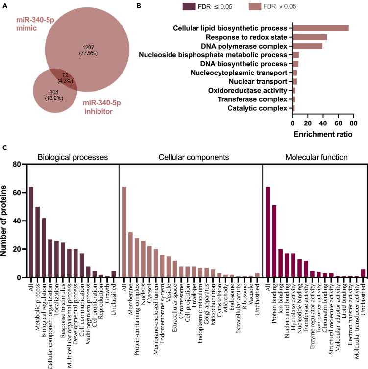
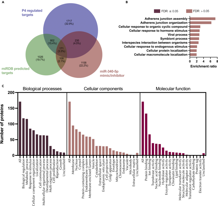
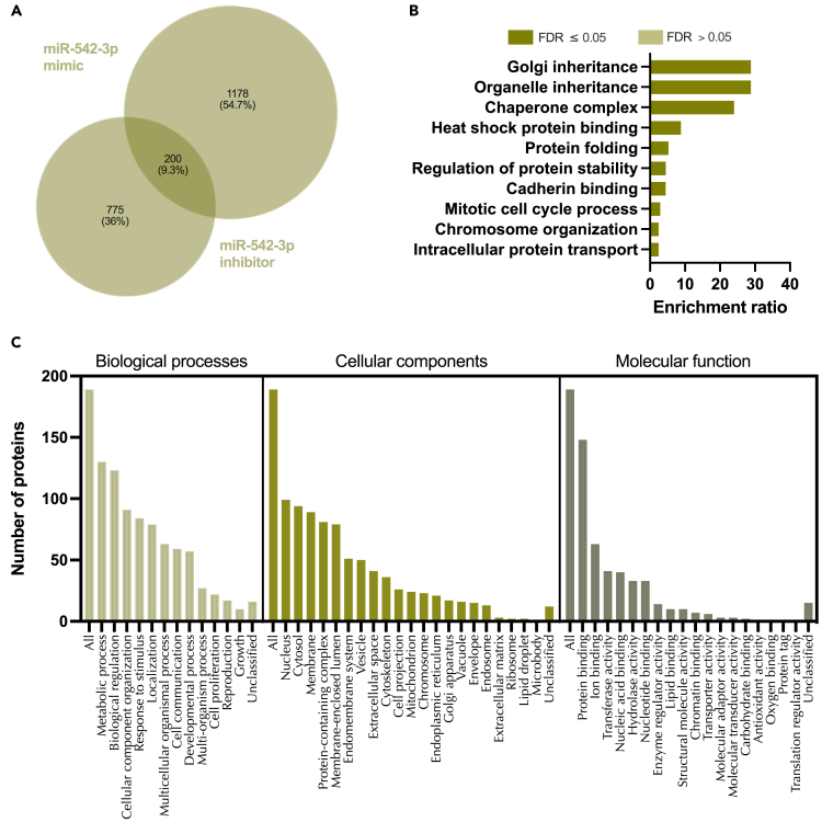
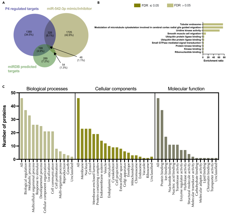
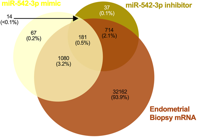
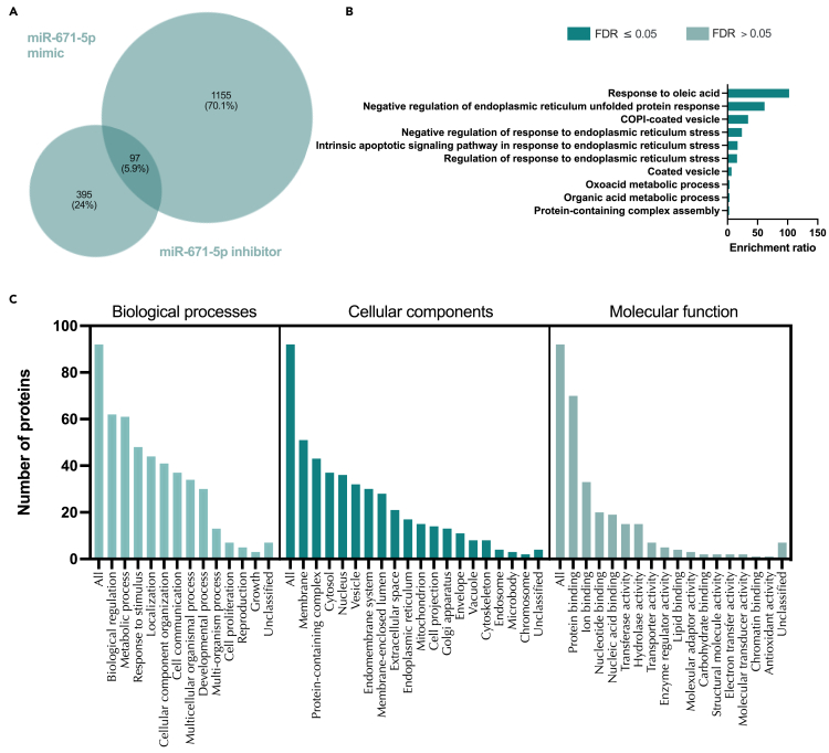
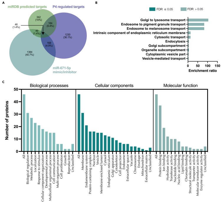
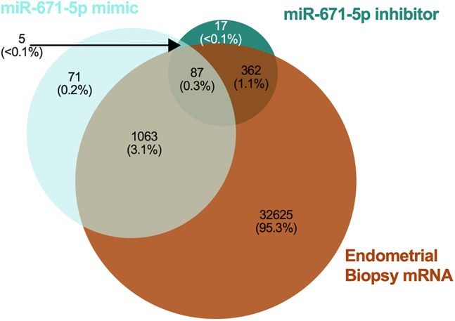
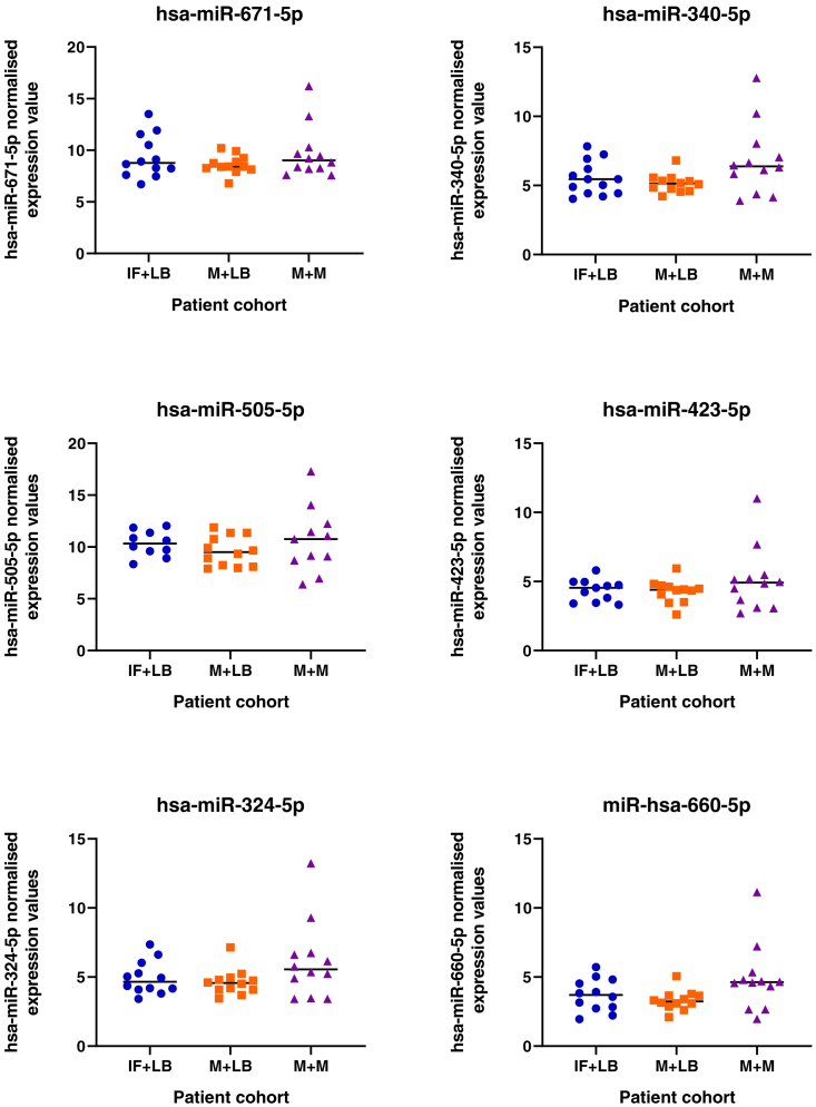
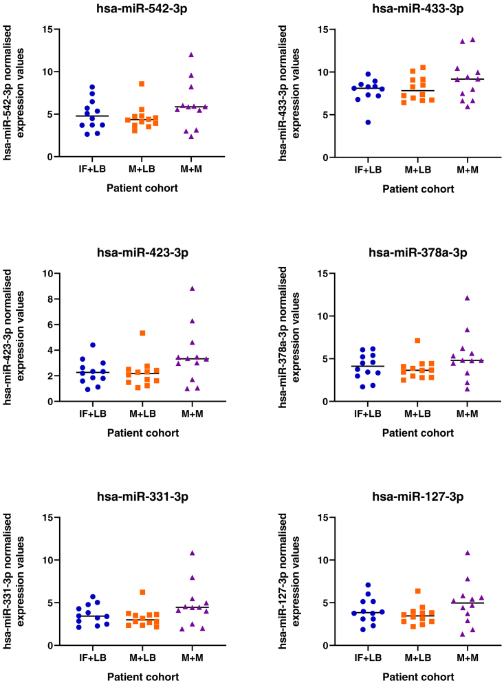
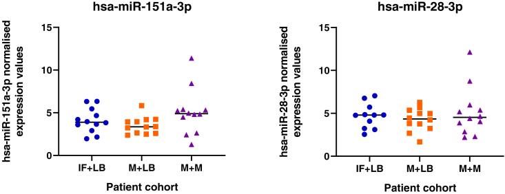
We tested the hypothesis that conserved placental mammal-specific microRNAs and their targets facilitate endometrial receptivity to implantation. Expression of miR-340-5p, -542-3p, and -671-5p was regulated by exposure of endometrial epithelial cells to progesterone (10 μg/ml) for 24 h coordinate with 1,713 of their predicted targets. Proteomic analysis of cells transfected with miRNA mimic/inhibitor (48 h: n = 3) revealed 1,745 proteins altered by miR-340-5p (mimic; 1,369, inhibitor; 376) of which 171 were predicted targets and P4-regulated. MiR-542-3p altered 2,353 (mimic; 1,378, inhibitor; 975) 100 which were mirDB predicted, including 46 P4-regulated. MiR-671-5p altered 1,744 proteins (mimic; 1,252, inhibitor; 492) 95 of which were predicted targets and 46 P4-regulated. All miRNAs were detected in luteal phase endometrial biopsies, irrespective of pregnancy outcomes. miR-340-5p expression increased in biopsies from individuals suffering previous and subsequent miscarriage compared to those with subsequent live birth. Dysfunction of these miRNAs and their targets contribute to endometrial-derived recurrent pregnancy loss.

Keywords: Biological sciences; Developmental biology; Embryology; Molecular biology.


Conflict of interest statement

The authors declare no competing interests.

Figures

None
Graphical abstract
Figure 1
Figure 1
Expression of selected miRNAs in human endometrial epithelial (Ishikawa) cells Cells were cultured for 24 h with: (1) Control (open circle), (2) Vehicle Control (10% EtOH: gray circle), (3) 0.1ug/ml P4 (light blue circle), (4) 1.0 μg/ml P4 (royal blue circle), or (5) 10 μg/ml P4 (dark blue circle: n = 3 biological replicates). Expression values of miRNAs were determined using LNA Sybr Green method and the relative expression values following normalization. Differences in expression were determined relative to vehicle control using an ANOVA and considered statistically different when p< 0.05. Differences between groups are denoted by difference in letters (a, b, c, d).
Figure 1
Figure 1
Expression of selected miRNAs in human endometrial epithelial (Ishikawa) cells Cells were cultured for 24 h with: (1) Control (open circle), (2) Vehicle Control (10% EtOH: gray circle), (3) 0.1ug/ml P4 (light blue circle), (4) 1.0 μg/ml P4 (royal blue circle), or (5) 10 μg/ml P4 (dark blue circle: n = 3 biological replicates). Expression values of miRNAs were determined using LNA Sybr Green method and the relative expression values following normalization. Differences in expression were determined relative to vehicle control using an ANOVA and considered statistically different when p< 0.05. Differences between groups are denoted by difference in letters (a, b, c, d).
Figure 2
Figure 2
Progesterone regulated microRNA targets (A)Venn diagram representing overlap in predicted mRNA targets of the three miRNAs altered following 24 h treatment of endometrial epithelial cells with P4 in vitro. These were associated with the Gene Ontology Biological Process of Reproductive Biology. (B) RT-qPCR analysis of predicted targets of all three P4-regulated miRNAs that are associated with the Gene Ontology Biological Process of Reproductive Function. Expression was determined in Ishikawa cells treated with control (open circles), vehicle control (gray circles), 0.1 μg/mL (pale red circle), 1.0 μg/mL (medium red circle), or 10.0 μg/mL (dark red circle) of P4 for 24 hin vitro (n = 3 independent cultures). Differences in expression compared were determined using ANOVA with differences depicted by different subscripts (a, b, c, d) when p < 0.05.
Figure 3
Figure 3
RNA-Seq analysis of Ishikawa cells treated with P4 in vitro (A) Principal component analysis depicting the overall transcriptional differences between cells via RNA sequencing of Ishikawa cells treated with (1) Control (orange circle), (2) Vehicle control (green circle), and (3) 10 μg/ml P4 (blue circle) for 24 h. Controls clearly separate on the left-hand side whereas P4 treatment clearly cluster together on the right-hand side of the graph (n = 3 biological replicates per group). (B) Bar chart of Biological Processes (red), Cellular Component (blue), and Molecular Function (green) associated with P4 modified transcripts.
Figure 4
Figure 4
Enriched Gene Ontology associated with P4 modified transcripts (A) Biological processes, (B) Cellular component, and (C) Molecular functions associated with P4 modified transcripts determined following RNA sequencing of human endometrial epithelial cells (Ishikawa cells) treated with 10 μg/ml P4 for 24 h. These are significantly enriched i.e. more transcripts associated with these ontologies than one would expect by chance (FDR <0.05).
Figure 5
Figure 5
Enriched pathways and networks associated with P4-modified transcripts (A) REACTOME pathways, (B) KEGG pathways, and (C) ontology network, associated with P4 modified transcripts determined following RNA sequencing of human endometrial epithelial cells (Ishikawa cells) treated with 10 μg/ml P4 for 24 h. These are significantly enriched i.e. more transcripts associated with these pathways than one would expect by chance (FDR <0.05).
Figure 6
Figure 6
Overlap of predicted miRNA targets and P4 altered transcripts Venn diagram analysis demonstrating the overlap in predicted targets (from miRBase) of the 3 miRNAs modified by treatment of Ishikawa cells with P4 for 24 hin vitro as well as those mRNAs that were identified as altered in the same cells following P4 treatment for 24 h compared to vehicle controls.
Figure 7
Figure 7
Enriched Gene Ontologies for P4-modified transcripts and miRNA targets (A) Biological processes, (B) Cellular component, and (C) Molecular functions associated with 473 P4 modified transcripts that are also predicted targets of the 3 miRNAs modified in human endometrial epithelial cells (Ishikawa cells) treated with 10 μg/ml P4 for 24 h. These are significantly enriched i.e. more transcripts associated with these ontologies than one would expect by chance (FDR <0.05).
Figure 8
Figure 8
Enriched pathways for P4-modified transcripts and miRNA targets (A) REACTOME pathways, and (B) KEGG pathways associated with 473 P4 modified transcripts that are also predicted targets of the 3 miRNAs modified in human endometrial epithelial cells (Ishikawa cells) treated with 10 μg/ml P4 for 24 h. These are significantly enriched, i.e., more transcripts associated with these ontologies than one would expect by chance when FDR <0.05.
Figure 9
Figure 9
Proteins altered in Ishikawa cells following treatment with miR-340-5p mimic and inhibitor (A) Venn diagram depicting total number of significantly differentially expressed proteins (p<0.05) following transfection of Ishikawa cells (n = 3 biological replicates) with miR-340-5p mimic (RHS) and inhibitor (LHS). (B) Enriched KEGG pathways associated with miR-340-5p mimic and inhibition regulated proteins. (C) WebGestalt overrepresentation analysis of biological process, cellular component and molecular function categories for identified significantly differentially expressed proteins (p<0.05) in response to miR-340-5p mimic and inhibition (total of 72).
Figure 10
Figure 10
Proteins altered in Ishikawa cells following treatment with miR-340-5p mimic and inhibitor compared to mirDB predicted targets and P4 regulated mRNAs (A) Venn diagram depicting total number of significantly differentially expressed proteins (p<0.05) following transfection of Ishikawa cells (n = 3 biological replicates) with miR-340-5p mimic or inhibitor vs non-targeting controls and mirDB predicted targets, or P4-regulated transcripts. (B) Enriched KEGG pathways associated with miR-340-5p mimic and inhibition regulated proteins, P4-regulated mRNAs and predicted target overlap. (C) WebGestalt overrepresentation analysis of biological process, cellular component and molecular function categories for identified significantly differentially expressed proteins (p<0.05) in response to miR-340-5p mimic and inhibition, predicted targets, and P4-regulated mRNAs overlap.
Figure 11
Figure 11
Comparison of miR-340-5p-regulated proteins to endometrial biopsy transcript expression Venn diagram showing overlap in proteins altered in abundance following transfection of Ishikawa cells (n = 3 biological replicates) with miR-340-5p mimic or inhibitor compared to RNA-Seq data from human endometrial biopsies (n = 36 biological replicates).
Figure 12
Figure 12
Proteins altered in Ishikawa cells following treatment with miR-542-3p mimic and inhibitor (A) Venn diagram depicting total number of significantly differentially expressed proteins (p<0.05) following transfection of Ishikawa cells (n = 3 biological replicates) with miR-542-3p mimic (RHS) and inhibitor (LHS). (B) Enriched KEGG pathways associated with miR-542-3p mimic and inhibition regulated proteins. (C) WebGestalt overrepresentation analysis of biological process, cellular component and molecular function categories for identified significantly differentially expressed proteins (p<0.05) in response to miR-542-3p mimic and inhibition (total of 200).
Figure 13
Figure 13
Proteins altered in Ishikawa cells following treatment with miR-542-3p mimic and inhibitor compared to mirDB predicted targets and P4 regulated mRNAs (A) Venn diagram depicting total number of significantly differentially expressed proteins (p<0.05) following transfection of Ishikawa cells (n = 3 biological replicates) with miR-542-3p mimic or inhibitor vs non-targeting controls and mirDB predicted targets, or P4-regulated transcripts. (B) Enriched KEGG pathways associated with miR-542-3p mimic and inhibition regulated proteins, P4-regulated mRNAs and predicted target overlap. (C) WebGestalt overrepresentation analysis of biological process, cellular component and molecular function categories for identified significantly differentially expressed proteins (p<0.05) in response to miR-542-3p mimic and inhibition, predicted targets, and P4-regulated mRNAs overlap.
Figure 14
Figure 14
Comparison of miR-542-3p-regulated proteins to endometrial biopsy transcript expression Venn diagram showing overlap in proteins altered in abundance following transfection of Ishikawa cells (n = 3 biological replicates) with miR-542-3p mimic or inhibitor compared to RNA-Seq data from human endometrial biopsies (n = 36 biological replicates).
Figure 15
Figure 15
Proteins altered in Ishikawa cells following treatment with miR-671-5p mimic and inhibitor (A) Venn diagram depicting total number of significantly differentially expressed proteins (p<0.05) following transfection of Ishikawa cells (n = 3 biological replicates) with miR-671-5p mimic (RHS) and inhibitor (LHS). (B) Enriched KEGG pathways associated with miR-671-5p mimic and inhibition regulated proteins. (C) WebGestalt overrepresentation analysis of biological process, cellular component and molecular function categories for identified significantly differentially expressed proteins (p<0.05) in response to miR-671-5p mimic and inhibition.
Figure 16
Figure 16
Proteins altered in Ishikawa cells following treatment with miR-671-5p mimic and inhibitor compared to mirDB predicted targets and P4 regulated mRNAs (A) Venn diagram depicting total number of significantly differentially expressed proteins (p<0.05) following transfection of Ishikawa cells (n = 3 biological replicates) with miR-671-5p mimic or inhibitor vs non-targeting controls and mirDB predicted targets, or P4-regulated transcripts. (B) Enriched KEGG pathways associated with miR-671-5p mimic and inhibition regulated proteins, P4-regulated mRNAs and predicted target overlap. (C) WebGestalt overrepresentation analysis of biological process, cellular component and molecular function categories for identified significantly differentially expressed proteins (p<0.05) in response to miR-671-5p mimic and inhibition, predicted targets, and P4-regulated mRNAs overlap.
Figure 17
Figure 17
Comparison of miR-671-5p-regulated proteins to endometrial biopsy transcript expression Venn diagram showing overlap in proteins altered in abundance following transfection of Ishikawa cells (n = 3 biological replicates) with miR-671-5p mimic or inhibitor compared to RNA-Seq data from human endometrial biopsies (n = 36 biological replicates).
Figure 18
Figure 18
Expression of miRNAs in endometrial biopsy samples Samples were collected from patients during the mid-luteal phase of the menstrual cycle (LH+6-LH+9), from patients (n = 12 per group) who have infertility and a subsequent live birth (IF + LB: blue dots), miscarriage and a subsequent live birth (M + LB: orange square), or miscarriage and a subsequent miscarriage (M + M: purple triangles). Following statistical analysis using an ANOVA, there was an overall effect on mir-340-5p expression (p <0.05) between groups.
Figure 18
Figure 18
Expression of miRNAs in endometrial biopsy samples Samples were collected from patients during the mid-luteal phase of the menstrual cycle (LH+6-LH+9), from patients (n = 12 per group) who have infertility and a subsequent live birth (IF + LB: blue dots), miscarriage and a subsequent live birth (M + LB: orange square), or miscarriage and a subsequent miscarriage (M + M: purple triangles). Following statistical analysis using an ANOVA, there was an overall effect on mir-340-5p expression (p <0.05) between groups.

Similar articles

Cited by

References

    1. Critchley H.O.D., Maybin J.A., Armstrong G.M., Williams A.R.W. Physiology of the endometrium and regulation of menstruation. Physiol. Rev. 2020;100:1149–1179. doi: 10.1152/physrev.00031.2019. - DOI - PubMed
    1. Macklon N.S., Brosens J.J. The human endometrium as a sensor of embryo quality. Biol. Reprod. 2014;91:98. - PubMed
    1. Weimar C.H.E., Kavelaars A., Brosens J.J., Gellersen B., de Vreeden-Elbertse J.M.T., Heijnen C.J., Macklon N.S. Endometrial stromal cells of women with recurrent miscarriage fail to discriminate between high-and low-quality human embryos. PLoS One. 2012;7:e41424. - PMC - PubMed
    1. Brosens J.J., Salker M.S., Teklenburg G., Nautiyal J., Salter S., Lucas E.S., Steel J.H., Christian M., Chan Y.-W., Boomsma C.M., et al. Uterine selection of human embryos at implantation. Sci. Rep. 2014;4:3894–3898. - PMC - PubMed
    1. Quenby S., Gallos I.D., Dhillon-Smith R.K., Podesek M., Stephenson M.D., Fisher J., Brosens J.J., Brewin J., Ramhorst R., Lucas E.S., et al. Miscarriage matters: the epidemiological, physical, psychological, and economic costs of early pregnancy loss. Lancet. 2021;397:1658–1667. - PubMed