

Deep learning-enabled fully automated pipeline system for segmentation and classification of single-mass breast lesions using contrast-enhanced mammography: a prospective, multicentre study
- PMID: 36969336
- PMCID: PMC10034267
- DOI: 10.1016/j.eclinm.2023.101913
Deep learning-enabled fully automated pipeline system for segmentation and classification of single-mass breast lesions using contrast-enhanced mammography: a prospective, multicentre study
Abstract
Background: Breast cancer is the leading cause of cancer-related deaths in women. However, accurate diagnosis of breast cancer using medical images heavily relies on the experience of radiologists. This study aimed to develop an artificial intelligence model that diagnosed single-mass breast lesions on contrast-enhanced mammography (CEM) for assisting the diagnostic workflow.
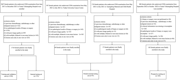
Methods: A total of 1912 women with single-mass breast lesions on CEM images before biopsy or surgery were included from June 2017 to October 2022 at three centres in China. Samples were divided into training and validation sets, internal testing set, pooled external testing set, and prospective testing set. A fully automated pipeline system (FAPS) using RefineNet and the Xception + Pyramid pooling module (PPM) was developed to perform the segmentation and classification of breast lesions. The performances of six radiologists and adjustments in Breast Imaging Reporting and Data System (BI-RADS) category 4 under the FAPS-assisted strategy were explored in pooled external and prospective testing sets. The segmentation performance was assessed using the Dice similarity coefficient (DSC), and the classification was assessed using heatmaps, area under the receiver operating characteristic curve (AUC), sensitivity, and specificity. The radiologists' reading time was recorded for comparison with the FAPS. This trial is registered with China Clinical Trial Registration Centre (ChiCTR2200063444).
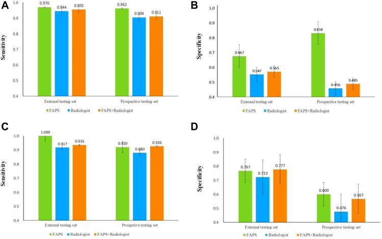
Findings: The FAPS-based segmentation task achieved DSCs of 0.888 ± 0.101, 0.820 ± 0.148 and 0.837 ± 0.132 in the internal, pooled external and prospective testing sets, respectively. For the classification task, the FAPS achieved AUCs of 0.947 (95% confidence interval [CI]: 0.916-0.978), 0.940 (95% [CI]: 0.894-0.987) and 0.891 (95% [CI]: 0.816-0.945). It outperformed radiologists in terms of classification efficiency based on single lesions (6 s vs 3 min). Moreover, the FAPS-assisted strategy improved the performance of radiologists. BI-RADS category 4 in 12.4% and 13.3% of patients was adjusted in two testing sets with the assistance of FAPS, which may play an important guiding role in the selection of clinical management strategies.
Interpretation: The FAPS based on CEM demonstrated the potential for the segmentation and classification of breast lesions, and had good generalisation ability and clinical applicability.
Funding: This study was supported by the Taishan Scholar Foundation of Shandong Province of China (tsqn202211378), National Natural Science Foundation of China (82001775), Natural Science Foundation of Shandong Province of China (ZR2021MH120), and Special Fund for Breast Disease Research of Shandong Medical Association (YXH2021ZX055).
Keywords: Breast lesions; Classification; Contrast-enhanced mammography; Deep learning; Full automated pipeline system; Segmentation.
© 2023 The Author(s).
Conflict of interest statement
HX received funding from 10.13039/501100007129Natural Science Foundation of Shandong Province of China (ZR2021MH120). NM received funding from Taishan Scholar Foundation of Shandong Province of China (tsqn202211378), 10.13039/501100001809National Natural Science Foundation of China (82001775), and Special Fund for Breast Disease Research of Shandong Medical Association (YXH2021ZX055). All other authors declare no competing interests.
Figures


References
-
- Sung H., Ferlay J., Siegel R.L., et al. Global cancer statistics 2020: GLOBOCAN estimates of incidence and mortality worldwide for 36 cancers in 185 countries. CA Cancer J Clin. 2021;71(3):209–249. - PubMed
-
- Siegel R.L., Miller K.D., Fuchs H.E., Jemal A. Cancer statistics, 2021. CA Cancer J Clin. 2021;71(1):7–33. - PubMed
-
- Pace L.E., Keating N.L. A systematic assessment of benefits and risks to guide breast cancer screening decisions. JAMA. 2014;311(13):1327–1335. - PubMed
-
- Kim H.-E., Kim H.H., Han B.-K., et al. Changes in cancer detection and false-positive recall in mammography using artificial intelligence: a retrospective, multireader study. Lancet Digit Health. 2020;2(3):e138–e148. - PubMed
LinkOut - more resources
Full Text Sources
