. 2023 May 1:382:133549.
doi: 10.1016/j.snb.2023.133549. Epub 2023 Feb 21.

Wireless in vivo Recording of Cortical Activity by an Ion-Sensitive Field Effect Transistor

Affiliations

Wireless in vivo Recording of Cortical Activity by an Ion-Sensitive Field Effect Transistor

Suyash Bhatt et al. Sens Actuators B Chem. .

Abstract

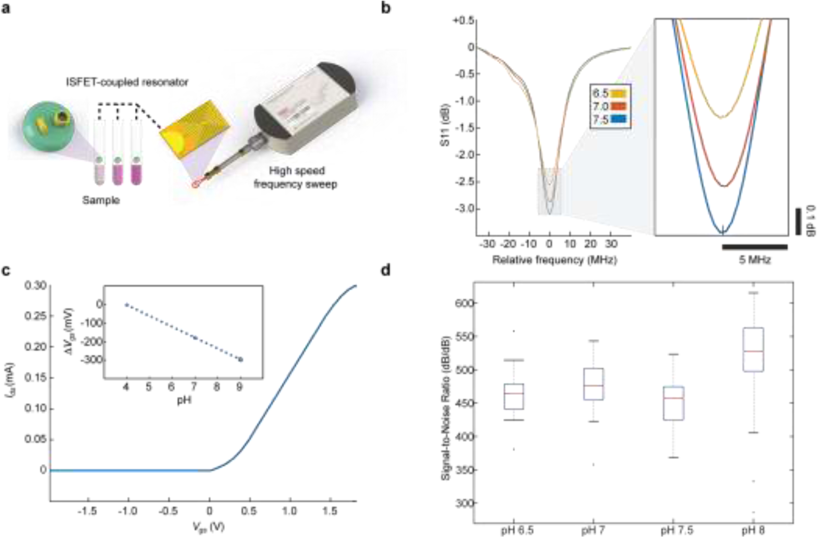
Wireless brain technologies are empowering basic neuroscience and clinical neurology by offering new platforms that minimize invasiveness and refine possibilities during electrophysiological recording and stimulation. Despite their advantages, most systems require on-board power supply and sizeable transmission circuitry, enforcing a lower bound for miniaturization. Designing new minimalistic architectures that can efficiently sense neurophysiological events will open the door to standalone microscale sensors and minimally invasive delivery of multiple sensors. Here we present a circuit for sensing ionic fluctuations in the brain by an ion-sensitive field effect transistor that detunes a single radiofrequency resonator in parallel. We establish sensitivity of the sensor by electromagnetic analysis and quantify response to ionic fluctuations in vitro. We validate this new architecture in vivo during hindpaw stimulation in rodents and verify correlation with local field potential recordings. This new approach can be implemented as an integrated circuit for wireless in situ recording of brain electrophysiology.

Keywords: Brain recording; ISFET; Ion-sensitive field effect transistor; Wireless.


Conflict of interest statement

COMPETING INTERESTS The authors declare no competing interests. Conflict of interest The authors declare that they have no known competing financial interests or personal relationships that could have appeared to influence the work reported in this paper.

Figures

Fig. 1
Fig. 1. Wireless ion sensitive field effect transistor (ISFET) for in vivo cortical recordings of ionic fluctuations.
a Source and drain of ISFET device are connected to top and bottom plates of capacitor, linking in parallel to circuit. b Q of resonator is dependent on ionic concentrations local to ISFET gate electrode. c Active site of ISFET is embedded through cranial window on surface of somatosensory cortex. d Ion fluctuations detected wirelessly represented in time domain by S11 minima between resonator and antenna over 60 s window.
Fig. 2
Fig. 2. Electromagnetic simulations of ISFET-coupled resonator response.
a Simulation arena of RLC resonator coupled to ISFET model. S-parameter frequency response is evaluated at a near field receiver. b E-field is maximized at resonance. c Changes in ionic concentrations at ISFET gate decrease impedance, Q, and e-field. d Left - representative ISFET model connected to resonator. Right - current field density at 0.5 V overdrive voltage (Vov). e Drain-source current (Ids) as a function of Vov. f Small signal transconductance (gm) as a function of Vov. g Frequency response modulation at physiological pH range. Inset - closeup surrounding resonance.
Fig. 3
Fig. 3. Benchtop assessment of wireless ISFET sensitivity.
a Experimental configuration: readouts of ISFET-coupled resonator immersed in different pH samples are received by near field antenna. A series of frequency response sweeps is acquired by high-speed vector network analyzer. b Examples of frequency response curves for physiological pH levels. c Current-voltage (IV) characteristic curve. Inset: Change in drain-source voltage (Vds) with pH levels. d Arithmetic mean of signal-to-noise ratio per pH level (red: outliers, included in mean, n = 10, all error bars denote s.e.m.).
Fig. 4
Fig. 4. Wireless ISFET somatosensory cortical recordings during hindpaw stimulation.
a Pre-stimulus readouts of spontaneous activity. b Readouts from S1HL somatosensory cortex during a 2 Hz electrical stimulus of contralateral hindpaw. c Representative maximum single pulse responses in wireless ISFET recording. d Peak differential LFP recordings in response to stimulation. e Heatmap depicting maximum peaks of LFP recordings in d. f Average amplitude of response to stimulation normalized to baseline. Asterix denotes t-test p-values < .05, error bars are standard errors, n = 5 for all conditions, error bars denote s.e.m.
Fig. 5
Fig. 5. A repertoire of positive and negative phase wireless ISFET responses correlating with delta LFP activity band.
a ISFET traces can be sorted by both duration and amplitude of response. b Majority of ISFET fluctuations are of duration < 500 ms, with positive sustained fluctuations displaying durations of up to 2.5 s. Negative phase responses are of duration < 100 ms. c Spectrograms of ISFET wireless response following stimulation onset reveal excitatory response centered around 0.1 – 5 Hz. Duration and intensity of response are inversely proportional to frequency of stimulation. ISFET activity at frequencies greater than the delta wave band was minimal. d Response normalized to baseline for both wireless ISFET and LFP electrode recordings show maximal excitatory response at 2Hz stimulation frequency, and a reduced response at frequencies > 5Hz in the delta wave band. Asterix denotes t-test p-values < .05, error bars are standard errors, n = 5 for all conditions, error bars denote s.e.m..

Update of

References

    1. Jun JJ, Steinmetz NA, Siegle JH, Denman DJ, Bauza M, Barbarits B, Lee AK, Anastassiou CA, Andrei A, Aydın Ç, Barbic M, Blanche TJ, Bonin V, Couto J, Dutta B, Gratiy SL, Gutnisky DA, Häusser M, Karsh B, Ledochowitsch P, Lopez CM, Mitelut C, Musa S, Okun M, Pachitariu M, Putzeys J, Rich PD, Rossant C, Sun W, Svoboda K, Carandini M, Harris KD, Koch C, O’Keefe J, Harris TD, Fully integrated silicon probes for high-density recording of neural activity, Nature. 551 (2017) 232–236. 10.1038/nature24636. - DOI - PMC - PubMed
    1. Frank JA, Antonini M-J, Anikeeva P, Next-generation interfaces for studying neural function, Nat Biotechnol. 37 (2019) 1013–1023. 10.1038/s41587-019-0198-8. - DOI - PMC - PubMed
    1. Nurmikko A, Challenges for Large-Scale Cortical Interfaces, Neuron. 108 (2020) 259–269. 10.1016/j.neuron.2020.10.015. - DOI - PubMed
    1. Steinmetz NA, Aydin C, Lebedeva A, Okun M, Pachitariu M, Bauza M, Beau M, Bhagat J, Böhm C, Broux M, Chen S, Colonell J, Gardner RJ, Karsh B, Kloosterman F, Kostadinov D, Mora-Lopez C, O’Callaghan J, Park J, Putzeys J, Sauerbrei B, van Daal RJJ, Vollan AZ, Wang S, Welkenhuysen M, Ye Z, Dudman JT, Dutta B, Hantman AW, Harris KD, Lee AK, Moser EI, O’Keefe J, Renart A, Svoboda K, Häusser M, Haesler S, Carandini M, Harris TD, Neuropixels 2.0: A miniaturized high-density probe for stable, long-term brain recordings, Science. 372 (2021) eabf4588. 10.1126/science.abf4588. - DOI - PMC - PubMed
    1. Borton DA, Yin M, Aceros J, Nurmikko A, An implantable wireless neural interface for recording cortical circuit dynamics in moving primates, J Neural Eng. 10 (2013) 026010. 10.1088/1741-2560/10/2/026010. - DOI - PMC - PubMed

LinkOut - more resources