Review
. 2023 Mar 9:15:1129036.
doi: 10.3389/fnsyn.2023.1129036. eCollection 2023.

Alzheimer's disease as a synaptopathy: Evidence for dysfunction of synapses during disease progression

Affiliations
Review

Alzheimer's disease as a synaptopathy: Evidence for dysfunction of synapses during disease progression

Soraya Meftah et al. Front Synaptic Neurosci. .

Abstract

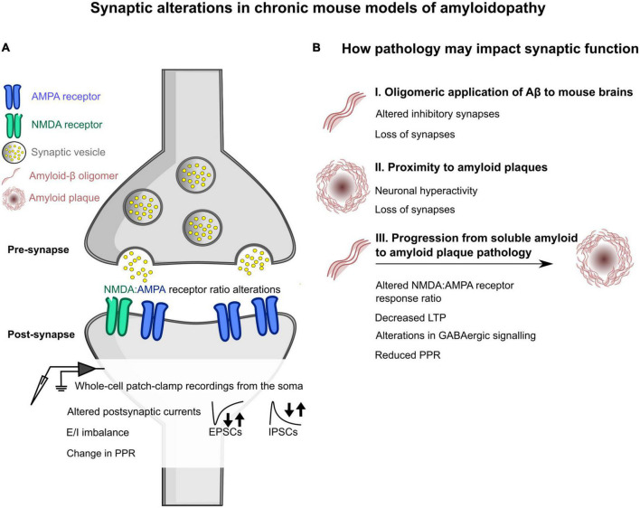
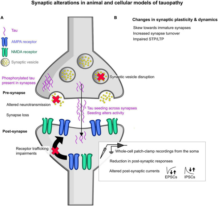
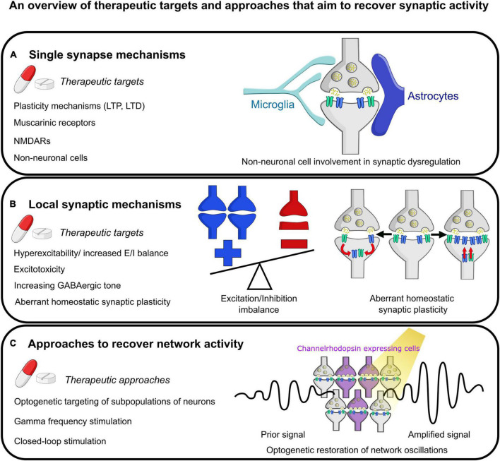
The synapse has consistently been considered a vulnerable and critical target within Alzheimer's disease, and synapse loss is, to date, one of the main biological correlates of cognitive decline within Alzheimer's disease. This occurs prior to neuronal loss with ample evidence that synaptic dysfunction precedes this, in support of the idea that synaptic failure is a crucial stage within disease pathogenesis. The two main pathological hallmarks of Alzheimer's disease, abnormal aggregates of amyloid or tau proteins, have had demonstrable effects on synaptic physiology in animal and cellular models of Alzheimer's disease. There is also growing evidence that these two proteins may have a synergistic effect on neurophysiological dysfunction. Here, we review some of the main findings of synaptic alterations in Alzheimer's disease, and what we know from Alzheimer's disease animal and cellular models. First, we briefly summarize some of the human evidence to suggest that synapses are altered, including how this relates to network activity. Subsequently, animal and cellular models of Alzheimer's disease are considered, highlighting mouse models of amyloid and tau pathology and the role these proteins may play in synaptic dysfunction, either in isolation or examining how the two pathologies may interact in dysfunction. This specifically focuses on neurophysiological function and dysfunction observed within these animal models, typically measured using electrophysiology or calcium imaging. Following synaptic dysfunction and loss, it would be impossible to imagine that this would not alter oscillatory activity within the brain. Therefore, this review also discusses how this may underpin some of the aberrant oscillatory patterns seen in animal models of Alzheimer's disease and human patients. Finally, an overview of some key directions and considerations in the field of synaptic dysfunction in Alzheimer's disease is covered. This includes current therapeutics that are targeted specifically at synaptic dysfunction, but also methods that modulate activity to rescue aberrant oscillatory patterns. Other important future avenues of note in this field include the role of non-neuronal cell types such as astrocytes and microglia, and mechanisms of dysfunction independent of amyloid and tau in Alzheimer's disease. The synapse will certainly continue to be an important target within Alzheimer's disease for the foreseeable future.

Keywords: Alzheimer’s disease; amyloid; dysfunction; electrophysiology; oscillations; synapse; synaptic transmission; tau.


Conflict of interest statement

The authors declare that the research was conducted in the absence of any commercial or financial relationships that could be construed as a potential conflict of interest.

Figures

FIGURE 1
FIGURE 1
Schematic overview of some of the changes induced by amyloidopathy at the synapse, as seen in chronic mouse models of amyloidopathy. (A) General changes in synaptic function at the pre- and post- synapse in chronic mouse models of amyloidopathy. Alterations at the post-synapse in amyloidopathy include a shift in the ratio between AMPA and NMDA receptors expressed at the post-synapse, measured using whole-cell patch-clamp electrophysiological recordings. Whole-cell patch-clamp recordings also showed either increases or decreases in the frequency of spontaneous and miniature excitatory post-synaptic currents (EPSCs) and inhibitory post-synaptic currents, denoted by arrows. The ratio of these two currents (E/I balance) was also skewed in some mouse models. There were also changes in the paired-pulse ratio (PPR), with either increased or decreased ratios measured. (B) An outline of how amyloid pathology can impact synaptic function. (B I) Changes observed when oligomeric amyloid-β is applied to the brains of mice by microinjection or by topical application to brain slices. This includes altered inhibitory synapses and an overall loss of synapses. (B II) How distance to amyloid plaques may alter synapses within chronic mouse models of amyloidopathy. Two phenomena that have been observed are increases in neuronal hyperexcitability when in close proximity to plaques, and an overall loss of synapses as well. (B III) How different phenotypes emerge with the progression of pathology in chronic mouse models of amyloidopathy. These experiments investigated changes with age in mouse models that will progressively develop amyloid plaque pathology. Some alterations that were observed at older ages, where plaque pathology is present, included a change in NMDA:AMPA receptor ratio as determined by whole-cell patch-clamp electrophysiology, decreased long term plasticity (LTP), changes in GABAergic signaling, and reduced PPR.
FIGURE 2
FIGURE 2
Schematic overview of some of the changes induced by tauopathy in animal and cellular models at the synapse. (A) General changes in synaptic function at the pre- and post- synapse in animal models of tauopathy. In the pre-synapse, synaptic vesicle disruption has been observed, as denoted by a red cross over the synaptic vesicle. Altered neurotransmission has also been seen, denoted by a red cross over the neurotransmitters shown as yellow dots. The detection of mislocalized phosphorylated tau has been seen at both the pre- and post-synapse, highlighted as purple waves in each synaptic space. Tau is also thought to seed across synapses by multiple hypothesized mechanisms, with this seeding shown to alter activity in the brain. At the post-synapse, glutamatergic receptor trafficking is impaired showing a loss of receptors at the post-synaptic domain. In addition, measures using whole-cell patch-clamp electrophysiological recordings revealed reductions in post-synaptic responses to stimulation and increased or decreased frequency of spontaneous and miniature excitatory post-synaptic currents (EPSCs) and inhibitory post-synaptic currents (IPSCs), denoted by the arrows. (B) Changes that have been observed in synapse plasticity and dynamics include a skew in synapse shape toward “immature” synapses, increased synapse turnover, and impairments in short-term and long-term plasticity (STP and LTP, respectively).
FIGURE 3
FIGURE 3
Summary of some future therapeutic targets aimed at restoring normal synaptic physiology. (A) Mechanisms targeting individual synapses. This includes targeting short- and long- term plasticity (STP and LTP, respectively) at the synapse, modulation of muscarinic receptor and NMDA receptor activity, and targeting aberrant activity of non-neuronal cells such as microglia and astrocytes on synapses. (B) Therapeutic mechanisms at the local synaptic level. This includes reducing hyperexcitability or restoring E/I imbalance, preventing excitotoxicity, increasing GABAergic tone, and evaluating the effect of aberrant homeostatic synaptic plasticity. The example images show an imbalance of excitation and inhibition with more excitation as has been described in the literature. Next shows one mechanism of homeostatic synaptic plasticity, synaptic scaling, with either abnormal increases or decreases in synaptic strength by altering the number of receptors present at the post-synaptic density. (C) Therapeutic mechanisms that could be utilized to rescue network activity including optogenetic modulation of the activity of subpopulations of neurons (as shown in the pictogram), stimulation of neurons at gamma-band frequency, and more sophisticated closed-loop network modulation to attempt to recover activity to normal physiological set points following deviations.

References

    1. Abuhassan K., Coyle D., Belatreche A., Maguire L. (2014). Compensating for synaptic loss in Alzheimer’s disease. J. Comput. Neurosci. 36 19–37. 10.1007/s10827-013-0462-8 - DOI - PubMed
    1. Adaikkan C., Tsai L. (2020). Gamma entrainment: Impact on neurocircuits, glia, and therapeutic opportunities. Trends Neurosci. 43 24–41. 10.1016/j.tins.2019.11.001 - DOI - PubMed
    1. Adams I. (1987). Plasticity of the synaptic contact zone following loss of synapses in the cerebral cortex of aging humans. Brain Res. 424 343–351. 10.1016/0006-8993(87)91479-x - DOI - PubMed
    1. Adler G., Brassen S., Jajcevic A. (2003). EEG coherence in Alzheimer’s dementia. J. Neural Transm. 110 1051–1058. 10.1007/s00702-003-0024-8 - DOI - PubMed
    1. Ahmed Z., Cooper J., Murray T., Garn K., McNaughton E., Clarke H., et al. (2014). A novel in vivo model of tau propagation with rapid and progressive neurofibrillary tangle pathology: The pattern of spread is determined by connectivity, not proximity. Acta Neuropathol. 127 667–683. 10.1007/s00401-014-1254-6 - DOI - PMC - PubMed