Review
. 2023 Mar;13(3):998-1013.
doi: 10.1016/j.apsb.2022.09.020. Epub 2022 Oct 3.

Roles of focal adhesion proteins in skeleton and diseases

Affiliations
Review

Roles of focal adhesion proteins in skeleton and diseases

Sheng Chen et al. Acta Pharm Sin B. 2023 Mar.

Abstract

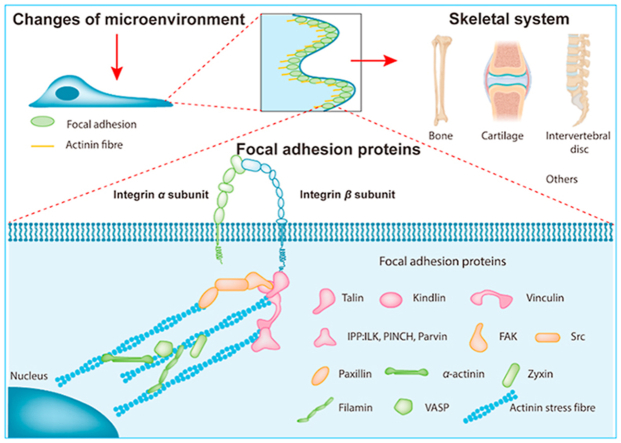
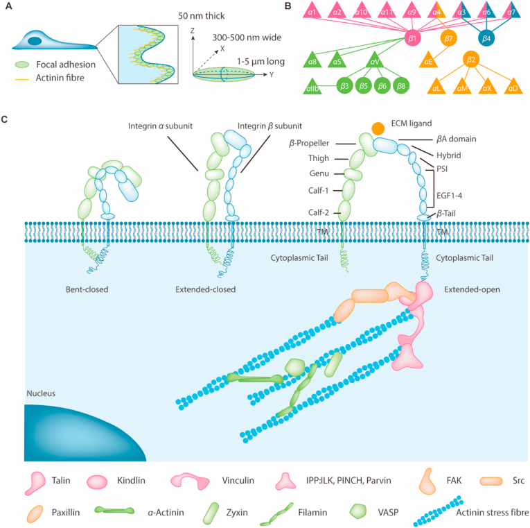
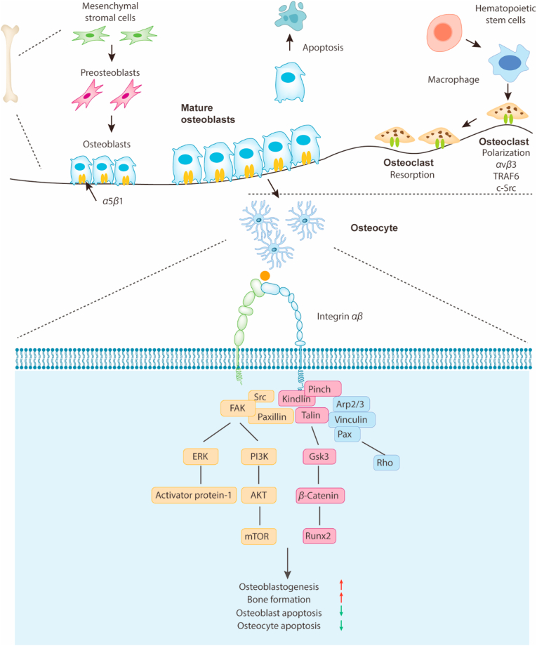
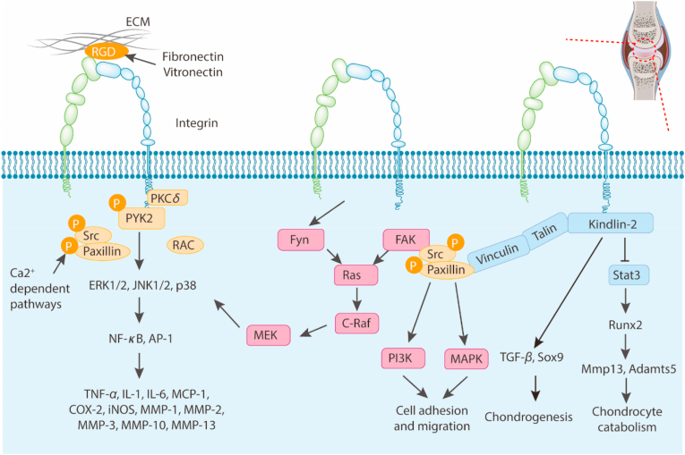
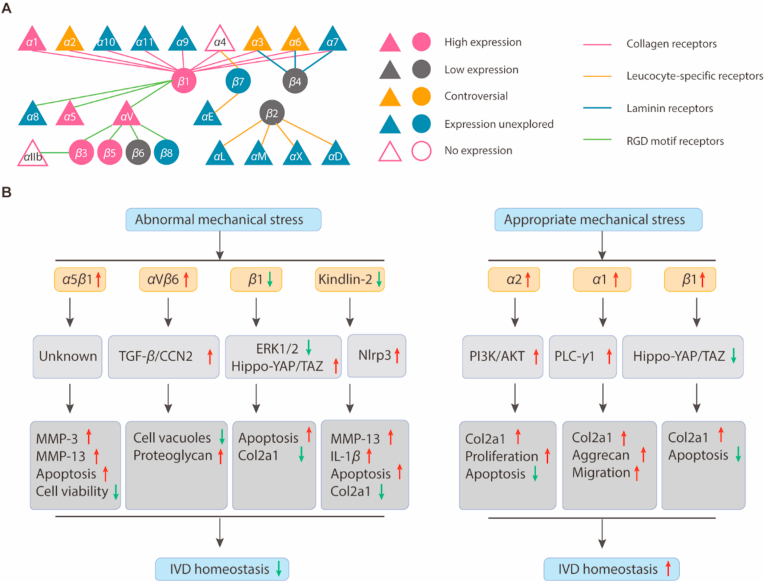
The skeletal system, which contains bones, joints, tendons, ligaments and other elements, plays a wide variety of roles in body shaping, support and movement, protection of internal organs, production of blood cells and regulation of calcium and phosphate metabolism. The prevalence of skeletal diseases and disorders, such as osteoporosis and bone fracture, osteoarthritis, rheumatoid arthritis, and intervertebral disc degeneration, increases with age, causing pain and loss of mobility and creating a huge social and economic burden globally. Focal adhesions (FAs) are macromolecular assemblies that are composed of the extracellular matrix (ECM), integrins, intracellular cytoskeleton and other proteins, including kindlin, talin, vinculin, paxillin, pinch, Src, focal adhesion kinase (FAK) and integrin-linked protein kinase (ILK) and other proteins. FA acts as a mechanical linkage connecting the ECM and cytoskeleton and plays a key role in mediating cell-environment communications and modulates important processes, such as cell attachment, spreading, migration, differentiation and mechanotransduction, in different cells in skeletal system by impacting distinct outside-in and inside-out signaling pathways. This review aims to integrate the up-to-date knowledge of the roles of FA proteins in the health and disease of skeletal system and focuses on the specific molecular mechanisms and underlying therapeutic targets for skeletal diseases.

Keywords: Cartilage; Focal adhesion; Integrin; Intervertebral disc; Mechanotransduction; Signal transduction; Skeletal diseases; Skeleton.


Figures

Image 1
Graphical abstract
Figure 1
Figure 1
FA proteins and signaling. (A) Schematic illustration of FA in a cell. FA is an elongated and flat structure (about 50 nm thick, 300–500 nm wide, 1–5 μm long). (B) In vertebrates, there are 18 α and 8 β subunits, which are combined into 24 different integrins and can be classified into four subgroups: collagen (pink), RGD motif (green), leucocyte-specific (brown) and laminin receptors (blue). (C) After conformation transition from the bent-closed/extended-closed form to the extended-open form, integrin is activated, followed in order by recruitments of FA proteins at three layers: (1) the structural layer, containing talin, kindlin, vinculin, integrin-linked kinase (ILK), particularly interesting new cysteine-histidine-rich protein (pinch) and parvin; (2) the signaling layer, containing focal adhesion kinase (FAK), Src and paxillin; (3) the actin cross-linking layer, containing α-actinin, vasodilator-stimulated phosphoprotein (VASP), filamin and zyxin.
Figure 2
Figure 2
Bone homeostasis and FA signaling. Bone homeostasis is achieved through the activity of cells of the osteoblastic and osteoclast lineages. Osteoblastogenesis is controlled by integrins. In bone, bone formation is characterized by the differentiation of mesenchymal stromal cells into pre-osteoblasts, which become osteoblasts and synthesize the bone matrix, and ultimately die by apoptosis. Some integrins, such as α5β1, have important roles in cell attachment to the extracellular matrix and in osteoblast function and survival. Osteoclasts originate from the hematopoietic lineage and differentiate into mature osteoclasts through the interaction of macrophage-colony stimulating factor (M-CSF) and RANKL. The αvβ3 integrin is involved in osteoclast polarization and resorption function. As FA is essential for skeletal development and homeostasis. In the FA complex, kindlin-2, talin, and other structural proteins are directly linked to the cytoplasmic tail of the integrin β subunit, which is further linked to pinch, paxillin, vinculin, and Arp 2/3 proteins, which in turn activate downstream signaling pathways, such as Erk, PI3K, Gsk3 and Rho pathways and, ultimately, enhances osteogenesis and bone remodeling and inhibits cell apoptosis.
Figure 3
Figure 3
FA signaling in cartilage. Integrins are transmembrane proteins connecting chemical and mechanical stimuli between the outside ECMs and inside of chondrocytes. The integrin-mediated signaling pathway is critical for osteoarthritis (OA) development. Peptides and fibronectin fragments containing the RGD cell binding sequence can bind to integrins and other the FA proteins which promotes expression of a number of pro-inflammatory mediators and matrix degrading enzymes. The combined effects of increased ligands as a result of cartilage damage may enable cells within OA joints to elicit pathogenic responses, and thereby drive the progression of OA. In response to integrin activation, PYK2 and/or FAK are recruited to a signaling complex that consists of Src, paxillin, vinculin, talin and kindlin, which result in activation of PI3K and/or MAPK that may lead to cell adhesion and migration. Further, development of OA may result from chronic signaling of inflammatory, catabolic, and hypertrophic molecules as well as other integrin effectors as a result of the increased levels of both ligand and receptor components following joint injury. Furthermore, kindlin-2 is related to mitochondrial oxidative stress and chondrocyte catabolism. In addition, kindlin-2 serves as a major controlling factor in the regulation of the chondrogenesis during prenatal and postnatal skeletal development by regulating TGF-β signalling and Sox 9 expression.
Figure 4
Figure 4
FA proteins in intervertebral disc (IVD). (A) In human IVDs, integrin subunits can be divided into five groups: high expression group (red), low expression group (grey), controversial expression group (brown), no expression group (white) and expression unexplored group (blue). (B) In IVDs, abnormal mechanical stress can excessively activate (α5β1, αvβ6) or decrease (β1, kindlin-2) FA proteins, and regulate TGF-β/CCN2, ERK1/2, Hippo–YAP/TAZ and Nlrp3 pathways to disturb IVD homeostasis; while appropriate mechanical stress can increase FA proteins (α1, α2, β1), and regulate PI3K/AKT, PLC-γ1 and Hippo–YAP/TAZ pathways to maintain IVD homeostasis.

Similar articles

Cited by

References

    1. Balestri W., Morris R.H., Hunt J.A., Reinwald Y. Current advances on the regeneration of musculoskeletal interfaces. Tissue Eng B Rev. 2021;27:548–571. - PubMed
    1. Baldwin M.J., Cribbs A.P., Guilak F., Snelling S.J.B. Mapping the musculoskeletal system one cell at a time. Nat Rev Rheumatol. 2021;17:247–248. - PubMed
    1. Roberts S., Colombier P., Sowman A., Mennan C., Rolfing J.H., Guicheux J., et al. Ageing in the musculoskeletal system. Acta Orthop. 2016;87:15–25. - PMC - PubMed
    1. Schumacher S., Vazquez Nunez R., Biertumpfel C., Mizuno N. Bottom-up reconstitution of focal adhesion complexes. FEBS J. 2022;289:3360–3373. - PMC - PubMed
    1. Revach O.Y., Grosheva I., Geiger B. Biomechanical regulation of focal adhesion and invadopodia formation. J Cell Sci. 2020;133:jcs244848. - PubMed