. 2023 Mar 9:11:1134086.
doi: 10.3389/fbioe.2023.1134086. eCollection 2023.

The lens capsule significantly affects the viscoelastic properties of the lens as quantified by optical coherence elastography

Affiliations

The lens capsule significantly affects the viscoelastic properties of the lens as quantified by optical coherence elastography

Taye Mekonnen et al. Front Bioeng Biotechnol. .

Abstract

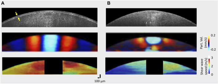
The crystalline lens is a transparent, biconvex structure that has its curvature and refractive power modulated to focus light onto the retina. This intrinsic morphological adjustment of the lens to fulfill changing visual demands is achieved by the coordinated interaction between the lens and its suspension system, which includes the lens capsule. Thus, characterizing the influence of the lens capsule on the whole lens's biomechanical properties is important for understanding the physiological process of accommodation and early diagnosis and treatment of lenticular diseases. In this study, we assessed the viscoelastic properties of the lens using phase-sensitive optical coherence elastography (PhS-OCE) coupled with acoustic radiation force (ARF) excitation. The elastic wave propagation induced by ARF excitation, which was focused on the surface of the lens, was tracked with phase-sensitive optical coherence tomography. Experiments were conducted on eight freshly excised porcine lenses before and after the capsular bag was dissected away. Results showed that the group velocity of the surface elastic wave, V , in the lens with the capsule intact ( V = 2.55 ± 0.23 m / s ) was significantly higher (p < 0.001) than after the capsule was removed ( V = 1.19 ± 0.25 m / s ). Similarly, the viscoelastic assessment using a model that utilizes the dispersion of a surface wave showed that both Young's modulus, E, and shear viscosity coefficient, η, of the encapsulated lens ( E = 8.14 ± 1.10 k P a , η = 0.89 ± 0.093 P a s ) were significantly higher than that of the decapsulated lens ( E = 3.10 ± 0.43 k P a , η = 0.28 ± 0.021 P a s ). These findings, together with the geometrical change upon removal of the capsule, indicate that the capsule plays a critical role in determining the viscoelastic properties of the crystalline lens.

Keywords: acoustic radiation force; lens biomechanical properties; lens capsule; optical coherence elastography (OCE); viscoelastic properties.


Conflict of interest statement

MS and KVL have a financial interest in ElastEye LLC., which is not directly related to this work. The remaining authors declare that the research was conducted in the absence of any commercial or financial relationships that could be construed as a potential conflict of interest.

Figures

FIGURE 1
FIGURE 1
(A) Schematic of the experimental setup comprising a phase sensitive spectral domain OCT system for imaging and acoustic radiation force system for excitation. C: collimator, CCD: charge-coupled device (line scan camera), FG: function generator, G: grating, GS: 2D galvo scanner, L: lens, M: reference mirror, P: pinhole, PC: polarization controller, SL: scan lens, SLD: superluminescent diode, US: ultrasound transducer. (B) Ultrasound transducer producing acoustic radiation force excitation at the apex of the lens. Propagating elastic waves were imaged and analyzed along the orthogonal x and y axes.
FIGURE 2
FIGURE 2
OCT structural images and elastic wave propagation characteristics of a typical porcine lens (A) with the capsule intact and (B) after the capsule was removed. Top: OCT structural images acquired before (left) and after removal (right) of the capsule; middle: wave propagation snapshots indicating instantaneous particle velocity in an encapsulated (left) and decapsulated (right) lens; bottom: spatial shear wave speed map in the encapsulated (left) and decapsulated (right) lens. The capsule layer is indicated by the yellow arrow in the top left structural image. For all samples, the excitation location was roughly at the apex.
FIGURE 3
FIGURE 3
A box-whisker plot and measured data set distribution of the elastic group wave speed in the porcine lens before and after removal of capsule for N = 8 porcine lenses. The horizontal bar in the diamond box corresponds to the median of the data.
FIGURE 4
FIGURE 4
(A) Typical elastic wave dispersion curves in the porcine lens before and after capsule removal fitted with Rayleigh wave dispersion equation. The shaded region indicates the error band (standard deviation) of the OCE results. (B) Estimated Young’s modulus and viscosity coefficient using Rayleigh wave dispersion equation for encapsulated and decapsulated lens. N = 8 porcine eye lenses.
FIGURE 5
FIGURE 5
Elastic wave attenuation characteristics in a typical porcine eye lens. (A) The energy distribution map of laterally propagating elastic wave in encapsulated (top) and decapsulated (bottom) lens. (B) The normalized spatial distribution curve of the peak intensity and the exponential fitting before and after capsule removal. The peak intensity occurred at a center frequency of ∼706 Hz. At the center frequency, the mean attenuation coefficient is higher in the decapsulated lens ( α=1.21mm1) than in the encapsulated lens ( α=0.46mm1 ).
FIGURE 6
FIGURE 6
(A) A representative 3D OCT image of a dissected porcine lens. The anterior region is facing up (i.e., along the z-axis). Lens sagittal thickness represents the maximum thickness along the anterior-posterior direction (along the z-axis), while the equatorial diameter stands for the maximum thickness along the x-axis or the y-axis. (B) Porcine lens geometric features characterized by the sagittal thickness and equatorial diameter with and without the capsule. For each sample, multiple cross-sectional images (n = 4) extracted from 3D OCT images were used to quantify the mean geometric features. N = 3 for each group of lenses.

References

    1. Ambekar Y. S., Singh M., Zhang J., Nair A., Aglyamov S. R., Scarcelli G., et al. (2020). Multimodal quantitative optical elastography of the crystalline lens with optical coherence elastography and Brillouin microscopy. Biomed. Opt. Express 11 (4), 2041–2051. 10.1364/BOE.387361 - DOI - PMC - PubMed
    1. Avetisov K. S., Bakhchieva N. A., Avetisov S. E., Novikov I. A., Frolova A. A., Akovantseva A. A., et al. (2020). Biomechanical properties of the lens capsule: A review. J. Mech. Behav. Biomed. Mater 103, 103600. 10.1016/j.jmbbm.2019.103600 - DOI - PubMed
    1. Avetisov K. S., Bakhchieva N. A., Avetisov S. E., Novikov I. A., Shitikova A. V., Frolova A. A., et al. (2021). Assessment of age-related changes in lens capsule biomechanics using atomic force microscopy. Vestn. Oftalmol. 137 (1), 28–34. 10.17116/oftalma202113701128 - DOI - PubMed
    1. Beers A. P., Van Der Heijde G. L. (1994). In vivo determination of the biomechanical properties of the component elements of the accommodation mechanism. Vis. Res. 34 (21), 2897–2905. 10.1016/0042-6989(94)90058-2 - DOI - PubMed
    1. Berggren C. C., Ameku K. A., Pedrigi R. M. (2021). Altered stress field of the human lens capsule after cataract surgery. J. Biomech. 115, 110127. 10.1016/j.jbiomech.2020.110127 - DOI - PMC - PubMed

LinkOut - more resources