. 2023 Jul 4;32(14):2269-2291.
doi: 10.1093/hmg/ddad047.

The HSPB1-p62/SQSTM1 functional complex regulates the unconventional secretion and transcellular spreading of the HD-associated mutant huntingtin protein

Affiliations

The HSPB1-p62/SQSTM1 functional complex regulates the unconventional secretion and transcellular spreading of the HD-associated mutant huntingtin protein

R Bonavita et al. Hum Mol Genet. .

Abstract

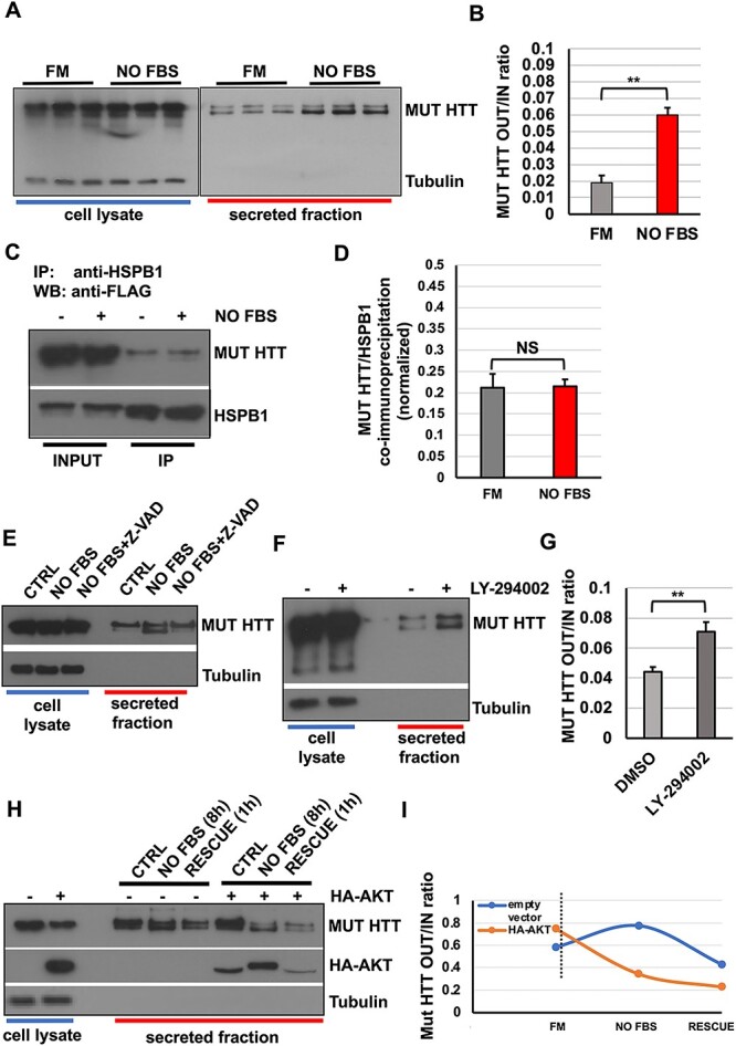
Conformational diseases, such as Alzheimer, Parkinson and Huntington diseases, are part of a common class of neurological disorders characterized by the aggregation and progressive accumulation of proteins bearing aberrant conformations. Huntington disease (HD) has autosomal dominant inheritance and is caused by mutations leading to an abnormal expansion in the polyglutamine (polyQ) tract of the huntingtin (HTT) protein, leading to the formation of HTT inclusion bodies in neurons of affected patients. Interestingly, recent experimental evidence is challenging the conventional view by which the disease pathogenesis is solely a consequence of the intracellular accumulation of mutant protein aggregates. These studies reveal that transcellular transfer of mutated huntingtin protein is able to seed oligomers involving even the wild-type (WT) forms of the protein. To date, there is still no successful strategy to treat HD. Here, we describe a novel functional role for the HSPB1-p62/SQSTM1 complex, which acts as a cargo loading platform, allowing the unconventional secretion of mutant HTT by extracellular vesicles. HSPB1 interacts preferentially with polyQ-expanded HTT compared with the WT protein and affects its aggregation. Furthermore, HSPB1 levels correlate with the rate of mutant HTT secretion, which is controlled by the activity of the PI3K/AKT/mTOR signalling pathway. Finally, we show that these HTT-containing vesicular structures are biologically active and able to be internalized by recipient cells, therefore providing an additional mechanism to explain the prion-like spreading properties of mutant HTT. These findings might also have implications for the turn-over of other disease-associated, aggregation-prone proteins.


Figures

Graphical Abstract
Graphical Abstract
Figure 1
Figure 1
Screening for the identification of HSPB family members unconventionally secreted. (AB) HeLa cells were transiently transfected with constructs encoding the different V5-tagged HSPBs. Twenty four hours after transfection, cells were cultured in DMEM with 1% FBS for 8 h to allow protein secretion. Both secreted fraction and cell lysate were collected and processed by western blot analysis. All the proteins were present in the cell lysate, but only HSPB1, HSPB4 and HSPB5 were detected in the secreted fraction. (CD) EVs were isolated from conditioned media of HeLa cells transiently transfected with the GFP-CD63 over-expression construct by differential ultracentrifugation. P100 fractions were processed and ultrathin sections (60 nm) prepared as detailed in Materials and Methods section and the grid observed by transmission electron microscopy (C, scale bar: 50 nm). For the immunogold staining of sections, an anti-CD63 specific antibody and 10-nm gold particles were used (D, scale bar: 100 nm). (EF) HeLa cells were kept in culture in 10-cm dishes for 48 h. In the last 8 h, a secretion assay was performed. Secreted fractions were subjected to ultracentrifugation at 100000xg for 2 h. Pellet (P100) fractions were subjected to nanoparticle tracking analysis. The plots in (E) and (F) report the concentration and particles size average distribution of EVs. (GH) HeLa cells were placed in DMEM 1% FBS for 8 h in the presence or not of 5-mg brefeldin A, then secreted fraction and cell lysate were collected. Secreted fractions were centrifuged at 100 000×g for 2 h. Pellet (P100) was resuspended in lysis buffer and processed for western blot analysis using an HSPB1 antibody. HSPB1 is present in the P100 fraction and in the cell lysate but not in the S100, also upon brefeldin A treatment, confirming that HSPB1 is secreted in EVs unconventionally. (IK) Western blot analysis of ATG16L1 WT and KO cells, which are unable to conjugate LC3 and form autophagosomes (E). Parental and ATG16L1 KO HeLa cells were transfected with V5-HSPB1 over-expression construct. After 24 h, cells were placed in DMEM 1% FBS for 8 h, then secreted fraction and cell lysate were collected. Secreted fraction was centrifuged at 100 000 g for 2 h. Pellet (P100) fractions were lysed and processed for western blot analysis using a HSPB1 antibody. HSPB1 is present in the P100 fraction and in the cell lysate but not in the S100, also in ATG16L1 KO Hela cells, suggesting that HSPB1 secretion is autophagy independent. The graphs in (B), (D) and (G) report the quantitative analysis of protein secretion relative to loading control from at least three independent experiments. The y-axis values are shown as the OUT/IN relative ratio and the error bars denote standard deviations. The P-values for the densitometric analyses were determined by Student’s t-test using STATVIEW v4.53 (Abacus Concepts) (n = 3, NS: non-significant).
Figure 2
Figure 2
HSPB1 interacts with p62/SQSTM1 and supports synergistically its unconventional secretion. (A) HeLa cells were transiently transfected with V5-HSPB1-V5 and HA-p62/SQSTM1. Twenty four hours after transfection, cell lysates were prepared and immunoprecipitation was performed using either anti-V5 or anti-HA antibody. HSPB1 and p62/SQSTM1 interaction was detected in both orientations. In addition, an unconjugated control IgG antibody was used to verify the specificity of the interaction (lane 4 of the top and bottom panels). (BC) HeLa cells were transiently transfected with GFP-HSPB1 and HA-p62/SQSTM1. After 24 h, cells were semi-permeabilized by digitonin treatment, fixed, subjected to immunofluorescence and analysed by confocal microscopy. Magnifications (10×) of the merged channels are shown in the insets. The co-localization analysis was performed using an ImageJ plug-in (JaCoP). The graph shows Mander’s coefficient (fraction of HSPB1-positive structures overlapping with p62-positive structures and reverse, grey bars). Scale bars: 10 μm. (D) HeLa cells were placed in DMEM 1% FBS for 8 h, then secreted fraction and cell lysate were collected. Secreted fraction was centrifuged at 100 000 g for 2 h. Pellet (P100) was resuspended in lysis buffer and processed for western blot analysis using a p62/SQSTM1 antibody. P62/SQSTM1 is present in the P100 fraction and in the cell lysate but not in the S100. (EF) HeLa cells were transfected with HSPB1-V5, P62-HA or both. After 24 h, the cells were placed in 1% FBS for 8 h to allow protein secretion. Both cell lysate and secreted fraction were collected and analysed. p62/SQSTM1 secretion is increased by HSPB1 over-expression as shown also in the quantification (F). (GH) HeLa cells were co-transfected with V5-HSPB1 and HA-p62/SQSTM1. Twenty four hours after transfection, cells were washed three times and placed in serum-free DMEM (NO FBS) for the indicated times (from 0 to 24 h). Secreted fractions were collected from 0.5 up to 24 h. Both HSPB1 and p62/SQSTM1 secretion increases over time, but the amount of p62/SQSTM1 is decreased after 24 h. (IJ) Schematic diagram depicting the experimental protocol deployed to assess the dynamic variations of HSPB1 and p62/SQSTM1 secretion upon serum deprivation. HeLa cells were serum starved or left in complete medium. Then cells were placed in 1% FBS or NO FBS for 8 h to allow protein secretion and 1 h before collection 1% FBS was added to the rescue condition. Cell lysates were collected and analysed using the indicated antibodies. AKT phosphorylation is reduced by serum deprivation and is increased again after re-addition of FBS. (KL) HeLa cells were transiently transfected with V5-HSPB1 and HA-p62/SQSTM1. After 24 h, cells were serum starved or left in complete medium. Then, cells were placed in 1% FBS for 8 h to allow protein secretion, and 1 h before collection, FBS was added back to the rescue condition. p62/SQSTM1 secretion is increased after serum starvation and the secretion is reversible. The graphs in (F), (H) and (L) report the quantitative analysis of HSPB1 and p62/SQSTM1 protein secretion relative to loading control from at least three independent experiments. The y-axis values are shown as the OUT/IN relative ratio and the error bars denote standard deviations. The P-values for the densitometric analyses were determined by factorial ANOVA or Student’s t-test using STATVIEW v4.53 (Abacus Concepts) (n = 3, **P < 0.001; NS: non-significant).
Figure 3
Figure 3
Effect of HSPB1 mutants on the unconventional secretion of p62/SQSTM1. (AB) HeLa cells were transiently transfected with 3A-HSPB1 and 3D-HSPB1 phosphorylation mutants. After 24 h, cells were placed in DMEM 1% FBS for 8 h and secreted fractions were collected, pre-cleared and centrifuged at 100 000×g for 2 h. Pellet (P100) fractions were lysed and processed for western blot analysis using an anti-FLAG antibody. The graph in (B) reports the quantitative analysis of 3A-HSPB1 and 3D-HSPB1 protein levels contained in the P100 fractions, relative to the inputs, from at least three independent experiments. (CD) HeLa cells were transiently transfected with HA-p62/SQSTM1 and either 3A-HSPB1 or 3D-HSPB1 expression constructs. After 24 h, cell extracts were collected and 3A-HSPB1, 3D-HDSPB1 (E) or p62/SQSTM1 (F) was immunoprecipitated using the indicated antibodies. Co-immunoprecipitation was then evaluated with specific antibodies, as indicated. 3A-HSPB1 and 3D-HSPB1 are both able to co-immunoprecipitate with p62/SQSTM1, but the phospho-mimetic mutant (3D) shows a higher affinity for p62/SQSTM1 compared with the 3A. (EF) HeLa cells were co-transfected with p62-HA and either 3A-HSPB1 or 3D-HSPB1. Twenty four hours after transfection, cells were serum starved overnight or left in complete medium. Then, cells were washed and placed in 1% FBS or serum-free DMEM (NO FBS) to allow protein secretion, and 1 h before collection, FBS was added to the rescue condition. Both cell lysates and secreted fractions were collected. 3D-HSPB1 strongly increases p62/SQSTM1 secretion both in steady state and in serum starvation. The graph in (F) and the inset report the quantitative analysis and the best-fit functional curve of HSPB1 and p62/SQSTM1 protein secretion, respectively, relative to loading control from three independent experiments. The y-axis values in the graph are shown as the OUT/IN relative ratio and the error bars denote standard deviations. The P-values for the densitometric analyses were determined by factorial ANOVA or Student’s t-test using STATVIEW v4.53 (Abacus Concepts) (n = 3, *P < 0.05; **P < 0.01; NS: non-significant).
Figure 4
Figure 4
The HSPB1 over-expression induces the clearance and secretion of mutant huntingtin. (A) Schematic representation of HTT WT and mutant (MUT) huntingtin constructs. (B) HeLa cells were transiently transfected with V5-HSPB1 and WT or mut HTT. Cells were placed in 1% FBS for 8 h and cell lysate and secreted fractions were analysed with the indicated antibodies. (CE) HeLa cells were transfected with mutant HTT with or without V5-HSPB1. Cells were placed in 1% FBS for 8 h and secreted fractions were collected and processed as previously described. Cell lysates were subjected to solubility fractionation, as detailed in Supplementary Material, Figure S5(B) and analysed by western blot. HSPB1 over-expression reduces both the soluble and insoluble fractions and increases HTT mutant secretion. The graph in (D) and (E) report the quantitative analysis of mut HTT protein levels and secretion relative to loading control from three independent experiments. The y-axis values are shown as the OUT/IN relative ratio and the error bars denote standard deviations. (FG) HeLa cells were transiently transfected with the GFP-HTT Q74 over-expression construct with or without V5-HSPB1 for 24 h. Cells were placed in 1% FBS for 8 h. Cell lysate and secreted fraction were collected and analysed by western blot. The graph in (G) reports the quantitative analysis of GFP-HTT Q74 protein secretion relative to loading control. The y-axis values are shown as the OUT/IN relative ratio and the error bars denote standard deviations. (HI) HeLa cells were transfected with V5-HSPB1 and WT or mutant huntingtin. Cell lysates were collected and HSPB1 was immunoprecipitated using an anti-V5 antibody. Both WT and mut HTT co-immunoprecipitate with HSPB1. The graph in (I) reports the quantitative analysis of WT and mut HTT co-immunoprecipitated by HSPB1. The y-axis values are shown as the mean values with standard deviations deriving from three independent experiments. The P-values for the densitometric analyses were determined by factorial ANOVA using STATVIEW v4.53 (Abacus Concepts) (n = 3, *P < 0.05; **P < 0.01; NS: non-significant).
Figure 5
Figure 5
Serum starvation acts upstream of HSPB1-p62/SQSTM1 and triggers the secretion of mutant huntingtin. (AB) HeLa cells were transiently transfected with mutant HTT. After 24 h, cells were serum starved or left in complete medium. Secreted fractions were collected and analysed by western blot. Serum starvation increases mut HTT secretion. (CD) HeLa cells were co-transfected with V5-HSPB1 and mut HTT. After 24 h, cells were either kept in full medium or serum starved overnight. Cell lysates were collected and HSPB1 was immunoprecipitated using an anti-V5 antibody. The graph in (D) reports the quantitative analysis of mut HTT co-immunoprecipitated by HSPB1. Serum starvation does not affect the HSPB1/mut HTT interaction. (E) HeLa cells were transiently transfected with mut HTT for 24 h. Cells were left in complete medium or serum starved overnight in the presence or absence of 10-mm Z-VAD, and then secretion assays were performed in the same conditions. Secreted fractions were collected and analysed by western blot. Z-VAD treatment inhibit the proteolytic cleavage of mut HTT, without interfering with its secretion. (FG) HeLa cells were transiently transfected with mut HTT. After 24 h, cells were treated or not with 10 μm LY-294002 overnight. Then cells were placed in 1% FBS (replenishing the LY-294002) for 8 h and both cell lysates and secreted fraction were collected and analysed. LY-294002 is able to mimic serum starvation and increases mut HTT secretion. (HI) HeLa cells were transiently transfected with mut HTT with or without HA-AKT. Cells were kept in complete medium or serum starved overnight. Then, cells were placed in 1% FBS to allow protein secretion and 1 h before collection FBS was added to the rescue condition. The over-expression of AKT reduces mut HTT secretion both at steady state and in response to serum starvation/rescue conditions. The P-values for the densitometric analyses were determined by factorial ANOVA or Student’s t-test using STATVIEW v4.53 (Abacus Concepts) (n = 3, **P < 0.01; NS: non-significant).
Figure 6
Figure 6
The formation of the HSPB1-p62/SQSTM1-MUT HTT ternary complex is instrumental for the unconventional secretion of mutant huntingtin. (A) HeLa cells were transfected with mut HTT, HA-p62/SQSTM1 with or without V5-HSPB1. Twenty four hours after transfection, cells were placed in DMEM 1% FBS and cell lysate and secreted fraction were collected. Mutant HTT was immunoprecipitated using anti-FLAG antibody and Co-IP assessed with the indicated antibodies. Mutant HTT is able to interact with HSPB1 both intracellularly and in the secreted fractions, whereas p62/SQSTM1 interacts with mut HTT only intracellularly. (B) HeLa cells were transiently transfected with mut HTT and p62/SQSTM1. Twenty four hours after transfection, cells were kept in full medium or serum starved overnight, cell lysates were prepared and mut HTT immunoprecipitated with an anti-FLAG antibody. Serum starvation does reduce the interaction between mut HTT and p62/SQSTM1. (CD) HeLa cells were silenced for scramble CTRL, HSPB1 or p62/SQSTM1 with 100-nm siRNA mix. HSPB1 and p62/SQSTM1 depletion were confirmed using specific antibodies. After 72 h, cells were re-transfected with the same mix, supplemented with 2 μg of FLAG-tagged mut HTT over-expression construct. After 48 h, cell lysates were prepared and mut HTT was immunoprecipitated using an anti-FLAG antibody and co-immunoprecipitation analysis performed with the indicated antibodies. (EF) HeLa cells were silenced for Ctrl, HSPB1 or p62/SQSTM1 and transfected with mutant HTT as detailed in (C–D). Cells were semi-permeabilized with digitonin immediately before fixation. Indirect immunofluorescence was performed and analysed by confocal microscopy. Co-localization analysis was performed by using ImageJ software. Thirty cells per experimental condition were analysed and mean values with standard deviations are reported in the graph. Scale bar: 10 μm. (G) Schematic representation of the hierarchy dictating the assembly and function of the HSPB1-p62-mut HTT ternary complex. (HI) HeLa cells were silenced for scramble control, HSPB1 or p62/SQSTM1 with 100-nm siRNA and transfected with 2 μg of FLAG mut HTT, as detailed above. Cell lysate and secreted fraction were collected and analysed by western blot. The P-values for the co-localization and densitometric analyses were determined by Student’s t-test using STATVIEW v4.53 (Abacus Concepts) (n = 3, **P < 0.01; ***P < 0.001).
Figure 7
Figure 7
The spreading of mutant huntingtin is facilitated by HSPB1 over-expression and is regulated in trans by the PI3K/AKT signalling axis. (AD) Feeder cells were prepared by transiently transfecting HeLa cells with FLAG-tagged mut HTT, with or without V5-HSPB1. Twenty four hours after transfection, cells were placed in DMEM 1% FBS for 12 h to allow protein secretion. Cell lysate and secreted fractions were collected and analysed (A). In addition, secreted fractions were used as conditioned medium for assessing the EV-mediated spreading of secreted proteins on recipient cells, as detailed in Supplementary Material, Figure S7. HSPB1 over-expression increases the uptake of mut HTT in recipient cells (B). The graph in (C) reports the relative levels of HSPB1, mut HTT alone or in the presence of HSPB1, internalized over time by recipient cells, whereas in (D), the cumulative value (area under curve, AUC) of the total uptake, for each experimental condition, is reported. In particular, for the quantification of the internalized mut HTT, it was taken in consideration and factored in the increased amount of protein present in the conditioned medium derived from HSPB1 over-expressing cells (fold increase: 2.267 ± 0.017) compared with empty vector transfected cells (A). The integral amount of the total mut HTT uptake (AUC, area under the curve) reported in (D) represents a normalized value (fold increase: 2.851 ± 0.5627) compared with empty vector transfected cells. The same approach has been applied to determine the AUC reported in (H) and in the Supplementary Figure S11(C). Histograms indicate mean values with standard deviations from three independent experiments. (EH) Feeder cells were prepared by transiently co-transfecting HeLa cells with FLAG-tagged mut HTT and V5-HSPB1. Cell lysates and secreted fractions were collected and conditioned medium, prepared as already detailed in (A–B), was used for the spreading experiment on recipient cells. In order to infer about the potential effect in trans of the PI3K/AKT signalling pathway, recipient cells were kept at steady state (Ctrl), transfected with HA-AKT or pre-treated with 10 μM LY-294002, 24 h before receiving the conditioned medium obtained from feeder cells for the indicated time points (0.5 to 8 h). The graph in (G) reports the relative levels of mut HTT internalized over time by recipient cells, whereas in (H) the cumulative value (area under curve, AUC) of the total uptake, for each experimental condition, is reported. Histograms indicate mean values with standard deviations from three independent experiments. The P-values for the densitometric analyses were determined by factorial ANOVA using STATVIEW v4.53 (Abacus Concepts) (n = 3, *P < 0.05; **P < 0.01).
Figure 8
Figure 8
Proposed model for the HSPB1/p62-mediated regulation of the unconventional secretion and transcellular spreading of mutant huntingtin. In response to both physiological and pathological conditions, such as variation of nutrient availability or the accumulation of aggregation-prone proteins, cells can activate a number of pathways, including the PI3K/AKT signalling axis, autophagy and unconventional secretion, aimed at counteracting cellular stress (1). The HSPB1/p62 functional complex can act as cargo selection platform, allowing the loading of cellular components, such as mutant HTT and other aggregation-prone proteins, destined to secretion (2). The activity of this complex can allow the generation and the regulated secretion of diverse extracellular vesicles (i.e. EVs and exosomes), which can display a different content and composition (3). These vesicular transport carriers are biologically active and capable of being taken up by recipient cells, contributing to the prion-like transcellular spreading of aggregation-prone proteins. Such a phenomenon can represent an additional pathophysiological mechanism underlying the onset of neurodegenerative diseases, such as Huntington disease (HD) (4).

References

    1. Camussi, G., Deregibus, M.C., Bruno, S., Grange, C., Fonsato, V. and Tetta, C. (2011) Exosome/microvesicle-mediated epigenetic reprogramming of cells. Am. J. Cancer Res., 1, 98–110. - PMC - PubMed
    1. Del Conde, I., Shrimpton, C.N., Thiagarajan, P. and Lopez, J.A. (2005) Tissue-factor-bearing microvesicles arise from lipid rafts and fuse with activated platelets to initiate coagulation. Blood, 106, 1604–1611. - PubMed
    1. Gatti, S., Bruno, S., Deregibus, M.C., Sordi, A., Cantaluppi, V., Tetta, C. and Camussi, G. (2011) Microvesicles derived from human adult mesenchymal stem cells protect against ischaemia-reperfusion-induced acute and chronic kidney injury. Nephrol. Dial. Transplant., 26, 1474–1483. - PubMed
    1. Lai, R.C., Arslan, F., Lee, M.M., Sze, N.S., Choo, A., Chen, T.S., Salto-Tellez, M., Timmers, L., Lee, C.N., El Oakley, R.M. et al. (2010) Exosome secreted by MSC reduces myocardial ischemia/reperfusion injury. Stem Cell Res., 4, 214–222. - PubMed
    1. Segura, E., Nicco, C., Bèrangère, L., Vèron, P., Raposo, G., Batteaux, F., Amigorena, S. and Théry, C. (2005) ICAM-1 on exosomes from mature dendritic cells is critical for efficient naive T-cell priming. Blood, 106, 216–223. - PubMed

Publication types

MeSH terms