. 2023 Apr 25;14(2):e0321822.
doi: 10.1128/mbio.03218-22. Epub 2023 Mar 27.

Scaffold-Scaffold Interaction Facilitates Cell Polarity Development in Caulobacter crescentus

Affiliations

Scaffold-Scaffold Interaction Facilitates Cell Polarity Development in Caulobacter crescentus

Ning Lu et al. mBio. .

Abstract

Cell polarity development is the prerequisite for cell differentiation and generating biodiversity. In the model bacterium Caulobacter crescentus, the polarization of the scaffold protein PopZ during the predivisional cell stage plays a central role in asymmetric cell division. However, our understanding of the spatiotemporal regulation of PopZ localization remains incomplete. In the current study, a direct interaction between PopZ and the new pole scaffold PodJ is revealed, which plays a primary role in triggering the new pole accumulation of PopZ. The coiled-coil 4-6 domain in PodJ is responsible for interacting with PopZ in vitro and promoting PopZ transition from monopolar to bipolar in vivo. Elimination of the PodJ-PopZ interaction impairs the PopZ-mediated chromosome segregation by affecting both the positioning and partitioning of the ParB-parS centromere. Further analyses of PodJ and PopZ from other bacterial species indicate this scaffold-scaffold interaction may represent a widespread strategy for spatiotemporal regulation of cell polarity in bacteria. IMPORTANCE Caulobacter crescentus is a well-established bacterial model to study asymmetric cell division for decades. During cell development, the polarization of scaffold protein PopZ from monopolar to bipolar plays a central role in C. crescentus asymmetric cell division. Nevertheless, the spatiotemporal regulation of PopZ has remained unclear. Here, we demonstrate that the new pole scaffold PodJ functions as a regulator in triggering PopZ bipolarization. The primary regulatory role of PodJ was demonstrated in parallel by comparing it with other known PopZ regulators, such as ZitP and TipN. Physical interaction between PopZ and PodJ ensures the timely accumulation of PopZ at the new cell pole and the inheritance of the polarity axis. Disruption of the PodJ-PopZ interaction impaired PopZ-mediated chromosome segregation and may lead to a decoupling of DNA replication from cell division during the cell cycle. Together, the scaffold-scaffold interaction may provide an underlying infrastructure for cell polarity development and asymmetric cell division.

Keywords: Caulobacter crescentus; PodJ; PopZ; asymmetric cell division; cell polarity; chromosome segregation; scaffold proteins.


Conflict of interest statement

The authors declare no conflict of interest.

Figures

FIG 1
FIG 1
PopZ polarizes at the new cell pole through de novo protein synthesis. (A) Schematics of cell polarity development in C. crescentus. PopZ organizes distinct signaling proteins at opposite cell poles during the cell cycle. As the new pole assembles, PopZ protein accumulates gradually upon initiation of replication, which is associated with the formation of the new pole signaling hub. The polarization of PopZ results in asymmetric cell division that generates daughter cells with distinct cell fates. (B) Construction of the tandem fluorescent-tagged PopZ. PopZ fused to two tandem fluorescent proteins: one matures rapidly (sfGFP, t50 = 19 min at 32°C), and one matures slowly (mCherry, t50 = 45 min at 32°C). The presence of high sfGFP fluorescence and weak mCherry fluorescence represents newly synthesized protein (32). (C) PopZ accumulates at the new pole through de novo synthesis. The tandem mCherry-sfGFP-PopZ is expressed under the native popZ promoter in the C. crescentus NA1000 genome. Time-lapse microscopy was used to detect the fluorescent signal of PopZ in cells after synchronization at 28°C on the PYE agarose pad containing kanamycin. Newly synthesized PopZ appears green, followed by a transition to “white” as the “magenta” mCherry matures, which is indicated with white arrows. One representative cell is shown here (see more cells in Fig. S1B). (D) The constitutively expressed RpoC (32) displays a constant sfGFP-to-mCherry ratio during the cell cycle. (E) Quantification of the average sfGFP-to-mCherry fluorescence ratios for PopZ during the cell cycle. The fluorescent signals of mCherry-sfGFP-PopZ at both cell poles were measured. The background-subtracted sfGFP and mCherry intensities were counted using Fiji/ImageJ (49). The sfGFP-to-mCherry ratio was calculated for each cell and the average sfGFP-to-mCherry ratios were measured for each time point. Each experiment was performed in four biological replicates. At least 50 cells (n) in total were calculated in each sample. ***, P < 0.01; *, P < 0.05; ns, P ≥ 0.05 by Welch’s unpaired t test. All scale bars = 1 μm.
FIG 2
FIG 2
PodJ promotes the new pole accumulation of PopZ in C. crescentus. (A) Deletion of podJ results in decreased bipolar accumulation of mCherry-PopZ. In the predivisional cell stage after synchronization, the subcellular localization of mCherry-PopZ was monitored in JP468 (NA1000, PpopZ-mcherry-popZ, and PparB-cfp-parB) (23), LN101 (NA1000 ΔpodJ, PpopZ-mcherry-popZ, and PparB-cfp-parB), and LN102 (NA1000 ΔpodJ, PpodJ-podJ, PpopZ-mcherry-popZ, and PparB-cfp-parB) strains. A sole copy of popZ in these strains was expressed under the control of the native promoter. Quantification of the cells with different mCherry-PopZ localization patterns is shown on the right. The observation of the stalk at the cell pole is considered the old cell pole. White arrows indicate the new cell poles. (B) Time-lapse microscopy indicates that ΔpodJ causes a significant delay in mCherry-PopZ accumulation at the new pole. Cells of JP468, ΔpodJ, ΔzitP, and ΔtipN were synchronized and transferred onto an M2G agarose pad before observation. The mCherry-PopZ signal was visualized at 15 min intervals at 30°C. The white arrow indicates the time points when the signal of mCherry-PopZ was first detected. The localization patterns of mCherry-PopZ over time are shown as schematics on the right. (C) Quantitative analyses suggest that PodJ plays a primary role in triggering bipolar PopZ. Cells were synchronized and transferred to a liquid M2G medium for continuous culture before observation. At least 50 cells (n) were calculated in each sample. (D) Quantitative analyses of bipolar mCherry-PopZ cells using cell length as the marker of cell cycle progression. All cells from all time points were sorted by cell length, and the fraction of cells with bipolar mCherry-PopZ is shown for each bin of 0.4 μm. (E) Disruption of the PodJ-PopZ interaction results in a failure to inherit PopZ in ~80% of swarmer cells. The schematic of mCherry-PopZ localization in ΔpodJ is shown on the right. Each experiment was performed in three biological replicates. Measurements are shown as mean ± standard deviations. All scale bars = 1 μm.
FIG 3
FIG 3
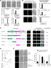
PopZ binds directly to the coiled-coil 4-6 region (CC4-6) of PodJ. (A) Heterologous expression of mCherry-PopZ alone exhibits a monopolar localization pattern, while expression of YFP-PodJ alone exhibits a bipolar localization pattern in E. coli. YFP-ZitP and YFP-TipN exhibit diffuse and bipolar localization patterns, respectively. The quantification of cells with different localization patterns is shown at the bottom. (B) PopZ interacts with PodJ and ZitP in E. coli. The mCherry-PopZ protein was coexpressed with YFP-PodJ, YFP-ZitP, or YFP-TipN. Quantitative analyses revealed that both YFP-PodJ and YFP-ZitP trigger the bipolar accumulation of mCherry-PopZ in E. coli, whereas YFP-TipN does not. (C) CC4-6 is shown as the interaction region between PodJ and PopZ in E. coli. Six coiled-coil domains (CC1-6) were predicted at the N-terminal of PodJ. Schematic diagrams of the truncated PodJ proteins are shown on the left. The colocalization values between PopZ and PodJ variants were calculated as a percentage (colocalized cell numbers/total cell numbers) and are shown on the right. When coexpressed with PopZ, for variants of PodJ that when expressed alone exhibited bipolar accumulation, we calculated the percentage of cells that also had bipolar PopZ foci and that for variants of PodJ that when expressed alone exhibited diffuse localization, we calculated the percentage of cells that exhibited colocalized PodJ foci. At least 200 cells (n) were counted in each sample. AU, arbitrary units; NTD, N-terminal domain; IDR, intrinsically disordered region; CTD, C-terminal domain; TM, transmembrane domain. (D) PopZ binds specifically to the CC4-6 domain of PodJ in vitro. A fluorescence polarization binding assay was performed using 100 nM BODIPY dye-labeled PodJIDR or PodJCC4-6 mixed with 10 μM PopZ. Bovine serum albumin was used as a negative control. Three independent experiments were analyzed. (E) CC4-6 is responsible for the new pole accumulation of PopZ in C. crescentus. In the predivisional cell stage after synchronization, the subcellular localization of mCherry-PopZ was observed in the LN102 (NA1000 ΔpodJ, PpodJ-podJ, PpopZ-mcherry-popZ, and PparB-cfp-parB), LN103 (NA1000 ΔpodJ, PpodJ-podJΔIDR, PpopZ-mcherry-popZ, and PparB-cfp-parB), and LN104 (NA1000 ΔpodJ, PpodJ-podJΔCC4-6, PpopZ-mcherry-popZ, and PparB-cfp-parB) strains. The observation of the stalk at the cell pole is considered the old cell pole. White arrows indicate the new cell poles in LN104. Quantification of the cells with different mCherry-PopZ localization patterns is shown on the right. Each experiment was performed in three biological replicates. All scale bars = 1 μm.
FIG 4
FIG 4
C. crescentus cells that lack PodJ exhibit chromosome segregation defects. (A) Deletion of podJ shows different morphology and different CFP-ParB localization compared to wild-type C. crescentus. Microscopy images of JP468 (wild type [WT]) and LN110 (ΔpodJ) cells after cultivation in liquid PYE medium for 2 days. All scale bars = 1 μm. (B) The CFP-ParB focus has higher mobility at the new pole of ΔpodJ than in wild-type cells. Kymographs of CFP-ParB fluorescence intensity (FI) along the cell length were recorded in the representative WT and ΔpodJ cells over time. Images were acquired every 2 min for cells on the PYE pad. Analysis of the average displacements of ParB foci to the cell poles in WT versus ΔpodJ cells is shown on the right. Each point refers to an average value of displacements for a focus during 36 min. At least 40 ParB foci were calculated for each sample. (C) Flow cytometry analyses suggest that deletion of podJ results in an increased proportion of > 2N cells and a decreased proportion of 1N cells (N indicates the chromosome ploidy in C. crescentus cells). Each experiment was performed in three biological replicates. A minimum of 10,000 cells were counted per experiment. The results are quantified and shown on the right. (D) C. crescentus cells that lack PodJ exhibit elongated cell lengths. Histograms of cell lengths for the indicated strains are shown, with frequency plotted versus cell length. At least 70 cells were calculated in each sample. (E) The C. crescentus cells lacking PodJ display comparable numbers of ParB foci per unit cell length. The normalized frequency of ParB foci number per micrometer of cell length is plotted for the indicated strains. The distributions of ParB foci per normalized cell length are similar between the wild-type and ΔpodJ cells. ***, P < 0.001; **, P < 0.01; ns, P ≥ 0.05 by Welch’s unpaired t test.
FIG 5
FIG 5
The interaction between PopZ and PodJ is conserved among alphaproteobacteria. The orthologs of C. crescentus PodJ and PopZ were selected from Xanthobacter autotrophicus, Sinorhizobium meliloti, Hyphomonas neptunium, and Agrobacterium fabrum and expressed alone (A) or coexpressed (B) in E. coli. All mCherry-PopZ tend to accumulate at one cell pole when expressed alone. However, the coexpression of mCherry-PopZ with YFP-PodJ results in colocalized PopZ-PodJ localization. Histograms on the right show the quantification of mCherry-PopZ localization patterns. Each experiment was performed in four biological replicates. All scale bars = 1 μm. The corresponding PopZ and PodJ in alphaproteobacteria were obtained on the NCBI database using the BLASTP program with C. crescentus PopZ and PodJ as the query sequences, respectively. X. autotrophicus PopZ, Xaut_4236; X. autotrophicus PodJ, Xaut_3064; S. meliloti PopZ, SMc02081; S. meliloti PodJ, SMc02230; H. neptunium PopZ, HNE_1677; H. neptunium PodJ, HNE_0666; A. fabrum PopZ, Atu1720; A. fabrum PodJ, Atu0499.
FIG 6
FIG 6
PodJ-PopZ interaction regulates cell polarity development in C. crescentus. In C. crescentus, scaffold-scaffold interactions between PopZ and PodJ facilitate monopolar to bipolar accumulation of PopZ during the predivisional cell stage. PodJ ensures timely accumulation of the polar PopZ, and the PodJ-PopZ interaction is essential for anchoring the ParB-parS centromere at the new pole. After cell division, the daughter cells inherit distinct polarity signaling hubs. In cells lacking PodJ, PopZ fails to accumulate at the new pole at the appropriate time. Disruption of the PodJ-PopZ interaction impairs the PopZ-mediated tethering of the ParB-parS centromere at the new cell pole. Moreover, abnormal cells with elongated cell lengths and increased chromosome copy numbers are generated, possibly due to the improper cell division and the ongoing DNA replication and cell growth (23).

References

    1. Lasker K, Mann TH, Shapiro L. 2016. An intracellular compass spatially coordinates cell cycle modules in Caulobacter crescentus. Curr Opin Microbiol 33:131–139. doi:10.1016/j.mib.2016.06.007. - DOI - PMC - PubMed
    1. Piroli ME, Blanchette JO, Jabbarzadeh E. 2019. Polarity as a physiological modulator of cell function. Front Biosci (Landmark Ed) 24:451–462. doi:10.2741/4728. - DOI - PMC - PubMed
    1. Aakre CD, Laub MT. 2012. Asymmetric cell division: a persistent issue? Dev Cell 22:235–236. doi:10.1016/j.devcel.2012.01.016. - DOI - PMC - PubMed
    1. Liu Z, Yang Y, Gu A, Xu J, Mao Y, Lu H, Hu W, Lei QY, Li Z, Zhang M, Cai Y, Wen W. 2020. Par complex cluster formation mediated by phase separation. Nat Commun 11:2266. doi:10.1038/s41467-020-16135-6. - DOI - PMC - PubMed
    1. Chau AH, Walter JM, Gerardin J, Tang C, Lim WA. 2012. Designing synthetic regulatory networks capable of self-organizing cell polarization. Cell 151:320–332. doi:10.1016/j.cell.2012.08.040. - DOI - PMC - PubMed

Publication types

Substances