. 2023 Mar 27;11(2):e0387822.
doi: 10.1128/spectrum.03878-22. Online ahead of print.

The Photoactive Photosynthetic Reaction Center of a Rhodobacter sphaeroides Mutant Lacking 3-Vinyl (Bacterio)Chlorophyllide a Hydratase Contains 3-Vinyl Bacteriochlorophyll a

Affiliations

The Photoactive Photosynthetic Reaction Center of a Rhodobacter sphaeroides Mutant Lacking 3-Vinyl (Bacterio)Chlorophyllide a Hydratase Contains 3-Vinyl Bacteriochlorophyll a

June Kim et al. Microbiol Spectr. .

Abstract

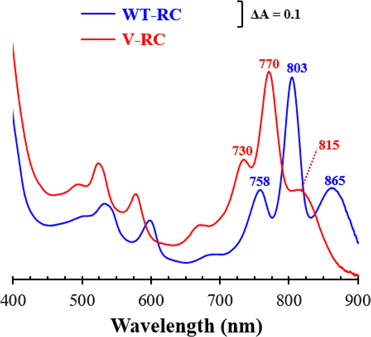
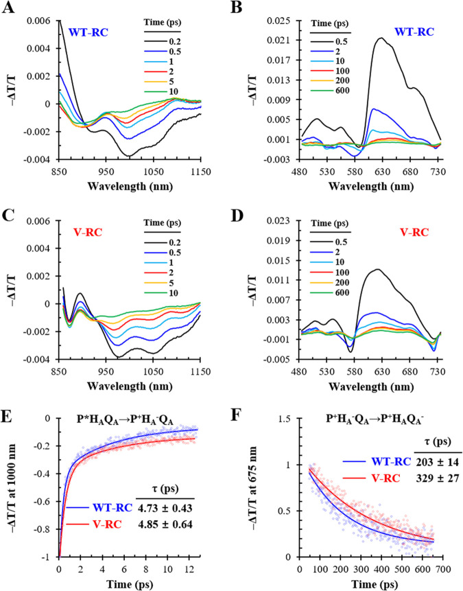
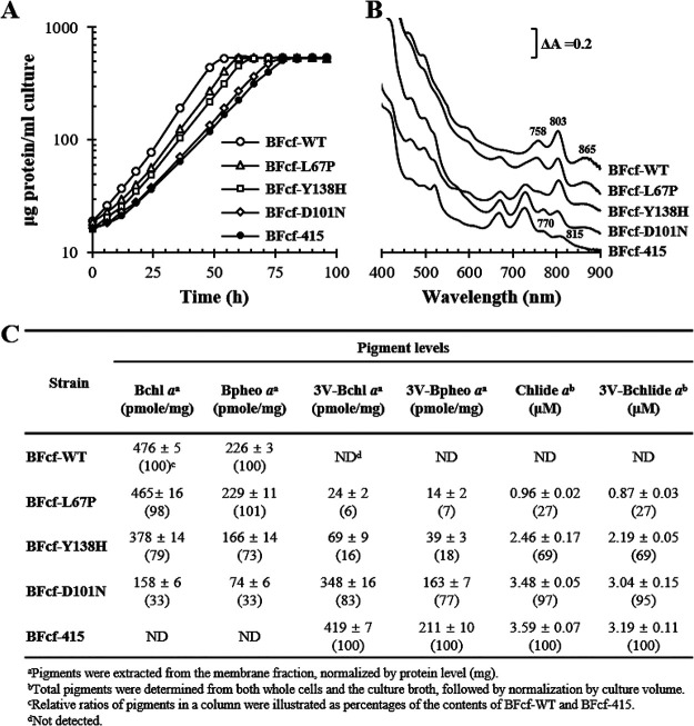
Rhodobacter sphaeroides mutant BF-lacking 3-vinyl (bacterio)chlorophyllide a hydratase (BchF)-accumulates chlorophyllide a (Chlide a) and 3-vinyl bacteriochlorophyllide a (3V-Bchlide a). BF synthesizes 3-vinyl bacteriochlorophyll a (3V-Bchl a) through prenylation of 3V-Bchlide a and assembles a novel reaction center (V-RC) using 3V-Bchl a and Mg-free 3-vinyl bacteriopheophytin a (3V-Bpheo a) at a molar ratio of 2:1. We aimed to verify whether a bchF-deleted R. sphaeroides mutant produces a photochemically active RC that facilitates photoheterotrophic growth. The mutant grew photoheterotrophically-implying a functional V-RC-as confirmed by the emergence of growth-competent suppressors of bchC-deleted mutant (BC) under irradiation. Suppressor mutations in BC were localized to bchF, which diminished BchF activity and caused 3V-Bchlide a accumulation. bchF expression carrying the suppressor mutations in trans resulted in the coproduction of V-RC and wild-type RC (WT-RC) in BF. The V-RC had a time constant (τ) for electron transfer from the primary electron donor P (a dimer of 3V-Bchl a) to the A-side containing 3V-Bpheo a (HA) similar to that of the WT-RC and a 60% higher τ for electron transfer from HA to quinone A (QA). Thus, the electron transfer from HA to QA in the V-RC should be slower than that in the WT-RC. Furthermore, the midpoint redox potential of P/P+ of the V-RC was 33 mV more positive than that of the WT-RC. R. sphaeroides, thus, synthesizes the V-RC when 3V-Bchlide a accumulates. The V-RC can support photoheterotrophic growth; however, its photochemical activity is inferior to that of the WT-RC. IMPORTANCE 3V-Bchlide a is an intermediate in the bacteriochlorophyll a (Bchl a)-specific biosynthetic branch and prenylated by bacteriochlorophyll synthase. R. sphaeroides synthesizes V-RC that absorbs light at short wavelengths. The V-RC was not previously discovered because 3V-Bchlide a does not accumulate during the growth of WT cells synthesizing Bchl a. The levels of reactive oxygen species increased with the onset of photoheterotrophic growth in BF, resulting in a long lag period. Although the inhibitor of BchF is unknown, the V-RC may act as a substitute for the WT-RC when BchF is completely inhibited. Alternatively, it may act synergistically with WT-RC at low levels of BchF activity. The V-RC may broaden the absorption spectra of R. sphaeroides and supplement its photosynthetic ability at various wavelengths of visible light to a greater extent than that by the WT-RC alone.

Keywords: 3-vinyl bacteriochlorophyll a; Rhodobacter sphaeroides; bchF mutation; reaction center.


Conflict of interest statement

The authors declare no conflict of interest.

Figures

FIG 1
FIG 1
Biosynthesis of 3V-Bchl a in R. sphaeroides mutant BF lacking BchF. Bchl a is synthesized from Chlide a via BchF, BchXYZ, BchC, BchG, and BchP and used to form the wild-type reaction center (WT-RC) and light-harvesting (LH) complexes. BF (see Table S1 in the supplemental material) was constructed after deletion of bchF (black crosses), which resulted in cellular accumulation of Chlide a and 3V-Bchlide a. 3V-Bchlide a is phytylated by BchG and BchP, and the resulting 3V-Bchl a can form the novel RC (V-RC). Carbon numbering and pyrrole-ring designation are indicated with the structure of 3V-Bchlide a. Differences at C-3 between 3V-Bchlide a and Bchlide a are shown with red and blue lines, respectively. The metabolic pathways to form Bchlide a with 3HE-Chlide a and 3HE-Bchlide a are also illustrated.
FIG 2
FIG 2
Effect of DMSO (10 mM) on photoheterotrophic growth of BF. (A and B) Photoheterotrophic growth of WT (A) and BF (B) cells at 15 W/m2 was recorded based on the total cellular protein in the presence (closed symbols) or absence (open symbols) of DMSO. (C and D) General ROS levels were determined using aliquots of exponentially growing cells with H2DCFDA as an indicator and normalized to cellular proteins. Each point represents the mean ± standard deviation (SD) from three independent experiments. Note that time scales of panels A and C are different from those of panels B and D.
FIG 3
FIG 3
Photoheterotrophic growth and pigment analysis of mutants deficient in Bchl a synthesis genes. (A and B) WT, BF, BZ (ΔbchZ), BZF (ΔbchZF), and BC (ΔbchC) cells (see Table S1 in the supplemental material) were grown with (A) or without (B) light in the presence of 10 mM DMSO. Cell growth was estimated based on total cellular protein levels. (C) Absorption spectra of pigment accumulation in the culture supernatant. (D) Membranes were obtained and adjusted to equivalent protein levels. Absorption spectra of the membranes are illustrated with λmax values indicated above each peak. (E to J) Phytylated pigments were extracted from the membranes of WT (E) and BF (H) cells and subjected to HPLC analysis at 771 nm. Pigments comprising the major peaks (black arrows) were pooled, and the absorption spectra (F and I) were recorded with the λmax of the major Qy bands. Pooled pigments were further subjected to cold-spray-TOF-MS analysis, and the masses of molecular ion (M) of the pigments from WT (G) and BF (J) cells were determined. AU, arbitrary units.
FIG 4
FIG 4
Kinetic analysis of BchG for 3V-Bchlide a and inhibition of BchG by Chlide a. Kinetic parameters (Km and kcat) of BchG for 3V-Bchlide a as the second substrate were determined using geranylgeranyl pyrophosphate (GGPP) or phytyl pyrophosphate (PPP) as the first substrate. Kinetic analysis for Bchlide a was performed for comparison. (A to D) Inhibition of BchG by Chlide a (circle, 0; triangle, 10 μM; square, 20 μM; diamond, 40 μM) was determined in the presence of Bchlide a (A and C) and 3V-Bchlide a (B and D). GGPP (A and B) or PPP (C and D) was included at 50 μM. (E) Data sets of V0 in double reciprocal plots were fitted to a competitive inhibition model using nonlinear regression to calculate the kinetic parameters. Each point was determined using three independent experiments.
FIG 5
FIG 5
Photosynthetic complex formation using Bchl a and 3V-Bchl a. (A) Semiaerobically grown Wcfh-puhA, Wcfh-puf, Wcfh-puc1, and Wcfh-puc2 were used as controls for the formation of RC, LH1, LH2-1, and LH2-2, respectively. (B) Semiaerobically grown BFcfh-puhA, BFcfh-puf, BFcfh-puc1, and BFcfh-puc2 were used to examine RC, LH1, LH2-1, and LH2-2 formation, respectively, using 3V-Bchl a. Wcfh-415 and BFcfh-415 contain empty vectors. The photosynthetic complexes were purified by His tag affinity chromatography, and the absorption spectra are illustrated.
FIG 6
FIG 6
Ground-state absorption spectra of RCs. The WT-RC and V-RC were purified by His tag affinity chromatography from semiaerobically grown Wcfh-puhA and BFcfh-puhA, respectively. Ground-state absorption spectra were measured at 298 K. The Qy regions are illustrated with the λmax of the peaks. A770 of the V-RC was normalized to A803 of the WT-RC.
FIG 7
FIG 7
Pigment compositions of WT-RC and V-RC. (A to D) Pigments were extracted from WT-RC (A and B) and V-RC (C and D) produced from semiaerobically grown Wcfh-puhA and BFcfh-puhA, respectively. Bchl a (with Bpheo a) of the WT-RC (A), 3V-Bchl a (with 3V-Bpheo a) of the V-RC (C), and their carotenoids (B and D) were quantified using HPLC. (A) Bchl a and Bpheo a of the WT-RC were monitored at 751 nm (the two pigments had the same molar extinction coefficient). (C) 3V-Bchl a and 3V-Bpheo a of the V-RC were detected at 715 nm. (B and D) The carotenoid spheroidenone (SO) was monitored at 482 nm. The absorption spectrum of each pigment is illustrated with the λmax of the prominent peaks in the insets of panels A to D. (E) The amounts of RCs were calculated using Western immunoblot analysis (see Fig. S4C and D in the supplemental material). The pigment content per RC is shown as the mean ± standard deviation. AU, arbitrary unit; SE, spheroidene.
FIG 8
FIG 8
Broadband transient absorption spectroscopy of WT-RC and V-RC after photooxidation. (A to D) The WT-RC (A and B) and V-RC (C and D) were excited by an 800-nm laser, and the changes in absorption spectra were recorded in the near-infrared (A and C) and visible wavelength (B and D) regions in process of time. The absorbance changes at 1,000 nm and 675 nm were assigned to reflect the dynamics of the P*HAQA state and P+HA−QA state, respectively, in both RCs if the electron transfers follow the sequential transfer model. (E and F) Absorbance changes at 1,000 nm (E) and 675 nm (F) were tracked over time, where open circles and solid lines represent raw data points and fitting curves, respectively. The fitted time constants (τ) for P*HAQA→P+HA−QA (E) and P+HA−QA→P+HAQA− (F) are represented with fitting errors. T, transmittance.
FIG 9
FIG 9
Electrochemical titration of P/P+ redox pairs in WT-RC and V-RC. WT-RC and V-RC were oxidatively titrated with potassium permanganate in the presence of potassium ferrocyanide as a mediator. The data from three independent titrations (open circles) were fitted (lines) to the Nernst equation with n = 1 (the number of electrons transferred in redox pair). The midpoint potentials (Em) of P/P+ for the WT-RC and V-RC were determined and presented as mean ± standard deviation. Ambient potentials were presented as the values versus the standard hydrogen electrode (SHE).
FIG 10
FIG 10
Characterization of RC-only strains producing Bchl a, 3V-Bchl a, or both Bchl a and 3V-Bchl a. Recombinant BFcf-WT (BFcf expressing bchF), BFcf-L67P (BFcf expressing bchFL67P), BFcf-Y138H (BFcf expressing bchFY138H), BFcf-D101N (BFcf expressing bchFD101N), and BFcf-415 (BFcf carrying empty vector) were cultured photoheterotrophically in the presence of 10 mM DMSO. (A) Growth was determined based on the total protein content of the cells. (B) The membrane fractions were isolated and analyzed for absorption spectra with λmax values shown on the peaks. (C) Pigment analysis in the membrane fractions. Chlide a and 3V-Bchlide a were extracted from whole cells and the culture broth, and each pigment was spectrally quantified.

Similar articles

References

    1. Proctor MS, Sutherland GA, Canniffe DP, Hitchcock A. 2022. The terminal enzymes of (bacterio)chlorophyll biosynthesis. R Soc Open Sci 9:211903. doi:10.1098/rsos.211903. - DOI - PMC - PubMed
    1. Pudek MR, Richards WR. 1975. A possible alternate pathway of bacteriochlorophyll biosynthesis in a mutant of Rhodopseudomonas sphaeroides. Biochemistry 14:3132–3137. doi:10.1021/bi00685a015. - DOI - PubMed
    1. Bollivar DW, Suzuki JY, Beatty JT, Dobrowolski JM, Bauer CE. 1994. Directed mutational analysis of bacteriochlorophyll a biosynthesis in Rhodobacter capsulatus. J Mol Biol 237:622–640. doi:10.1006/jmbi.1994.1260. - DOI - PubMed
    1. Struck A, Beese D, Cmiel E, Fischer M, Müller A, Schäfer W, Scheer H. 1990. Modified bacterial reaction centers: 3. Chemically modified chromophores at sites BA, BB and HA, HB, p 313–326. In Michel-Beyerle M-E (ed), Reaction centers of photosynthetic bacteria. Springer, Berlin, Germany.
    1. Ortega-Ramos M, Canniffe DP, Radle MI, Hunter CN, Bryant DA, Golbeck JH. 2018. Engineered biosynthesis of bacteriochlorophyll gF in Rhodobacter sphaeroides. Biochim Biophys Acta Bioenerg 1859:501–509. doi:10.1016/j.bbabio.2018.02.006. - DOI - PubMed

LinkOut - more resources