. 2023;99(7):1096-1108.
doi: 10.1080/09553002.2023.2194404. Epub 2023 Apr 28.

Differences in radiation-induced heart dysfunction in male versus female rats

Affiliations

Differences in radiation-induced heart dysfunction in male versus female rats

Neal Andruska et al. Int J Radiat Biol. 2023.

Abstract

Purpose: Radiation therapy remains part of the standard of care for breast, lung, and esophageal cancers. While radiotherapy improves local control and survival, radiation-induced heart dysfunction is a common side effect of thoracic radiotherapy. Cardiovascular dysfunction can also result from non-therapeutic total body radiation exposures. Numerous studies have evaluated the relationship between radiation dose to the heart and cardiotoxicity, but relatively little is known about whether there are differences based on biological sex in radiation-induced heart dysfunction (RIHD).

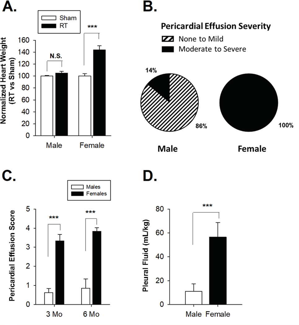
Materials and methods: We evaluated whether male and female inbred Dahl SS rats display differences in RIHD following delivery of 24 Gy in a single fraction to the whole heart using a 1.5 cm beam size (collimater). We also compared the 2.0 cm vs. 1.5 cm collimator in males. Pleural and pericardial effusions and normalized heart weights were measured, and echocardiograms were performed.

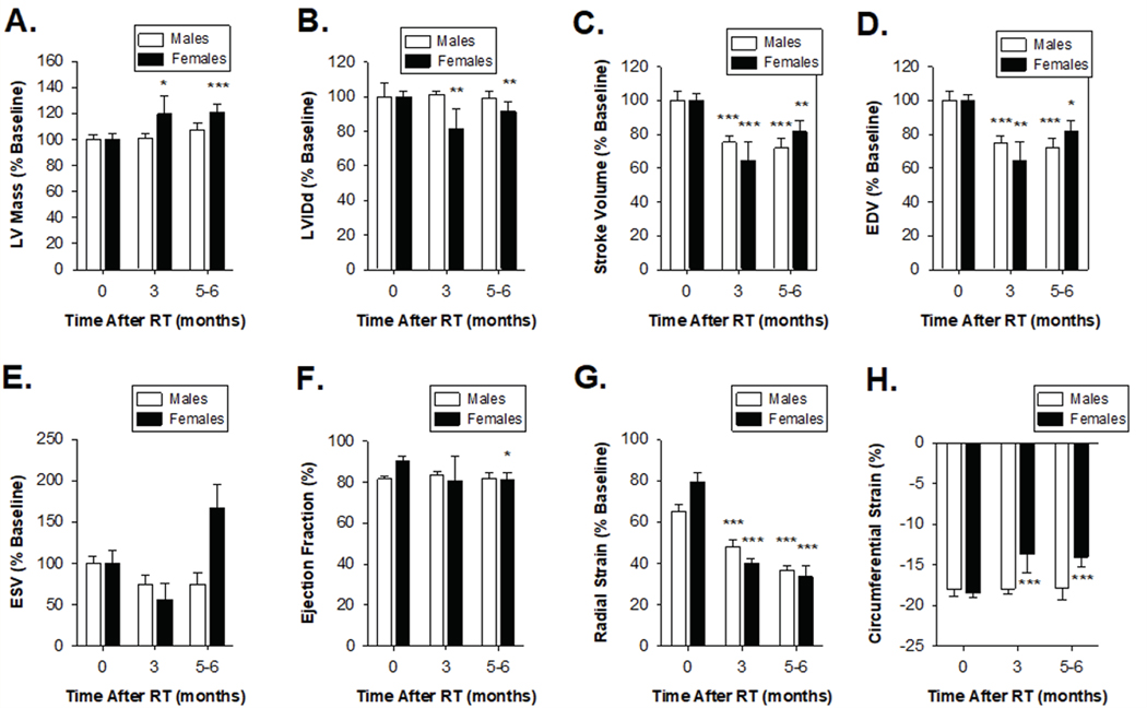
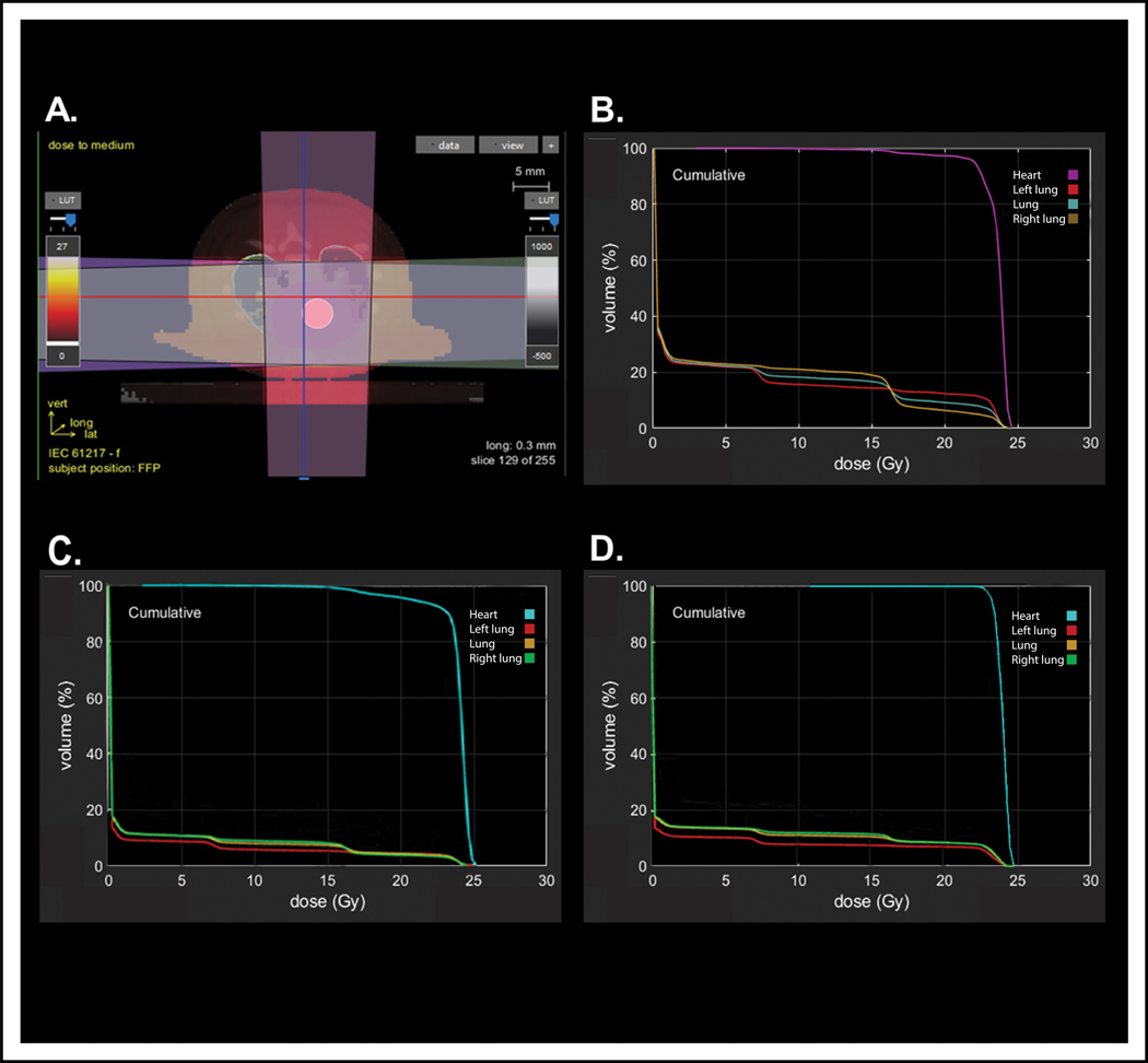
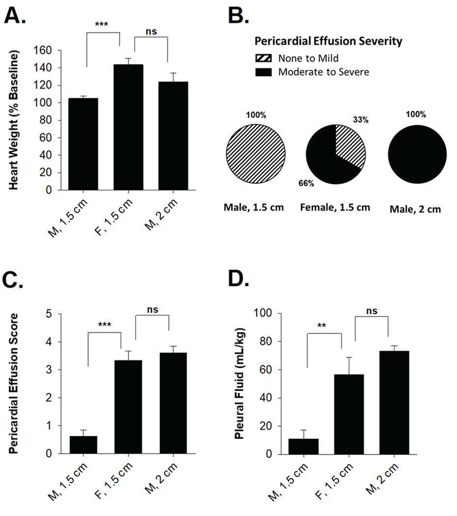
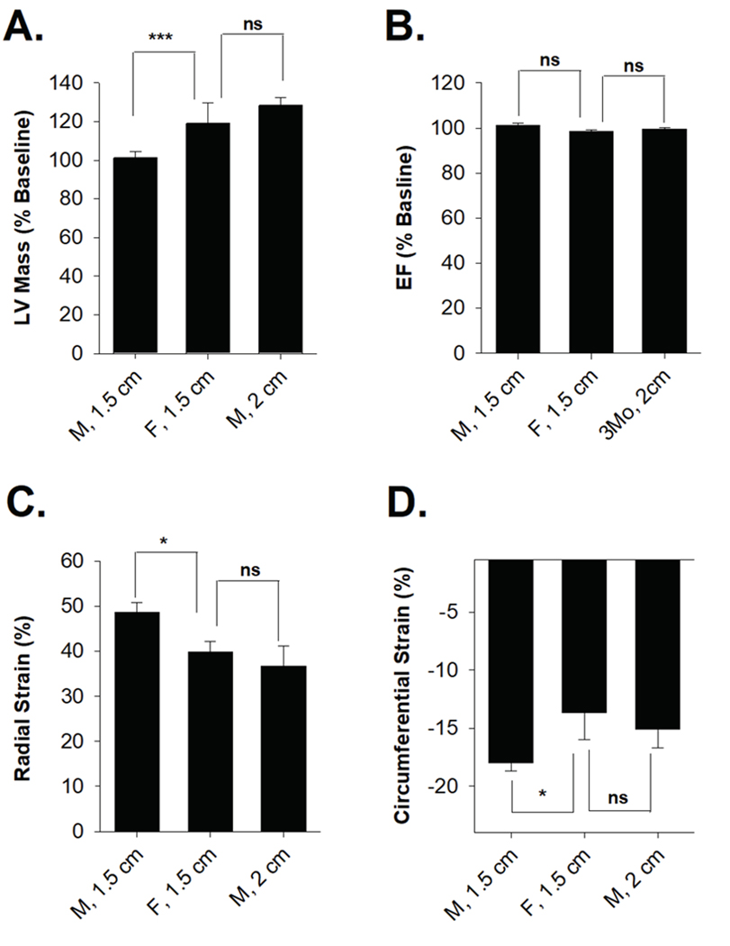
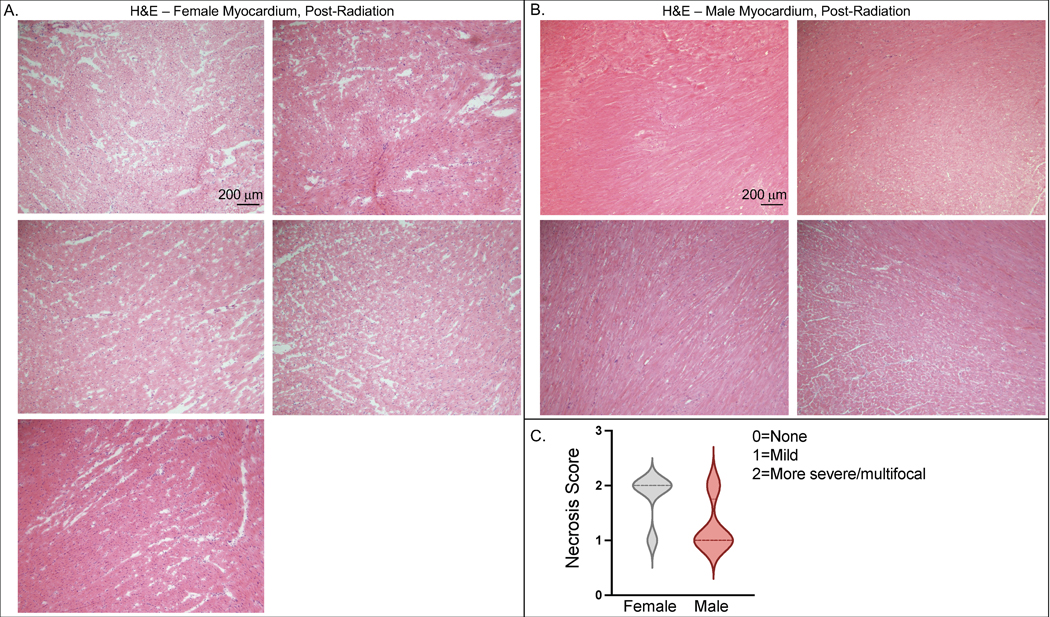
Results: Female SS rats displayed more severe RIHD relative to age-matched SS male rats. Normalized heart weight was significantly increased in females, but not in males. A total of 94% (15/16) of males and 55% (6/11) of females survived 5 months after completion of radiotherapy (p < .01). Among surviving rats, 100% of females and 14% of males developed moderate-to-severe pericardial effusions at 5 months. Females demonstrated increased pleural effusions, with the mean normalized pleural fluid volume for females and males being 56.6 mL/kg ± 12.1 and 10.96 mL/kg ± 6.4 in males (p = .001), respectively. Echocardiogram findings showed evidence of heart failure, which was more pronounced in females. Because age-matched female rats have smaller lungs, a higher percentage of the total lung was treated with radiation in females than males using the same beam size. After using a larger 2 cm beam in males which results in higher lung exposure, there was not a significant difference between males and females in terms of the development of moderate-to-severe pericardial effusions or pleural effusions. Treatment of males with a 2 cm beam resulted in comparable increases in LV mass and reductions in stroke volume to female rats treated with a 1.5 cm beam.

Conclusion: Together, these results illustrate that there are differences in radiation-induced cardiotoxicity between male and female SS rats and add to the data that lung radiation doses, in addition to other factors, may play an important role in cardiac dysfunction following heart radiation exposure. These factors may be important to factor into future mitigation studies of radiation-induced cardiotoxicity.

Keywords: Radiation; cardio-oncology; heart radiation; lung radiation; pericardial effusion; radiation-induced cardiotoxicity; sex differences.


Conflict of interest statement

Conflicts of Interest: The authors declare no relevant conflicts of interest. M.J.F. currently is a Principal Research Scientist at Abbvie, but at the time of his contributions to the manuscript, he was employed at the Medical College of Wisconsin.

Figures

Figure 1:
Figure 1:. Female SS (Dahl salt-sensitive/Mcwi) rats exhibit worse mortality and increased effusions after localized cardiac irradiation compared to males.
(A) Increased normalized heart weight in females compared to males after 24 Gy localized cardiac radiation therapy vs. sham treatment. (B) Increased frequency of moderate to severe pericardial effusions in female SS rats compared to males. (C) Average pericardial effusions severity was higher in females relative to males at 12 weeks and 5–6 months post-radiation. (D) Normalized pleural fluid volume was higher in females relative to males 5–6 months post-radiation. Values are the mean ± SE. *P<0.05, **P<0.01, ***P< 0.001; ns, nonsignificant.
Figure 2:
Figure 2:. Echocardiogram measurements indicate similar reductions in cardiac performance in male and female SS rats (Dahl salt-sensitive/Mcwi) at 24 Gy localized heart radiation.
M-mode echocardiogram measurements of SS male and female rats that received 24 Gy of localized heart radiation therapy at baseline, 3 mo, and 5–6 months post-RT. (A) Left ventricular mass was significantly increased at 3 months and 6 months post-RT female rats, but not in male rats. (B) There was no significant change in left ventricular internal diameter end diastole (LVIDd) following radiation in male and female rats. (C) Stroke volume and (D) end-diastolic volume (EDV) were decreased in male and female rats. (E) End-systolic volume (ESV) increased in females, but not in males, 5–6 following RT. (F) Ejection fraction (%EF) significantly decreased at 6 months in females, but not in males. (G) Radial strain was significantly lower in male and female rats 3- and 6-months following RT. (H) Circumferential strain showed male and female rats had a decreased ability to contract, indicated by a smaller negative percentage. Values are the mean ± SE. *P<0.05, **P< 0.01, ***P<0.001 for values compared to baseline.
Figure 3:
Figure 3:. Dosimetric comparisons of heart radiation plans.
(A) Representative axial slice demonstrating radiotherapy setup using a 3-beam arrangement to deliver 24 Gy in one fraction to the whole heart. Representative dose-volume histograms (DVH) for (B) a female rat treated with a 1.5 cm collimator, (C) a male rat treated with a 1.5 cm collimator, and (D) a male rat treated with a 2.0 cm collimator.
Figure 4:
Figure 4:. Male and female SS rats demonstrated similar increases in heart weight, pericardial effusion frequency and severity, and pleural effusions when both receive comparable total lung doses.
(A) Change in total heart weight relative to baseline was significantly increased and comparable between male rats treated with a 2 cm collimator and females treated with a 1.5 cm collimator. (B) No male rats treated with a 1.5 cm collimator developed moderate to severe pericardial effusions, whereas all male rats treated with a 2 cm collimator developed moderate to serve pericardial effusions. (C) Pericardial effusion index scores were similar between male rats treated with a 2 cm collimator and female rats treated with a 1.5 cm collimator. (D) Normalized pleural fluid volume was similar in male rats treated with a 2 cm collimator and female rats treated with a 1.5 cm collimator. Values are the mean ± SE. *P<0.05, **P< 0.01, ***P<0.001; ns, nonsignificant.
Figure 5:
Figure 5:. Male rats treated with a 2.0 cm beam size showed significant signs of heart failure on echocardiogram, which were comparable to female rats treated with a 1.5 cm beam size.
Female rats treated with a 1.5 cm collimator and male rats treated with a 2 cm collimator showed similar changes 3 months following radiation in terms of (A) left ventricular mass, (B) ejection fraction, (C) radial strain, and (D) circumferential strain. Values are mean ± SE. *P<0.05, **P< 0.01, ***P<0.001; ns, nonsignificant.
Figure 6.
Figure 6.. Female rats exhibit more histologic evidence of necrosis in the myocardium than male rats after 24Gy of localized cardiac RT.
Ten weeks after sham treatment or 24 Gy of localized cardiac RT, H&E staining was performed on fixed cardiac tissue. Female hearts (A, N=5) exhibited more pronounced necrosis than male rats (B, N=4), with a representative image from each rat shown (scale bar = 200 μm). There were 4 of 5 female hearts exhibiting more severe/multifocal necrosis and only 1 of 4 male rats exhibiting more severe/multifocal necrosis (C). Median value represented with hatched line.

References

    1. The 2007 Recommendations of the International Commission on Radiological Protection. ICRP publication Annal ICRP. 37:1–332. - PubMed
    1. Aleman BM, van den Belt-Dusebout AW, De Bruin ML, van ‘t Veer MB, Baaijens MH, de Boer JP, Hart AA, Klokman WJ, Kuenen MA, Ouwens GM, et al. 2007. Late cardiotoxicity after treatment for Hodgkin lymphoma. Blood. 109(5):1878–1886. - PubMed
    1. Anselmino M, De Ferrari GM, Massa R, Manca L, Tritto M, Molon G, Curnis A, Devecchi P, Sarzi Braga S, Bartesaghi G, et al. 2009. Predictors of mortality and hospitalization for cardiac causes in patients with heart failure and nonischemic heart disease: a subanalysis of the ALPHA study. Pacing Clin Electrophysiol. 32 Suppl 1:S214–218. - PubMed
    1. Antonia SJ, Villegas A, Daniel D, Vicente D, Murakami S, Hui R, Kurata T, Chiappori A, Lee KH, de Wit M, et al. 2018. Overall Survival with Durvalumab after Chemoradiotherapy in Stage III NSCLC. N Engl J Med. 379(24):2342–2350. - PubMed
    1. Bates JE, Howell RM, Liu Q, Yasui Y, Mulrooney DA, Dhakal S, Smith SA, Leisenring WM, Indelicato DJ, Gibson TM, et al. 2019. Therapy-Related Cardiac Risk in Childhood Cancer Survivors: An Analysis of the Childhood Cancer Survivor Study. J Clin Oncol. 37(13):1090–1101. - PMC - PubMed

Publication types

LinkOut - more resources