. 2023 Jun 13;7(11):2347-2359.
doi: 10.1182/bloodadvances.2023010100.

Mitochondrial pyruvate dehydrogenase kinases contribute to platelet function and thrombosis in mice by regulating aerobic glycolysis

Affiliations

Mitochondrial pyruvate dehydrogenase kinases contribute to platelet function and thrombosis in mice by regulating aerobic glycolysis

Gagan D Flora et al. Blood Adv. .

Abstract

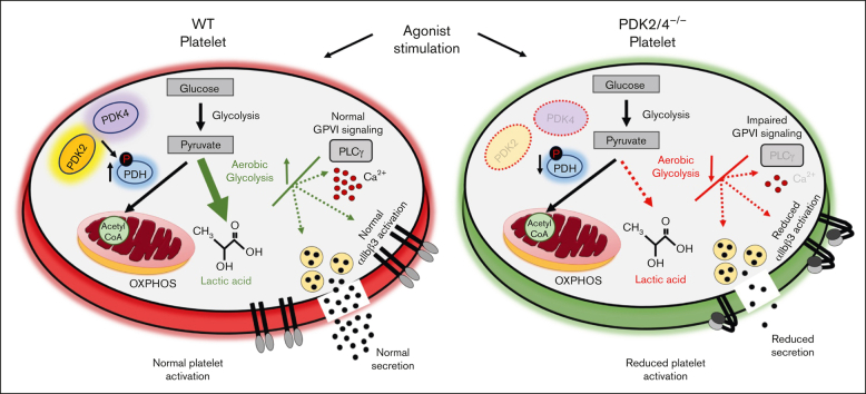
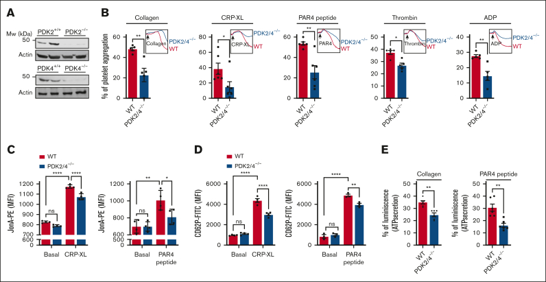
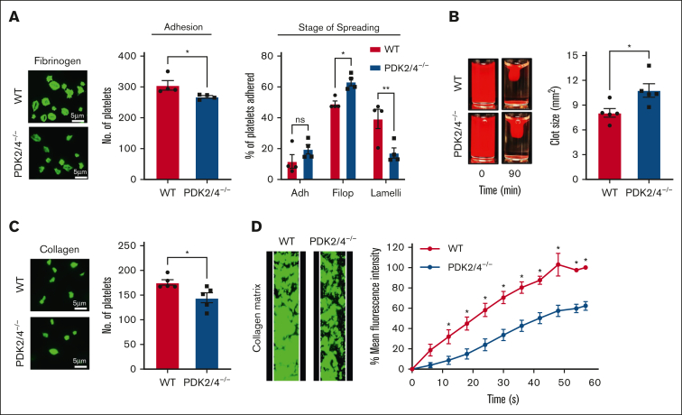
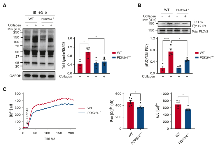
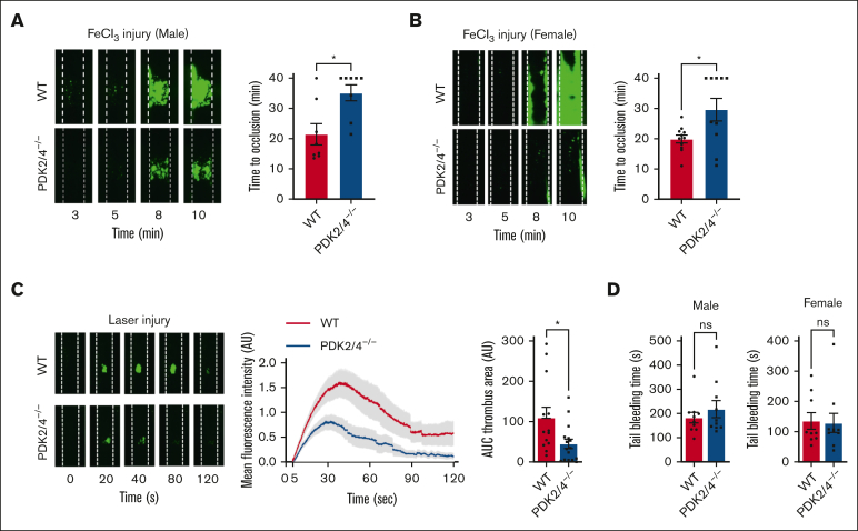
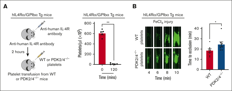
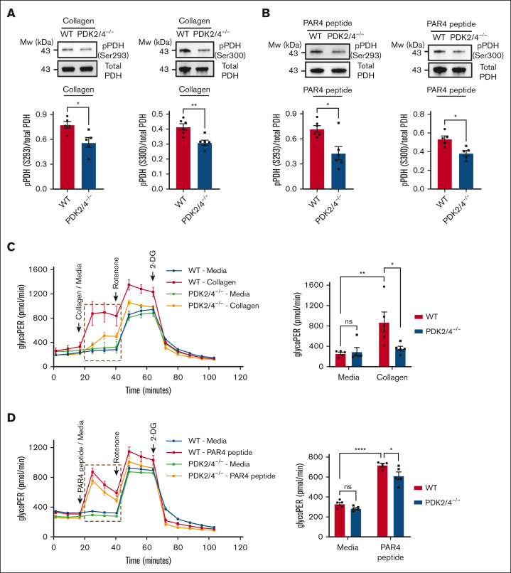
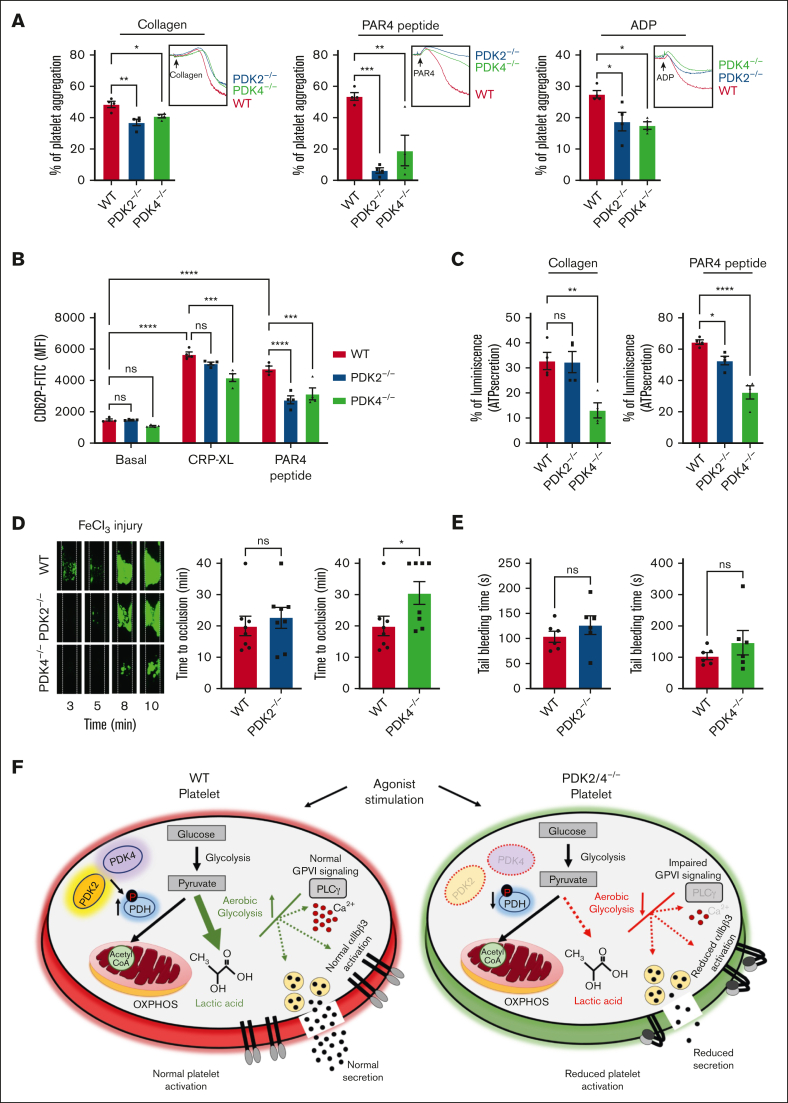
Resting platelets rely on oxidative phosphorylation (OXPHOS) and aerobic glycolysis (conversion of glucose to lactate in the presence of oxygen) for their energy requirements. In contrast, platelet activation exhibits an increased rate of aerobic glycolysis relative to OXPHOS. Mitochondrial enzymes pyruvate dehydrogenase kinases (PDKs) phosphorylate the pyruvate dehydrogenase (PDH) complex to inhibit its activity, thereby diverting the pyruvate flux from OXPHOS to aerobic glycolysis upon platelet activation. Of 4 PDK isoforms, PDK2 and PDK4 (PDK2/4) are predominantly associated with metabolic diseases. Herein, we report that the combined deletion of PDK2/4 inhibits agonist-induced platelet functions, including aggregation, integrin αIIbβ3 activation, degranulation, spreading, and clot retraction. In addition, collagen-mediated PLCγ2 phosphorylation and calcium mobilization were significantly reduced in PDK2/4-/- platelets, suggesting impaired GPVI signaling. The PDK2/4-/- mice were less susceptible to FeCl3-induced carotid and laser-induced mesenteric artery thrombosis without any effect on hemostasis. In adoptive transfer experiments, thrombocytopenic hIL-4Rα/GPIbα-transgenic mice transfused with PDK2/4-/- platelets exhibited less susceptibility to FeCl3 injury-induced carotid thrombosis compared with hIL-4Rα/GPIbα-Tg mice transfused with WT platelets, suggesting a platelet-specific role of PDK2/4 in thrombosis. Mechanistically, the inhibitory effects of PDK2/4 deletion on platelet function were associated with reduced PDH phosphorylation and glycoPER in activated platelets, suggesting that PDK2/4 regulates aerobic glycolysis. Finally, using PDK2 or PDK4 single KO mice, we identified that PDK4 plays a more prominent role in regulating platelet secretion and thrombosis compared with PDK2. This study identifies the fundamental role of PDK2/4 in regulating platelet functions and identifies the PDK/PDH axis as a potentially novel antithrombotic target.


Conflict of interest statement

Conflict-of-interest disclosure: The authors declare no competing financial interests.

Figures

None
Graphical abstract
Figure 1.
Figure 1.
PDK2/4-double-deficient mice exhibit reduced platelet functions. (A)The deletion of PDK2 and PDK4 was confirmed using western blotting. (B) Platelet-rich plasma (PRP) from WT or PDK2/4−/− mice was stimulated with collagen (0.4 μg/mL), CRP-XL (0.05 μg/mL), PAR4 peptide (40 μM), thrombin (0.013 U/mL; washed platelelets were used), or ADP (0.13 μM). Results are expressed as the percent change in light transmission with respect to the blank (buffer without platelets), set at 100%. The representative aggregation curves are shown. Values are mean ± SEM, n = 5 to 6 mice per group. Statistical analysis: Mann-Whitney U test; ∗P < .05 and ∗∗P < .01. (C) The extent of integrin αIIbβ3 activation (using JonA binding), and (D) α-granule secretion (using P-selectin exposure) in WT and PDK2/4−/− platelets (in PRP) stimulated with CRP-XL (0.1 μg/mL) or PAR4 peptide (100 μM) was determined using flow cytometry. Values are mean ± SEM, n = 3 to 4 mice per group. Statistical analysis: 2-way ANOVA followed by Tukey multiple comparisons test; ∗P < .05; ∗∗P < .01 and ∗∗∗∗P < .0001. (E) ATP secretion from the dense granules of WT and PDK2/4−/− platelets (in PRP) after stimulation with collagen (0.8 μg/mL) or PAR4 peptide (70 μM) was measured using Lumi-aggregometry. Values are mean ± SE n = 6 mice per group. Statistical analysis: Mann-Whitney U test; ∗∗P < .01.
Figure 2.
Figure 2.
PDK2/4−/− platelets exhibit reduced outside-in signaling and in vitro thrombus formation. (A) WT or PDK2/4−/− washed platelets (2 × 107 cells/mL) stained using Alexa-Fluor 488-conjugated phalloidin were stimulated with PAR4 peptide (70 μM) for 10 minutes and added onto fibrinogen-coated (100 μg/mL) coverslips for 120 minutes. Five images were captured of each sample at random locations. A representative image of platelet adhesion and spreading is shown. Spreading platelets were divided into 3 classes: adhered (Adh) but not spread, filopodia (Filop): spreading platelets and lamellipodia (Lamelli): fully spread. Results are expressed as a percentage of the total number of platelets adhered. Cumulative data of the number of platelets adhered in each sample is shown. Values are mean ± SEM, n = 4 mice per group. Statistical analysis: Mann-Whitney U test (adhesion) or 2-way ANOVA (stage of spreading) followed by Sidak multiple comparisons test; ∗P < .05 and ∗∗P < .01. (B) Clot retraction was measured for 90 minutes in PRP, supplemented with red blood cells, after adding 1 U/mL of thrombin. The left panel shows the representative images at 90 minutes, and the right panel shows the quantification of the clot size. Values are mean ± SEM, n = 5 mice per group. Statistical analysis: Mann-Whitney U test; ∗P < .05. (C) WT or PDK2−/− washed platelets (2 × 107 cells/mL) stained using Alexa-Fluor 488-conjugated phalloidin were added onto collagen-coated (100 μg/mL) coverslips for 45 minutes. Five images were captured of each sample at random locations. A representative image of platelet adhesion is shown. Cumulative data of the number of platelets adhered in each sample is shown. Values are mean ± SEM, n = 5 mice per group. Statistical analysis: Mann-Whitney U test; ∗P < .05. (D) DiOC6-loaded whole blood from WT or PDK2/4−/− mouse was perfused over a collagen-coated (100 μg/mL) matrix for 60 seconds at an arterial shear rate in a BioFlux Microfluidic flow chamber system. The left panel shows the representative image at the end of the assay, and the right panel shows the thrombus growth on the collagen matrix over time. Values are mean ± SEM, n = 3 mice per group. Statistical analysis: 2-way ANOVA followed by Sidak multiple comparisons test; ∗P < .05.
Figure 3.
Figure 3.
Platelets from PDK2/4 double-deficient mice exhibit impaired GPVI-mediated signaling. (A) Total tyrosine phosphorylation and (B) phospho-PLCγ2 phosphorylation (Tyr 1217) were measured in resting and collagen (25 μg/mL)–stimulated WT and PDK2/4−/− platelets. Representative western blots for total tyrosine phosphorylation and phospho-PLCγ2 (Tyr 1217) are shown. GAPDH or total PLCγ2 were used as loading controls. The bar graphs show densitometry analysis of immunoblots. Values are mean ± SEM, n = 4 mice per group. Statistical analysis: 2-way ANOVA followed by Tukey's multiple comparisons test; ∗P < .05 and ∗∗∗∗P < .0001. (C) Calcium mobilization was evaluated in Fura-2 LR loaded WT or PDK2/4−/− platelets washed platelets that were stimulated with CRP-XL (0.7 μg/mL) in the presence of 1.3 mM CaCl2. The left panel shows representative calcium mobilization after stimulation with CRP-XL. The middle and right panel shows the peak calcium level and the area under the curve (AUC), respectively. Values are mean ± SEM, n = 5 mice per group. Statistical analysis: Mann-Whitney U test; ∗P < .05.
Figure 4.
Figure 4.
PDK2/4−/− mice are less susceptible to experimental arterial thrombosis. (A) A representative image of carotid artery thrombus (5% FeCl3 injury for 2 minutes) and the time to occlusion in WT and PDK2/4−/− male mice is shown. The time to occlusion was measured until 40 minutes, the cutoff point at which the experiment was terminated. Values are mean ± SEM, n = 8 vessels from 8 mice per group. Statistical analysis: Mann-Whitney U test; ∗P < .05. (B) A representative image of carotid artery thrombus (5% FeCl3 injury for 2 minutes) and the time to occlusion in WT and PDK2/4−/− female mice is shown. Values are mean ± SEM, n = 10 to 11 vessels from 6 mice per group. Statistical analysis: Mann-Whitney U test; ∗P < .05. (C) A representative image of laser injury–induced mesenteric artery thrombus in WT and PDK2/4−/− male mice as visualized by upright intravital microscopy. The mean fluorescence intensity over time and AUC (thrombus area) is shown. Values are mean ± SEM, n = 13 to 16 thrombi from 4 mice per group. Statistical analysis: Mann-Whitney U test; ∗P < .05. (D) The tail-bleeding time in WT and PDK2/4−/− male and female mice was determined by measuring the time taken for the initial cessation of bleeding after the tail transection. Values are mean ± SEM, n = 10 mice per group. Statistical analysis: Mann-Whitney U test.
Figure 5.
Figure 5.
Platelet-specific PDK2/4 contributes to arterial thrombosis. (A) The left panel demonstrates the schematic depicting the technique for generating mice with platelet-specific PDK2 and PDK4 deficiency. Thrombocytopenia was induced in hIL-4Rα/GPIbα-Tg mice by injecting antibodies against human IL-4R and platelet count was measured after 2 hours. N = 5 mice per group. Statistical analysis: Mann-Whitney U test; ∗∗P < .01. This was followed by the adoptive transfer of WT or PDK2/4−/− platelets into hIL-4Rα/GPIbα-Tg mice. (B) The left panel shows a representative image of carotid artery thrombus (5% FeCl3 injury for 3 minutes) as visualized by intravital microscopy in hIL-4Rα/GPIbα-Tg male mice transfused with WT or PDK2/4−/− platelets. The right panel shows the time to occlusion. The time to occlusion was measured until 40 minutes, the cutoff point at which the experiment was terminated. Values are mean ± SEM, n = 8 vessels from 4 mice per group. Data represents left and right carotid artery injury from each mouse. Statistical analysis: Mann-Whitney U test; ∗P < .05.
Figure 6.
Figure 6.
Deletion of PDK2 and PDK4 inhibits PDH phosphorylation and aerobic glycolysis in activated platelets. PDH phosphorylation at Ser293 and Ser300 was measured in (A) collagen- (25 μg/mL) or (B) PAR4 peptide (70μM)–stimulated WT or PDK2/4−/− platelets. Representative western blots are shown. Total PDH was used as a loading control. The bar graphs show densitometry analysis of immunoblots. Values are mean ± SEM, n = 5 to 6 mice per group. Statistical analysis: Mann-Whitney U test; ∗P < .05 and ∗∗P < .01. The glycolytic proton efflux rate (glycoPER) was measured in WT or PDK2/4−/− platelets stimulated with (C) collagen (25 μg/mL) or (D) PAR4 peptide (50μM) using a Seahorse extracellular flux analyzer. The bar graph shows the quantified data (for the line graph values marked with a box). Values are mean ± SEM, with n = 4 to 5 mice per group. Statistical analysis: 2-way ANOVA followed by Tukey multiple comparisons test; ∗P < .05; ∗∗P < .01 and ∗∗∗∗P < .0001.
Figure 7.
Figure 7.
PDK4 is the major determinant contributing to thrombosis in the FeCl3 injury–induced carotid thrombosis model. (A) Platelet-rich plasma (PRP) from WT or PDK2−/− or PDK4−/− mice was stimulated with collagen (0.4 μg/mL), PAR4 peptide (40 μM), or ADP (0.13 μM). Results are expressed as the percent change in light transmission with respect to the blank (buffer without platelets), set at 100%. The representative aggregation curves are shown. Values are mean ± SEM, n = 4 mice per group. Statistical analysis: 1-way ANOVA followed by Tukey's multiple comparisons test ∗P < .05; ∗∗P < .01 and ∗∗∗P < .001 (B) The extent of α-granule secretion (using P-selectin exposure) in WT, PDK2−/− and PDK4−/− platelets (in PRP) stimulated with CRP-XL (0.1 μg/mL) or PAR4 peptide(100 μM) was determined using flow cytometry. Values are mean ± SEM, n = 4 mice per group. Statistical analysis: 2-way ANOVA followed by Tukey's multiple comparisons test; ∗∗∗P < .001 and ∗∗∗∗P < .0001. (C) ATP secretion from the dense granules of WT, PDK2−/−, and PDK4−/− platelets (in PRP) after stimulation with collagen (0.8 μg/mL) or PAR4 peptide (70 μM) was measured using Lumi-aggregometry. Values are mean ± SE n = 4 mice per group. Statistical analysis: 1-way ANOVA followed by Tukey's multiple comparisons test ∗P < .05; ∗∗P < .01 and ∗∗∗∗P < .0001. (D) A representative image of carotid artery thrombus (5% FeCl3 injury for 2 minutes) as visualized by intravital microscopy in WT, PDK2−/−, and PDK4−/− male mice. Platelets were labeled ex vivo with calcein green. The time to occlusion is shown. These experiments were performed in the same setup with the same WT mice serving as a control group for both PDK2−/− and PDK4−/− mice. Values are mean ± SEM, n = 8 mice per group. Statistical analysis: Mann-Whitney U test. ∗P < .05 (E) The tail-bleeding time in WT, PDK2−/−, and PDK4−/− male mice was determined by the time taken for the initial cessation of bleeding after the tail transection. Values are mean ± SEM, n = 6 mice per group. Statistical analysis: Mann-Whitney U test. (F) A schematic depicting the role of PDK/PDH axis in regulating platelet activation.

References

    1. Sanchez-Lopez E, Cheng A, Guma M. Can metabolic pathways be therapeutic targets in rheumatoid arthritis? J Clin Med. 2019;8(5):753. - PMC - PubMed
    1. Heidker RM, Emerson MR, LeVine SM. Metabolic pathways as possible therapeutic targets for progressive multiple sclerosis. Neural Regen Res. 2017;12(8):1262–1267. - PMC - PubMed
    1. Harris AL. Development of cancer metabolism as a therapeutic target: new pathways, patient studies, stratification and combination therapy. Br J Cancer. 2020;122(1):1–3. - PMC - PubMed
    1. Mazumdar C, Driggers EM, Turka LA. The untapped opportunity and challenge of immunometabolism: a new paradigm for drug discovery. Cell Metab. 2020;31(1):26–34. - PubMed
    1. Rizzieri D, Paul B, Kang Y. Metabolic alterations and the potential for targeting metabolic pathways in the treatment of multiple myeloma. J Cancer Metastasis Treat. 2019;5:26. - PMC - PubMed

Publication types

Substances