. 2023 Mar 27;13(1):4940.
doi: 10.1038/s41598-023-32176-5.

Natural history and predictors for progression in pediatric keratoconus

Affiliations

Natural history and predictors for progression in pediatric keratoconus

Rosalia Antunes-Foschini et al. Sci Rep. .

Abstract

We studied the demographic and clinical predictors associated with keratoconus progression in a pediatric population. Retrospective cohort study. We evaluated 305 eyes without previous surgeries from 168 patients, 9 to < 18 years old, and with a minimum 36-month follow-up in a hospital corneal ambulatory. We used Kaplan-Meyer survival curves; the dependent variable (main outcome measure) was the interval time (months) until the event, defined as an increase of 1.5 D in the maximum keratometry (Kmax), obtained with Pentacam. We evaluated the predictors: age (< or ≥ 14 years), sex, keratoconus familial history, allergy medical history, and the baseline tomographic parameters: mean keratometry (Km), Kmax (< or ≥ 55 D); and thinnest pachymetry (TP). We used log-rank tests and compared median survival times for right (RE)/left eyes (LE) and better (BE)/worse eyes (WE). A p value < 0.05 was considered significant. The patients' mean ± SD age was 15.1 ± 2.3 years old; 67% were boys, 30% were < 14 years, 15% had keratoconus familial history, and 70% were allergic. The general Kaplan-Meyer curves showed no differences between RE/LE or BE/WE. RE with allergy and LE with Kmax ≥ 55 D had smaller survival times ((95%CI 9.67-32.1), p 0.031 and (95%CI 10.1-44.1), p 0.042, respectively). For BE and WE, Kmax ≥ 55 D had smaller survival times ((95% CI 6.42- ), p 0.031 and (95%CI 8.75-31.8), p 0.043, respectively). Keratoconus progression was similar between RE/LE and BE/WE. Steepest corneas are predictors of faster progression. Allergy is also a predictor of keratoconus progression in RE.


Conflict of interest statement

The authors declare no competing interests.

Figures

Figure 1
Figure 1
Examples of patients who were or were not included in this survival analysis. Patient 1: analyzed from time points A (T0) to B; (event or censoring) Patient 2: censored after the first appointment; Patient 3: excluded due to not having the possibility of being evaluated during a minimum period of at least 36 months; Patient 4: analyzed from time points C (T0) to D (censoring); Patients 5 and 6: included.
Figure 2
Figure 2
Pediatric keratoconus: Distribution of the eyes, showing the reasons for exclusion and the final number included.
Figure 3
Figure 3
Kaplan Meyer empirical curves of time until progression, in months. (a) Right and left eyes, overall progression (Right Eyes (n = 156); 88 events; Median of 21.9 CI95% (14.5; 39.7). Left Eyes (n = 149); 76 events; Median of 22.4 CI95% (13.4; 50.0). p value = 0.76); (b) Right eyes, by the presence of allergy (p value 0.032); and (c). Left eyes, by Kmax severity (p value = 0.043).
Figure 4
Figure 4
Better and worse eyes: Kaplan Meyer empirical curves of time until progression, in months. The eye with the highest Km between both eyes was considered the worse eye. Better (n = 136); 72 events; Median of 24.9 CI95% (15.5, 61.1). Worse (n = 136); 77 events; Median of 19.0; CI95% (11.0, 31.8). p value = 0.301.
Figure 5
Figure 5
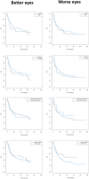
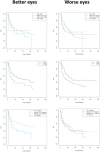
Better and worse eyes: Kaplan Meyer empirical curves of time until progression, in months, by sex, age, familial history of keratoconus, and the presence of allergy.
Figure 6
Figure 6
Better and worse eyes: Kaplan Meyer empirical curves of time until progression, in months, by Km, (divided into four groups), by Kmax severity (< or ≥ 55D), and by thinnest pachymetry (TP) (divided into four groups).

Similar articles

Cited by

References

    1. Rabinowitz YS. Keratoconus. Surv. Ophthalmol. 1998;42(4):297–319. doi: 10.1016/S0039-6257(97)00119-7. - DOI - PubMed
    1. McGhee CNJ, Kim BZ, Wilson PJ. Contemporary treatment paradigms in keratoconus. Cornea. 2015;34:S16–S23. doi: 10.1097/ICO.0000000000000504. - DOI - PubMed
    1. Kennedy RH, Bourne WM, Dyer JA. A 48-year clinical and epidemiologic study of keratoconus. Am. J. Ophthalmol. 1986;101(3):267–273. doi: 10.1016/0002-9394(86)90817-2. - DOI - PubMed
    1. Chatzis N, Hafezi F. Progression of keratoconus and efficacy of pediatric [corrected] corneal collagen cross-linking in children and adolescents. J. Refract. Surg. 2012;28(11):753–758. doi: 10.3928/1081597X-20121011-01. - DOI - PubMed
    1. Fujimoto H, Maeda N, Shintani A, et al. Quantitative evaluation of the natural progression of keratoconus using three-dimensional optical coherence tomography. Invest. Ophthalmol. Vis. Sci. 2016;57(9):OCT169–OCT175. doi: 10.1167/iovs.15-18650. - DOI - PubMed

Publication types