Review
. 2023 Oct;23(10):635-654.
doi: 10.1038/s41577-023-00849-x. Epub 2023 Mar 27.

The NLR gene family: from discovery to present day

Affiliations
Review

The NLR gene family: from discovery to present day

Wei-Chun Chou et al. Nat Rev Immunol. 2023 Oct.

Erratum in

Abstract

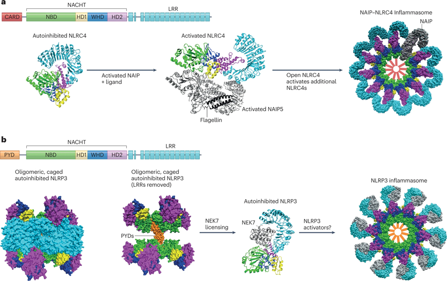
The mammalian NLR gene family was first reported over 20 years ago, although several genes that were later grouped into the family were already known at that time. Although it is widely known that NLRs include inflammasome receptors and/or sensors that promote the maturation of caspase 1, IL-1β, IL-18 and gasdermin D to drive inflammation and cell death, the other functions of NLR family members are less well appreciated by the scientific community. Examples include MHC class II transactivator (CIITA), a master transcriptional activator of MHC class II genes, which was the first mammalian NBD-LRR-containing protein to be identified, and NLRC5, which regulates the expression of MHC class I genes. Other NLRs govern key inflammatory signalling pathways or interferon responses, and several NLR family members serve as negative regulators of innate immune responses. Multiple NLRs regulate the balance of cell death, cell survival, autophagy, mitophagy and even cellular metabolism. Perhaps the least discussed group of NLRs are those with functions in the mammalian reproductive system. The focus of this Review is to provide a synopsis of the NLR family, including both the intensively studied and the underappreciated members. We focus on the function, structure and disease relevance of NLRs and highlight issues that have received less attention in the NLR field. We hope this may serve as an impetus for future research on the conventional and non-conventional roles of NLRs within and beyond the immune system.


Conflict of interest statement

Competing Interests

J. Ting is a co-founder of IMMvention Therapeutix.

Figures

Figure 1:
Figure 1:. Nucleotide-binding and oligomerizing sensors as a universal strategy for cellular defense.
(A) The STAND ATPase module consisting of NBD, helical domain 1 (HD1), and winged helix domain (WHD) is utilized for cellular defense from prokaryotes to eukaryotes. An additional helical domain 2 (HD2) is present in many STAND proteins including a NACHT-specific domain used in the NLR family of proteins. (B) The primary structural organization of human NLRs. Here, NLRs are grouped according to subfamily which is determined by the effector domain at the N-terminus. The variable domains associated with the NACHT domain are color coded and indicated in the domain legend below. CIITA exhibits cell-type specific alternative promoter usage, and this is designated with arrows. The oligomerized inflammasome for NAIP/NLRC4 is shown (PDB:3JBL). (C) Two representative members of the plant disease resistance proteins with either TIR or CC domains are shown. The NB-ARC module lacks the HD2 domain. The C-terminal sensor is an LRR domain for these proteins as well. The oligomerized resistosome for ZAR1 is shown (PDB:6J5T). (D) Two representative members of the recently described prokaryotic Avs protein family are shown. The effector domain in these proteins is a nuclease instead of a protein-recruitment domain, and the sensor domain is composed of tetratricopeptide repeats (TPR). The oligomerized complex for EcAvs4 is shown (PDB:8DGF). Cryo-EM structure representations in this figure were created with BioRender.com.
Figure 2:
Figure 2:. Key historical events in the NLR field
The timeline highlights some of the key discoveries and conceptual advances that have influenced the field over the last two decades. There are clearly many important contributions to the field which are not included here, and we apologize to those who have been left out due to spatial constraints.
Figure 3:
Figure 3:. Inflammasome activators and related disorders
The NLRP3, NLRP1, NLRC4, NLRP12 and NLRP6 inflammasomes with their intracellular mediators involved in activation are summarised. Dysregulated or chronically activated inflammasomes may lead to inflammatory diseases. NLRP3 is a sensor for numerous PAMPs and DAMPs responding to intracellular damage induced by pathogenic or sterile insults. NLRP1 is a sensor for ribotoxic stress and dsRNA. DPP8/9 inhibitors are activators of the NLRP1 inflammasome. The current model for NLRP1 inflammasome activation involves ASC-dependent recruitment of pro-caspase 1 by the UPA-CARD C-terminal fragment of NLRP1. The NLRC4 inflammasome detects T3SS bacterial proteins via NAIPs and can assemble an inflammasome with or without ASC. NLRP12 inflammasome is assembled in response to Yersinia pestis and Plasmodium chabaudi. NLRP6 inflammasome detects commensal‐derived metabolites, LTA derived from Gram‐positive bacteria. Abbreviations used: CINCA, chronic neurologic cutaneous and articular syndrome; NLRP3, NLR family pyrin domain containing 3; NEK7, NIMA related kinase 7; LRR, leucine rich repeats; NBD, nucleotide-binding domain; PYD, pyrin domain; ASC, Apoptosis-associated speck-like protein containing a CARD; CARD, caspase recruitment domain; ROS, reactive oxygen species; FIIND, function to find domain; UPA, conserved in UNC5, the death-domain-containing protein PIDD and proteins of the ankyrin family; NLRC4, NLR family CARD domain containing 4; T3SS, type 3 secretory system, NAIP; neuronal apoptosis inhibitory protein.
Figure 4:
Figure 4:. Regulatory Functions of NLRs
Different NLRs act as either positive or negative regulators in transcription, MAPK, NF-κB, and type I IFN signalling pathways and in autophagy. CIITA and NLRC5 induced by IFNγ acts as transactivators of MHC genes. NLRC5 also negatively regulates the NF-κB and type I IFN signalling pathways. In addition to NLRC5, several NLRs reduce either NF-κB or type I IFN signalling pathways or both. These NLRs are NLRC3, NLRX1, NLRP2, 4, 11, 12, and 14. On the other hand, NLRP7 may positively regulate NF-κB pathway, and NLRP2 promotes MAPK signalling. Likewise, NOD1 and NOD2 recognizes iE-DAP and MDP separately and interact with RIP2 to activate NF-κB and MAPK signalling pathway. Additionally, NOD2 recognizes MDP and recruits ATG16L1 to the plasma membrane to induce autophagy. The right shows that NLRX1 interacts with TUFM to induce autophagy while NLRP4 interacts with Beclin-1 to inhibit autophagy. The right box shows NLRs that regulate development in the reproductive system. NLRP14 is the only NLRP molecule that contributes to spermatogenesis while other NLRPs including NLRP2,4,5,7,9,11 may regulate oogenesis and embryogenesis. NLR, NOD-like receptors; MAPK, mitogen-activated protein kinase; NF-κB, nuclear factor kappa B; TRAF6, tumor necrosis factor (TNF) receptor-associated factor 6; MHC, major histocompatibility complexes; STING, Stimulator of interferon genes; TBK1, TANK-binding kinase 1; IRF3, Interferon Regulatory Factor 3; iE-DAP, γ-D-glutamyl-meso-diaminopimelic acid; MDP, muramyl dipeptide; RIP2, receptor interacting protein 2; TUFM, Tu translation elongation factor of mitochondria.
Figure 5:
Figure 5:. Structural Insights into NLR Activation
(A) Assembly of the NAIP/NLRC4 Inflammasome. The primary structure of NLRC4 is shown, and color coding for domains is replicated in the structural depictions. The crystal structure of closed, autoinhibited NLRC4 is adapted from PDB:4kxf. Interactions between the HD2 and LRR domains stabilize the closed conformation. The cryo-EM structure of activated NLRC4 and NAIP5 is adapted from PDB:6b5b. Activated NAIP5 (silver) with bound flagellin (black) initiates a conformational change in NLRC4 to an open, activated form which can activate downstream NLRC4 molecules. Finally, the cryo-EM structure of the assembled NAIP/NLRC4 inflammasome is adapted from PDB:3jbl. A top view of the surface representation shows a single NAIP (gray) present in the complex with ten NLRC4 molecules. Red bars are added in the center of the complex to show the location of the NLRC4 CARDs which will recruit pro-caspase 1. (B) NLRP3 structural regulation preceding inflammasome assembly. The primary structure of NLRP3 is shown, and color coding is replicated in the structural depictions. The cryo-EM structure for autoinhibited NLRP3 alone is adapted from PDB:7pzc. The left panel shows a surface representation of an oligomeric complex consisting of ten NLRP3 molecules. When the LRR domains are excluded from the image, the position of the PYDs within the “cage” shows how they are sequestered and prevented from initiating spurious inflammation. The cryo-EM structure of NLRP3 with bound NEK7 is adapted from PDB:6npy. NEK7 interaction with the LRR domain of NLRP3 prevents NLRP3 from forming caged oligomers. All NLRP3 structures shown here show NLRP3 in a closed, autoinhibited conformation bound to ADP. Presumably, NEK7 licenses NLRP3 to freely interact with activating signals or ligands to form an inflammasome. Upon activation, NLRP3 assembles into an inflammasome disc with 10–11 NLRP3 molecules. A cartoon representation is shown as the PDB file is currently unavailable. Structural representations were created using VMD.

References

    1. Baker B, Zambryski P, Staskawicz B & Dinesh-Kumar SP Signaling in plant-microbe interactions. Science 276, 726–733, doi: 10.1126/science.276.5313.726 (1997). - DOI - PubMed
    1. McHale L, Tan X, Koehl P & Michelmore RW Plant NBS-LRR proteins: adaptable guards. Genome Biol 7, 212, doi: 10.1186/gb-2006-7-4-212 (2006). - DOI - PMC - PubMed
    1. Barbary A, Djian-Caporalino C, Palloix A & Castagnone-Sereno P Host genetic resistance to root-knot nematodes, Meloidogyne spp., in Solanaceae: from genes to the field. Pest Manag Sci 71, 1591–1598, doi: 10.1002/ps.4091 (2015). - DOI - PubMed
    1. Steimle V, Otten LA, Zufferey M & Mach B Complementation cloning of an MHC class II transactivator mutated in hereditary MHC class II deficiency (or bare lymphocyte syndrome). Cell 75, 135–146 (1993). - PubMed
    1. Harton JA & Ting JP Class II transactivator: mastering the art of major histocompatibility complex expression. Mol Cell Biol 20, 6185–6194, doi: 10.1128/MCB.20.17.6185-6194.2000 (2000). - DOI - PMC - PubMed

Publication types

Substances