. 2023 Mar 27;13(1):4988.
doi: 10.1038/s41598-023-31979-w.

Magnetothermal-based non-invasive focused magnetic stimulation for functional recovery in chronic stroke treatment

Affiliations

Magnetothermal-based non-invasive focused magnetic stimulation for functional recovery in chronic stroke treatment

Hohyeon Kim et al. Sci Rep. .

Abstract

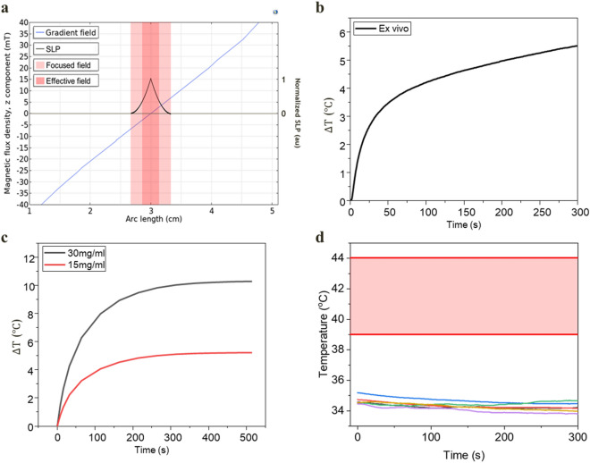
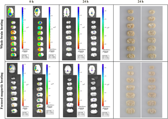
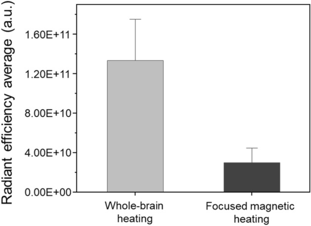
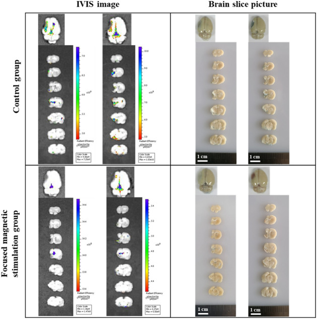
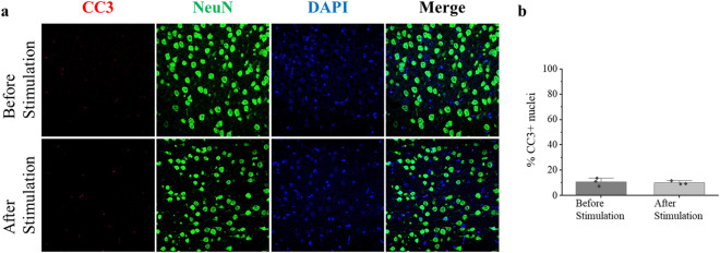
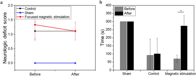
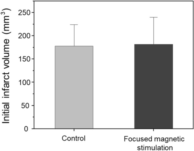
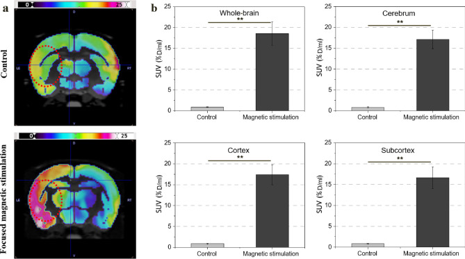
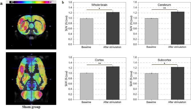
Magnetic heat-based brain stimulation of specific lesions could promote the restoration of impaired motor function caused by chronic stroke. We delivered localized stimulation by nanoparticle-mediated heat generation within the targeted brain area via focused magnetic stimulation. The middle cerebral artery occlusion model was prepared, and functional recovery in the chronic-phase stroke rat model was demonstrated by the therapeutic application of focused magnetic stimulation. We observed a transient increase in blood-brain barrier permeability at the target site of < 4 mm and metabolic brain activation at the target lesion. After focused magnetic stimulation, the rotarod score increased by 390 ± 28% (p < 0.05) compared to the control group. Standardized uptake value in the focused magnetic stimulation group increased by 2063 ± 748% (p < 0.01) compared to the control group. Moreover, an increase by 24 ± 5% (p < 0.05) was observed in the sham group as well. Our results show that non-invasive focused magnetic stimulation can safely modulate BBB permeability and enhance neural activation for chronic-phase stroke treatment in the targeted deep brain area.


Conflict of interest statement

The authors declare no competing interests.

Figures

Figure 1
Figure 1
Simulation and experimental results of temperature control. (a) FFP area, with the generated gradient field and SLP inside the area. The FFP is generated up to a distance where the gradient field and external field strength match, which is equivalent to the theoretical calculated distance of 7 mm. The theoretically expected size of the FFP can be calculated by the magnetic field H and gradient field G; dFFP2=HG. The magnetic heating that begins at the boundary becomes stronger as it approaches the center (SLPH2). An effective FFP can be expected to generate heat up to 3.5 mm in diameter, which is approximately half of its maximum size. (b) Ex vivo temperature increase with 15 mg/mL concentration. (c) Simulation results of temperature increase in vivo using different concentrations. (d) The brain baseline temperature before magnetic stimulation was maintained at 34 °C (n = 6), which was slightly lowered by anesthesia. The range of 39°–44° was set for the experiment to induce stimulation and change of BBB permeability.
Figure 2
Figure 2
Evans blue fluorescence. Each row shows the recovery of BBB permeability after whole-brain (n = 10) and focused heating (n = 10) in a normal rat. Each column shows BBB recovery immediately after focused heating and after 24 h, as well as a brain-slice image after 24 h. The IVIS image showed that the permeability does not change with a magnetic field alone in the right cerebral hemisphere where the particles are not spread. Only by the MNP to which the magnetic field was applied could affect the area, even in the left hemisphere region where the particles are spread, the BBB permeability change occurred only in that region where a focused magnetic field was applied.
Figure 3
Figure 3
Average IVIS fluorescence value within ROI. Evans blue leakage inside the ROIs of the whole-brain (n = 10) and focused heating (n = 10) group were quantitatively evaluated. IVIS fluorescence by Evans blue leak to which a focused magnetic field was applied showed a more localized area than when the magnetic field was applied to the entire area.
Figure 4
Figure 4
IVIS image and slice picture of stroke model. Brain damage due to stroke shows irreversible histological damage. Therefore, focused heating induces functional recovery by stimulating the remaining tissue around the lesion. By applying focused heating to the stroke model (n = 6), additional damage to the lesion while inducing nerve stimulation was evaluated. In stroke models, the BBB was destroyed as a symptom of the stroke; although some Evans blue fluorescence was observed after 48 h, magnetic stimulation did not result in further deterioration. In all groups, the transient increase in BBB permeability induced by hyperthermia was safely recovered over time.
Figure 5
Figure 5
Immunohistochemistry staining before and after stimulation of the sham group. Original magnification, ×200. (a) Each of the images is representative of a field of immuno-stained cells. Each merged image includes the nuclear marker DAPI, CC3, and NeuN. Damage by the surgical process before stimulation and damage by the stimulation protocol was not confirmed. No significant changes such as cell damage indicating apoptosis or morphological changes of neuronal nuclei were observed in CC3 and NeuN. (b) The bar charts show the percentage of CC3-positive cells relative to the percentage of DAPI stained nuclei, in cells before and after stimulation. Each data point represents a biological replicate (n = 3).
Figure 6
Figure 6
Behavioral assessment analysis and NDS for each group. (a) No deficit in the sham group (n = 9) was observed. The NDS value of the control group (n = 6) was maintained at 1 point. The NDS in the magnetic stimulation group (n = 9) was recovered after stimulation. (b) No decline or recovery of motor function was observed in the normal model of the sham group without stroke. In the control group, a stroke-induced decline in function was identified and did not recover over time. The magnetic stimulation group showed significant motor function recovery after stimulation (p < 0.05).
Figure 7
Figure 7
Initial infarct volume of stroke model prepared by MCAO method. The initial infarct size of stroke models prepared by the MCAO method was compared. The prepared stroke model (n = 15) was divided into a control group to which no magnetic heating was applied and a group to which focused magnetic heating was applied. Initial infarct size between the two groups showed no significant difference.
Figure 8
Figure 8
Changes in SUV index in the control (n = 6) and focused magnetic stimulation groups (n = 9). (a) PET/CT images of brain activation after heating in the control and focused magnetic stimulation groups. (b) Comparison of SUV index in the cerebrum, cortex, and subcortex, and whole-brain average with and without magnetic stimulation. In the group to which the focused magnetic field was applied to the stroke lesion, a significant increase in metabolic activity was observed (p < 0.005).
Figure 9
Figure 9
Changes in SUV index in the sham group (n = 9). (a) PET/CT images of brain activation before and after heating in the sham group. (b) SUV index at baseline and after heating in the cerebrum, cortex, and subcortex, and whole-brain average (p < 0.05). Increased metabolic activation of 24% was observed even in the same brain site without stroke.

Similar articles

Cited by

References

    1. Chollet F, et al. Fluoxetine for motor recovery after acute ischaemic stroke (FLAME): A randomised placebo-controlled trial. Lancet Neurol. 2011;10:123–130. doi: 10.1016/S1474-4422(10)70314-8. - DOI - PubMed
    1. Brenneman MM, Hylin MJ, Corwin JV. The time-dependent and persistent effects of amphetamine treatment upon recovery from hemispatial neglect in rats. Behav. Brain Res. 2015;293:153–161. doi: 10.1016/j.bbr.2015.07.032. - DOI - PubMed
    1. Tam RY, Fuehrmann T, Mitrousis N, Shoichet MS. Regenerative therapies for central nervous system diseases: A biomaterials approach. Neuropsychopharmacology. 2014;39:169–188. doi: 10.1038/npp.2013.237. - DOI - PMC - PubMed
    1. Nih LR, Carmichael ST. Hydrogels for brain repair after stroke: an emerging treatment option. Curr. Opin. Biotechnol. 2016;40:155–163. doi: 10.1016/j.copbio.2016.04.021. - DOI - PMC - PubMed
    1. Gu SY, Chang MC. The effects of 10-Hz repetitive transcranial magnetic stimulation on depression in chronic stroke patients. Brain Stimul. 2017;10:270–274. doi: 10.1016/j.brs.2016.10.010. - DOI - PubMed

Publication types