Review
. 2023 May;23(5):275-294.
doi: 10.1038/s41568-023-00557-7. Epub 2023 Mar 27.

Nucleotide metabolism: a pan-cancer metabolic dependency

Affiliations
Review

Nucleotide metabolism: a pan-cancer metabolic dependency

Nicholas J Mullen et al. Nat Rev Cancer. 2023 May.

Abstract

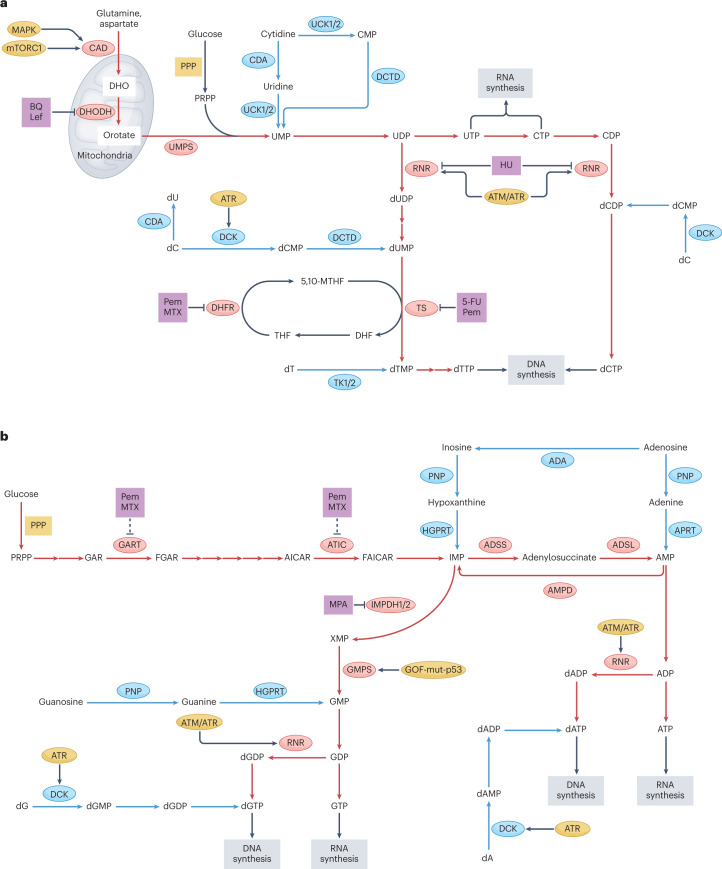
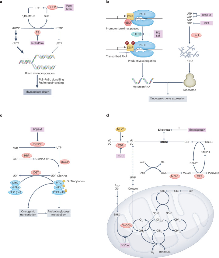
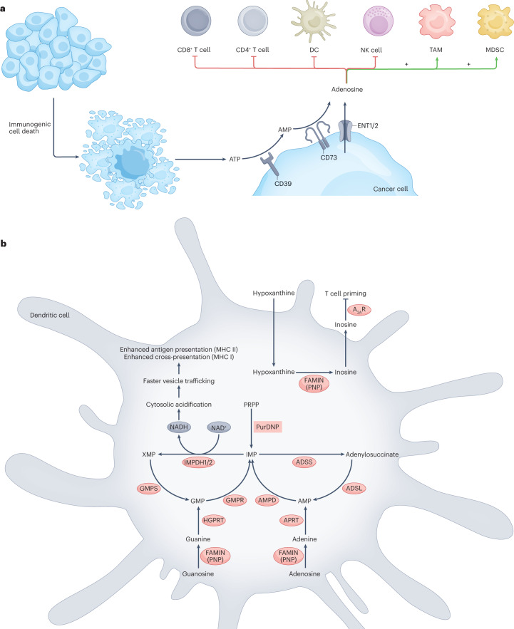
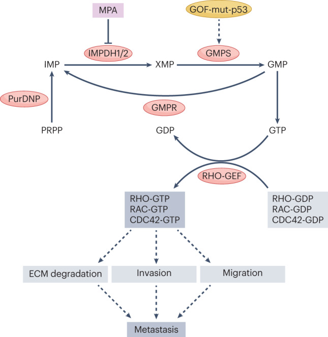
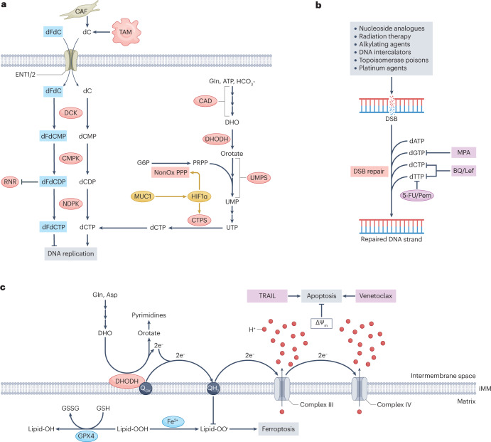
Metabolic alterations are a key hallmark of cancer cells, and the augmented synthesis and use of nucleotide triphosphates is a critical and universal metabolic dependency of cancer cells across different cancer types and genetic backgrounds. Many of the aggressive behaviours of cancer cells, including uncontrolled proliferation, chemotherapy resistance, immune evasion and metastasis, rely heavily on augmented nucleotide metabolism. Furthermore, most of the known oncogenic drivers upregulate nucleotide biosynthetic capacity, suggesting that this phenotype is a prerequisite for cancer initiation and progression. Despite the wealth of data demonstrating the efficacy of nucleotide synthesis inhibitors in preclinical cancer models and the well-established clinical use of these drugs in certain cancer settings, the full potential of these agents remains unrealized. In this Review, we discuss recent studies that have generated mechanistic insights into the diverse biological roles of hyperactive cancer cell nucleotide metabolism. We explore opportunities for combination therapies that are highlighted by these recent advances and detail key questions that remain to be answered, with the goal of informing urgently warranted future studies.


Conflict of interest statement

The authors declare no competing interests.

Figures

Fig. 1
Fig. 1. Biosynthetic pathways for pyrimidine and purine nucleotides, relevant inhibitors and oncogenic regulators.
The biosynthetic pathways leading to pyrimidine (part a) and purine (part b) nucleotides are shown. For both pyrimidines and purines, de novo synthesis entails a complex series of steps that transform amino acids and phosphoribosyl pyrophosphate (PRPP) into uridine monophosphate (UMP) or inosine monophosphate (IMP), respectively; salvage pathways recycle nucleosides and nucleobases to form nucleoside monophosphates (NMPs) or deoxy NMPs in a single step using ATP or PRPP, respectively. The de novo synthesis pathways are shown by red arrows and enzymes, and nucleotide salvage pathways are shown by blue arrows and enzymes. Inhibitors of these pathways are shown in purple boxes. Selected oncogenic regulators that influence these pathways are shown in yellow. In addition to the oncogenic regulators shown, the pentose phosphate pathway (PPP) can also be upregulated by the oncogenic factors hypoxia-inducible factor 1α (HIF1α), mucin 1 (MUC1), MYC and SREBP1. Dashed inhibitory arrows in part b indicate that the inhibitors pemetrexed (Pem) and methotrexate (MTX) inhibit GART and ATIC indirectly by disruption of the folic acid cycle. 5,10-MTHF, 5,10-methylenetetrahydrofolate; 5-FU, 5-fluorouracil; ADA, adenosine deaminase; ADSL, adenylosuccinate lyase; ADSS, adenylosuccinate synthetase; AICAR, 5-aminoimidazole-4-carboxamide-1-β-d-ribofuranoside; AMPD, AMP deaminase; APRT, adenine phosphoribosyltransferase; BQ, brequinar; CAD, carbamoyl phosphate synthetase II, aspartate transcarbamoylase and dihydroorotase; CDA, cytidine deaminase; CMP, cytidine monophosphate; CTP, cytidine triphosphate; dA, deoxyadenosine; dC, deoxycytidine; dCDP, deoxycytidine phosphate; DCK, deoxycytidine kinase; dCMP, deoxycytidine monophosphate; DCTD, deoxycytidylate deaminase; dCTP, deoxycytidine triphosphate; DHF, dihydrofolate; DHFR, DHF reductase; DHO, dihydroorotate; DHODH, DHO dehydrogenase; dT, thymidine; dTTP, deoxythymidine triphosphate; dU, deoxyuridine; dUMP, deoxyuridine monophosphate; FGAR, phosphoribosyl-N-formylglycineamide; FAICAR, 5-formamidoimidazole-4-carboxamide ribotide; GAR, glycineamide ribonucleotide; GMPS, GMP synthase; GOF-mut-p53, gain-of-function mutant p53; HGPRT, hypoxanthine–guanine phosphoribosyltransferase; HU, hydroxyurea; IMPDH1/2, IMP dehydrogenases 1 and 2; Lef, leflunomide; MAPK, mitogen-activated protein kinase; MPA, mycophenolic acid; mTOR, mammalian target of rapamycin; PNP, purine nucleoside phosphorylase; RNR, ribonucleotide reductase; THF, tetrahydrofolate; TK1/2, thymidine kinases 1 and 2; TS, thymidylate synthase; UCK1/2, uridine–cytidine kinases 1 and 2; UDP, uridine diphosphate; UMPS, UMP synthase; UTP, uridine triphosphate; XMP, xanthine monophosphate.
Fig. 2
Fig. 2. Nucleotides fuel cancer cell growth and proliferation.
a, Deoxythymidine triphosphate (dTTP) is required for the synthesis of DNA. Depletion of dTTP by the inhibition of thymidine synthase (TS) results in the accumulation of deoxyuridine triphosphate (dUTP) and an increase in the dUTP-to-dTTP ratio. As DNA polymerases cannot distinguish dUTP from dTTP, this leads to widespread misincorporation of uracil and a massive DNA damage response, ultimately resulting in thymineless death. b, Nucleotide triphosphates (NTPs) are required for the synthesis of RNA; inhibition of dihydroorotate dehydrogenase (DHODH) or inosine monophosphate dehydrogenase (IMPDH) depletes NTPs and disrupts RNA transcription. DHODH inhibition by brequinar (BQ) or leflunomide (Lef) impairs the productive elongation of RNA polymerase II (Pol II) by an unknown mechanism and thereby inhibits oncogenic transcription. Pol II pause release is normally triggered by positive transcription elongation factor B (P-TEFB), which phosphorylates DSIF and NELF; this causes DSIF to promote elongation and results in the dissociation of NELF (left). Pyrimidine depletion using BQ or Lef or purine depletion using mycophenolic acid (MPA) disrupt ribosomal RNA (rRNA) synthesis by starving RNA Pol I of NTP substrates and thereby hinder ribogenesis and the translation of oncogenic proteins (right). c, Uridine triphosphate (UTP) is required for the synthesis of uridine diphosphate N-acetylglucosamine (UDP-GlcNAc), the substrate for O-GlcNAcylation of proteins by O-GlcNAc transferase (OGT). O-GlcNAcylation of various proteins, including MYC, hypoxia-inducible factor 1α (HIF1α) and  phosphofructokinase 1 (PFK1), promotes cancer progression by diverse mechanisms. d, Generation of mitochondrial reactive oxygen species (mitoROS) requires high-energy electrons, either derived from α-ketoglutarate (αKG) via the TCA cycle or from DHODH via reduced coenzyme Q. DHODH-dependent mitoROS generation thus frees up αKG to produce cytosolic NADPH and reduced glutathione (GSH) via a pathway involving malate dehydrogenase 1 (MDH1) and malic enzyme 1 (ME1), allowing for control of cytosolic oxidative stress. Deoxyuridine (dU) produced downstream of DHODH serves as another cytosolic reactive oxygen species (ROS) quenching agent. Blockade of dU synthesis by inhibition of DHODH or cytidine deaminase (CDA) by the CDA inhibitor tetrahydrouridine (THU) accentuates oxidative and endoplasmic reticulum (ER) stress upon treatment with thapsigargin. 5,10-MTHF, 5,10-methylenetetrahydrofolate; 5-FU, 5-fluorouracil; CIII, complex III; CIV, complex IV; CTP, cytidine triphosphate; dC, deoxycytidine; DHF, dihydrofolate; DHFR, dihydrofolate reductase; DHO, dihydroorotate; dTMP, deoxythymidine monophosphate; dUMP, deoxyuridine monophosphate; GSSG, oxidized glutathione; MTX, methotrexate; MUC1, mucin 1; OAA, oxaloacetate; Pem, pemetrexed; PyrDNP, pyrimidine de novo pathway; rC, cytidine; rU, uridine; THF, tetrahydrofolate; UDP, uridine diphosphate; UGGP, UDP-glucose/galactose pyrophosphorylase; UMP, uridine monophosphate.
Fig. 3
Fig. 3. Altered nucleoside handling facilitates cancer cell immune evasion.
a, Cancer cell-directed adenosine build-up in the tumour microenvironment dampens anticancer immunity by signalling to various immune cell subsets. Extracellular ATP, which is a powerful immunostimulatory molecule, is cleared by CD39 and CD73, which are overexpressed in cancer cells. This process yields adenosine, which is immunosuppressive. Adenosine can also be directly exported from cancer cells through equilibrative nucleoside transporters 1 and 2 (ENT1/2). Extracellular adenosine dampens the antitumour activity of T cells, natural killer (NK) cells and dendritic cells (DCs); it also promotes the immunosuppressive activity of tumour-associated macrophages (TAMs) and myeloid-derived suppressor cells (MDSCs), thereby facilitating cancer cell immune evasion. b, The multifunctional purine catabolism enzyme fatty acid metabolism–immunity nexus (FAMIN) serves as a biochemical immune checkpoint by dampening DC-mediated T cell priming. Genetic inactivation of FAMIN in DCs accelerates antigen presentation downstream of cytosolic acidification resulting from increased NADH production by inosine monophosphate (IMP) dehydrogenases 1 and 2 (IMPDH1/2). FAMIN inactivation also abolishes DC secretion of inosine, which normally signals through adenosine receptors on T cells to depress T cell activation during priming. A2AR, adenosine A2A receptor; ADSL, adenylosuccinate lyase; ADSS, adenylosuccinate synthetase; AMPD, AMP deaminase; APRT, adenine phosphoribosyltransferase; GMPR, GMP reductase; GMPS, GMP synthase; HGPRT, hypoxanthine–guanine phosphoribosyltransferase; MHC I, MHC class I; PNP, purine nucleoside phosphorylase; PRPP, phosphoribosyl pyrophosphate; PurDNP, purine de novo pathway; XMP, xanthine monophosphate.
Fig. 4
Fig. 4. Supraphysiological GTP abundance promotes metastasis through the activation of RHO-family GTPases.
Small GTPase proteins of the RHO family support multiple behaviours necessary for metastasis, including extracellular matrix (ECM) degradation, basement membrane invasion and cell migration. These proteins are active in their GTP-bound state and inactivated by hydrolysis of bound GTP to GDP. Reactivation is catalysed by guanine nucleotide exchange factors (GEFs), which replace bound GDP with a new molecule of GTP. Depletion of GTP by genetic or pharmacological inhibition of inosine monophosphate (IMP) dehydrogenases 1 and 2 (IMPDH1/2) or GMP synthase (GMPS), or by overexpression of GMP reductase (GMPR), inhibits RHO-family GTPase activity by decreasing the GTP-bound fraction and blocks cancer metastasis in various animal models. GOF-mut-p53, gain-of-function mutant p53; MPA, mycophenolic acid; PRPP, phosphoribosyl pyrophosphate; PurDNP, purine de novo pathway; XMP, xanthine monophosphate.
Fig. 5
Fig. 5. Hyperactive nucleotide synthesis confers resistance to a range of therapeutic interventions.
a, Gemcitabine, or 2′,2′-difluorodeoxycytidine (dFdC), is in molecular competition with endogenous deoxycytidylate species at every stage of its metabolism, from uptake to phosphorylation to incorporation into elongating nascent DNA. The efficacy of gemcitabine can therefore be enhanced by depleting cellular deoxycytidylate nucleotides through either dihydroorotate dehydrogenase (DHODH) inhibition or disruption of oncogenic signalling through mucin 1 (MUC1) and hypoxia-inducible factor 1α (HIF1α) as this increases the proportion of gemcitabine-derived nucleotides relative to their endogenous counterparts. b, Various cytotoxic anticancer therapies operate by causing double-strand breaks (DSBs) in DNA. Adaptive resistance to these therapies emerges downstream of hyperactive DSB repair, which requires large amounts of deoxynucleotide triphosphates (dNTPs). Inhibition of dNTP synthesis therefore enhances the efficacy of these therapies in many contexts. c, Aside from its role in pyrimidine synthesis, DHODH also opposes ferroptosis by neutralizing mitochondrial lipid peroxide radicals through the generation of reduced ubiquinol and opposes apoptosis by contributing to mitochondrial membrane potential. DHODH inhibition therefore sensitizes cancer cells to ferroptosis and apoptosis-inducing agents such as GPX4 inhibitors and TNF-related apoptosis-inducing ligand (TRAIL), respectively. ΔΨm, mitochondrial membrane potential; 5-FU, 5-fluorouracil; BQ, brequinar; CAD, carbamoyl phosphate synthetase II, aspartate transcarbamoylase, dihydroorotase; CAF, cancer-associated fibroblast; CMPK, cytidine monophosphate kinase; CTPS, cytidine triphosphate synthase; dC, deoxycytidine; dCDP, deoxycytidine phosphate; DCK, deoxycytidine kinase; dCMP, deoxycytidine monophosphate; dCTP, deoxycytidine triphosphate; dFdCDP, gemcitabine diphosphate; dFdCMP, gemcitabine monophosphate; dFdCTP, gemcitabine triphosphate; DHO, dihydroorotate; dTTP, deoxythymidine triphosphate; ENT1/2, equilibrative nucleoside transporters 1 and 2; G6P, glucose 6-phosphate; GSH, reduced glutathione; GSSG, oxidized glutathione; IMM, inner mitochondrial membrane; Lef, leflunomide; MPA, mycophenolic acid; NDPK, nucleoside-diphosphate kinase; NonOx PPP, non-oxidative pentose phosphate pathway; Pem, pemetrexed; PRPP, phosphoribosyl pyrophosphate; RNR, ribonucleotide reductase; TAM, tumour-associated macrophage; UMP, uridine monophosphate; UMPS, UMP synthase; UTP, uridine triphosphate.

References

    1. Biancur DE, et al. Functional genomics identifies metabolic vulnerabilities in pancreatic cancer. Cell Metab. 2021;33:199–210. doi: 10.1016/j.cmet.2020.10.018. - DOI - PMC - PubMed
    1. Li L, et al. Identification of DHODH as a therapeutic target in small cell lung cancer. Sci. Transl Med. 2019;11:517. doi: 10.1126/scitranslmed.aaw7852. - DOI - PMC - PubMed
    1. Zhu XG, et al. Functional genomics in vivo reveal metabolic dependencies of pancreatic cancer cells. Cell Metab. 2021;33:211–221. doi: 10.1016/j.cmet.2020.10.017. - DOI - PMC - PubMed
    1. Sykes DB, et al. Inhibition of dihydroorotate dehydrogenase overcomes differentiation blockade in acute myeloid leukemia. Cell. 2016;167:171–186.e15. doi: 10.1016/j.cell.2016.08.057. - DOI - PMC - PubMed
    1. Koundinya M, et al. Dependence on the pyrimidine biosynthetic enzyme DHODH is a synthetic lethal vulnerability in mutant KRAS-driven cancers. Cell Chem. Biol. 2018;25:705–717.e11. doi: 10.1016/j.chembiol.2018.03.005. - DOI - PubMed

Publication types