

Effect of sclerostin inactivation in a mouse model of severe dominant osteogenesis imperfecta
- PMID: 36973504
- PMCID: PMC10043013
- DOI: 10.1038/s41598-023-32221-3
Effect of sclerostin inactivation in a mouse model of severe dominant osteogenesis imperfecta
Abstract
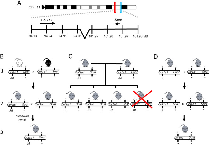
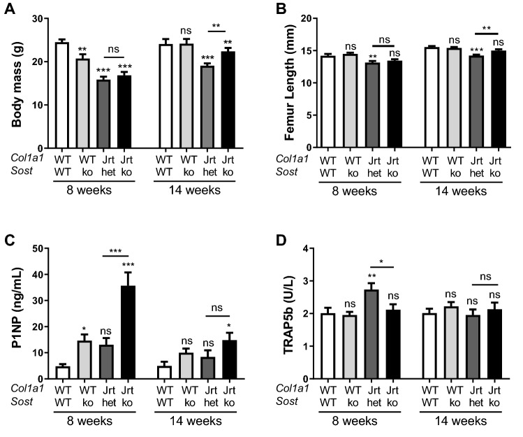
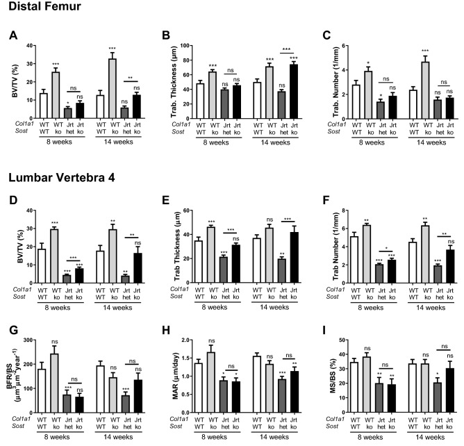
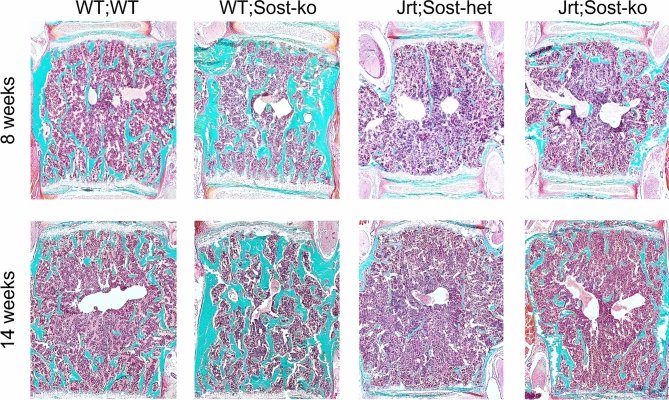
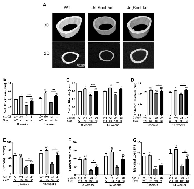
Osteogenesis imperfecta (OI) is a rare bone disease that is associated with fractures and low bone mass. Sclerostin inhibition is being evaluated as a potential approach to increase bone mass in OI. We had previously found that in Col1a1Jrt/+ mice, a model of severe OI, treatment with an anti-sclerostin antibody had a minor effect on the skeletal phenotype. In the present study, we assessed the effect of genetic sclerostin inactivation in the Col1a1Jrt/+ mouse. We crossed Col1a1Jrt/+ mice with Sost knockout mice to generate Sost-deficient Col1a1Jrt/+ mice and assessed differences between Col1a1Jrt/+ mice with homozygous Sost deficiency and Col1a1Jrt/+ mice with heterozygous Sost deficiency. We found that Col1a1Jrt/+ mice with homozygous Sost deficiency had higher body mass, femur length, trabecular bone volume, cortical thickness and periosteal diameter as well as increased biomechanical parameters of bone strength. Differences between genotypes were larger at the age of 14 weeks than at 8 weeks of age. Transcriptome analysis of RNA extracted from the tibial diaphysis revealed only 5 differentially regulated genes. Thus, genetic inactivation of Sost increased bone mass and strength in the Col1a1Jrt/+ mouse. It appears from these observations that the degree of Sost suppression that is required for eliciting a beneficial response can vary with the genetic cause of OI.
© 2023. The Author(s).
Conflict of interest statement
Frank Rauch: Ultragenyx Inc: Study grant to institution. Catabasis: Study grant to institution. Ibsen: Advisory Board. Sanofi: Advisory Board. The other authors declare no competing interests.
Figures
Similar articles
-
Effect of sclerostin antibody treatment in a mouse model of severe osteogenesis imperfecta.Bone. 2014 Sep;66:182-8. doi: 10.1016/j.bone.2014.06.015. Epub 2014 Jun 19. Bone. 2014. PMID: 24953712
-
Sclerostin-Antibody Treatment Decreases Fracture Rates in Axial Skeleton and Improves the Skeletal Phenotype in Growing oim/oim Mice.Calcif Tissue Int. 2020 May;106(5):494-508. doi: 10.1007/s00223-019-00655-5. Epub 2020 Feb 6. Calcif Tissue Int. 2020. PMID: 32025752
-
Effect of Anti-TGF-β Treatment in a Mouse Model of Severe Osteogenesis Imperfecta.J Bone Miner Res. 2019 Feb;34(2):207-214. doi: 10.1002/jbmr.3617. Epub 2018 Nov 29. J Bone Miner Res. 2019. PMID: 30357929
-
Osteogenesis imperfecta: prospects for molecular therapeutics.Mol Genet Metab. 2000 Sep-Oct;71(1-2):225-32. doi: 10.1006/mgme.2000.3039. Mol Genet Metab. 2000. PMID: 11001814 Review.
-
Sclerostin inhibition in rare bone diseases: Molecular understanding and therapeutic perspectives.J Orthop Translat. 2024 Jun 19;47:39-49. doi: 10.1016/j.jot.2024.05.004. eCollection 2024 Jul. J Orthop Translat. 2024. PMID: 39007037 Free PMC article. Review.
Cited by
-
Dickkopf-1 (DKK1) blockade mitigates osteogenesis imperfecta (OI) related bone disease.Mol Med. 2024 May 21;30(1):66. doi: 10.1186/s10020-024-00838-3. Mol Med. 2024. PMID: 38773377 Free PMC article.
-
Bone Quality and Mineralization and Effects of Treatment in Osteogenesis Imperfecta.Calcif Tissue Int. 2024 Dec;115(6):777-804. doi: 10.1007/s00223-024-01263-8. Epub 2024 Sep 4. Calcif Tissue Int. 2024. PMID: 39231826 Review.
-
Reduced orthodontic tooth movement in Ank knockout mice.JBMR Plus. 2025 Apr 17;9(6):ziaf064. doi: 10.1093/jbmrpl/ziaf064. eCollection 2025 Jun. JBMR Plus. 2025. PMID: 40390808 Free PMC article.
References
Publication types
MeSH terms
Substances
LinkOut - more resources
Full Text Sources
Medical
Molecular Biology Databases
Miscellaneous
