. 2023 Apr;26(4):673-681.
doi: 10.1038/s41593-023-01286-8. Epub 2023 Mar 27.

A consensus protocol for functional connectivity analysis in the rat brain

Joanes Grandjean  1   2 Gabriel Desrosiers-Gregoire  3   4 Cynthia Anckaerts  5   6 Diego Angeles-Valdez  7 Fadi Ayad  8   9   10 David A Barrière  11 Ines Blockx  5   6 Aleksandra Bortel  9   10   12 Margaret Broadwater  13   14   15 Beatriz M Cardoso  16 Marina Célestine  17 Jorge E Chavez-Negrete  18 Sangcheon Choi  19   20 Emma Christiaen  21 Perrin Clavijo  22 Luis Colon-Perez  23 Samuel Cramer  24 Tolomeo Daniele  25 Elaine Dempsey  26   27 Yujian Diao  28   29 Arno Doelemeyer  30 David Dopfel  24 Lenka Dvořáková  31 Claudia Falfán-Melgoza  32 Francisca F Fernandes  16 Caitlin F Fowler  3   8 Antonio Fuentes-Ibañez  18 Clément M Garin  17 Eveline Gelderman  33 Carla E M Golden  34 Chao C G Guo  33 Marloes J A G Henckens  33   35 Lauren A Hennessy  36   37 Peter Herman  38   39 Nita Hofwijks  33 Corey Horien  38 Tudor M Ionescu  40 Jolyon Jones  41 Johannes Kaesser  42 Eugene Kim  43 Henriette Lambers  44 Alberto Lazari  45 Sung-Ho Lee  13   14   15 Amanda Lillywhite  46   47 Yikang Liu  24 Yanyan Y Liu  48 Alejandra López-Castro  7 Xavier López-Gil  49 Zilu Ma  24 Eilidh MacNicol  43 Dan Madularu  8   50 Francesca Mandino  38 Sabina Marciano  40 Matthew J McAuslan  26 Patrick McCunn  51 Alison McIntosh  26   27 Xianzong Meng  33 Lisa Meyer-Baese  22 Stephan Missault  5   6 Federico Moro  52 Daphne M P Naessens  53 Laura J Nava-Gomez  54   55 Hiroi Nonaka  56 Juan J Ortiz  18 Jaakko Paasonen  31 Lore M Peeters  5   6 Mickaël Pereira  57 Pablo D Perez  24 Marjory Pompilus  58 Malcolm Prior  59 Rustam Rakhmatullin  60 Henning M Reimann  61 Jonathan Reinwald  62 Rodrigo Triana Del Rio  63 Alejandro Rivera-Olvera  33 Daniel Ruiz-Pérez  60 Gabriele Russo  64 Tobias J Rutten  33 Rie Ryoke  56 Markus Sack  32 Piergiorgio Salvan  45 Basavaraju G Sanganahalli  38   39 Aileen Schroeter  65 Bhedita J Seewoo  36   37   66 Erwan Selingue  67 Aline Seuwen  65 Bowen Shi  68 Nikoloz Sirmpilatze  69   70   71 Joanna A B Smith  72   73   74 Corrie Smith  22 Filip Sobczak  19   20 Petteri J Stenroos  75 Milou Straathof  76 Sandra Strobelt  42 Akira Sumiyoshi  56   77 Kengo Takahashi  19   20 Maria E Torres-García  18 Raul Tudela  78 Monica van den Berg  5   6 Kajo van der Marel  76 Aran T B van Hout  33 Roberta Vertullo  33 Benjamin Vidal  57 Roël M Vrooman  33 Victora X Wang  79 Isabel Wank  42 David J G Watson  46 Ting Yin  80 Yongzhi Zhang  81 Stefan Zurbruegg  82 Sophie Achard  83 Sarael Alcauter  18 Dorothee P Auer  59   84 Emmanuel L Barbier  75 Jürgen Baudewig  69 Christian F Beckmann  33   45 Nicolau Beckmann  30 Guillaume J P C Becq  85 Erwin L A Blezer  76 Radu Bolbos  86 Susann Boretius  69   70   71 Sandrine Bouvard  57 Eike Budinger  87   88 Joseph D Buxbaum  34 Diana Cash  43 Victoria Chapman  46   47   84 Kai-Hsiang Chuang  89 Luisa Ciobanu  67 Bram F Coolen  53 Jeffrey W Dalley  41 Marc Dhenain  17 Rick M Dijkhuizen  76 Oscar Esteban  90 Cornelius Faber  44 Marcelo Febo  58 Kirk W Feindel  66 Gianluigi Forloni  91 Jérémie Fouquet  3 Eduardo A Garza-Villarreal  7 Natalia Gass  32 Jeffrey C Glennon  92 Alessandro Gozzi  93 Olli Gröhn  31 Andrew Harkin  26   27 Arend Heerschap  94 Xavier Helluy  64   95 Kristina Herfert  40 Arnd Heuser  96 Judith R Homberg  33 Danielle J Houwing  33 Fahmeed Hyder  38   39 Giovanna Diletta Ielacqua  96 Ileana O Jelescu  28 Heidi Johansen-Berg  45 Gen Kaneko  97 Ryuta Kawashima  56 Shella D Keilholz  22 Georgios A Keliris  5   6 Clare Kelly  27   98   99 Christian Kerskens  27   100 Jibran Y Khokhar  51 Peter C Kind  72   73   74   101 Jean-Baptiste Langlois  86 Jason P Lerch  45   102 Monica A López-Hidalgo  55 Denise Manahan-Vaughan  64 Fabien Marchand  103 Rogier B Mars  33   45 Gerardo Marsella  104 Edoardo Micotti  91 Emma Muñoz-Moreno  49 Jamie Near  3   105 Thoralf Niendorf  61   106 Willem M Otte  76   107 Patricia Pais-Roldán  19   108 Wen-Ju Pan  22 Roberto A Prado-Alcalá  18 Gina L Quirarte  18 Jennifer Rodger  36   37 Tim Rosenow  109 Cassandra Sampaio-Baptista  45   110 Alexander Sartorius  62 Stephen J Sawiak  111 Tom W J Scheenen  94   112 Noam Shemesh  16 Yen-Yu Ian Shih  13   14   15   113 Amir Shmuel  8   9   10   12   114 Guadalupe Soria  115 Ron Stoop  63 Garth J Thompson  68 Sally M Till  72   73   74 Nick Todd  81 Annemie Van Der Linden  5   6 Annette van der Toorn  76 Geralda A F van Tilborg  76 Christian Vanhove  21 Andor Veltien  94 Marleen Verhoye  5   6 Lydia Wachsmuth  44 Wolfgang Weber-Fahr  32 Patricia Wenk  87 Xin Yu  19   116 Valerio Zerbi  117   118 Nanyin Zhang  24 Baogui B Zhang  48 Luc Zimmer  57   86   119 Gabriel A Devenyi  3   120 M Mallar Chakravarty  3   8   120 Andreas Hess  42
Affiliations

A consensus protocol for functional connectivity analysis in the rat brain

Joanes Grandjean et al. Nat Neurosci. 2023 Apr.

Erratum in

  • Author Correction: A consensus protocol for functional connectivity analysis in the rat brain.
    Grandjean J, Desrosiers-Gregoire G, Anckaerts C, Angeles-Valdez D, Ayad F, Barrière DA, Blockx I, Bortel A, Broadwater M, Cardoso BM, Célestine M, Chavez-Negrete JE, Choi S, Christiaen E, Clavijo P, Colon-Perez L, Cramer S, Daniele T, Dempsey E, Diao Y, Doelemeyer A, Dopfel D, Dvořáková L, Falfán-Melgoza C, Fernandes FF, Fowler CF, Fuentes-Ibañez A, Garin CM, Gelderman E, Golden CEM, Guo CCG, Henckens MJAG, Hennessy LA, Herman P, Hofwijks N, Horien C, Ionescu TM, Jones J, Kaesser J, Kim E, Lambers H, Lazari A, Lee SH, Lillywhite A, Liu Y, Liu YY, López-Castro A, López-Gil X, Ma Z, MacNicol E, Madularu D, Mandino F, Marciano S, McAuslan MJ, McCunn P, McIntosh A, Meng X, Meyer-Baese L, Missault S, Moro F, Naessens DMP, Nava-Gomez LJ, Nonaka H, Ortiz JJ, Paasonen J, Peeters LM, Pereira M, Perez PD, Pompilus M, Prior M, Rakhmatullin R, Reimann HM, Reinwald J, Del Rio RT, Rivera-Olvera A, Ruiz-Pérez D, Russo G, Rutten TJ, Ryoke R, Sack M, Salvan P, Sanganahalli BG, Schroeter A, Seewoo BJ, Selingue E, Seuwen A, Shi B, Sirmpilatze N, Smith JAB, Smith C, Sobczak F, Stenroos PJ, Straathof M, Strobelt S, Sumiyoshi A, Takahashi K, Torres-García ME, Tudela R, van den Berg M, van der Marel K, … See abstract for full author list ➔ Grandjean J, et al. Nat Neurosci. 2023 Jun;26(6):1127-1128. doi: 10.1038/s41593-023-01328-1. Nat Neurosci. 2023. PMID: 37072562 No abstract available.

Abstract

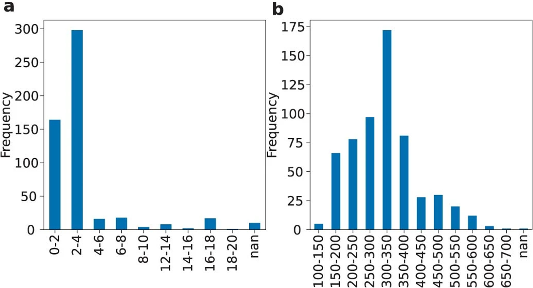
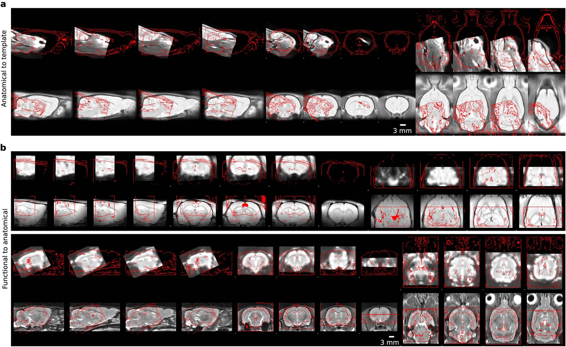
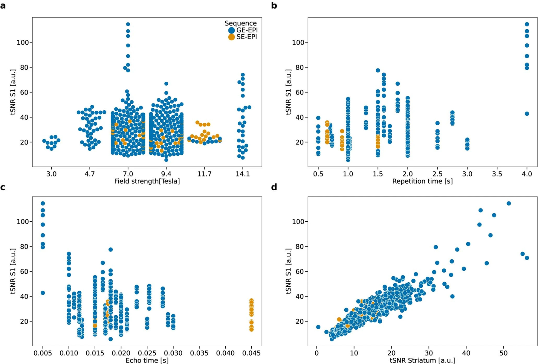
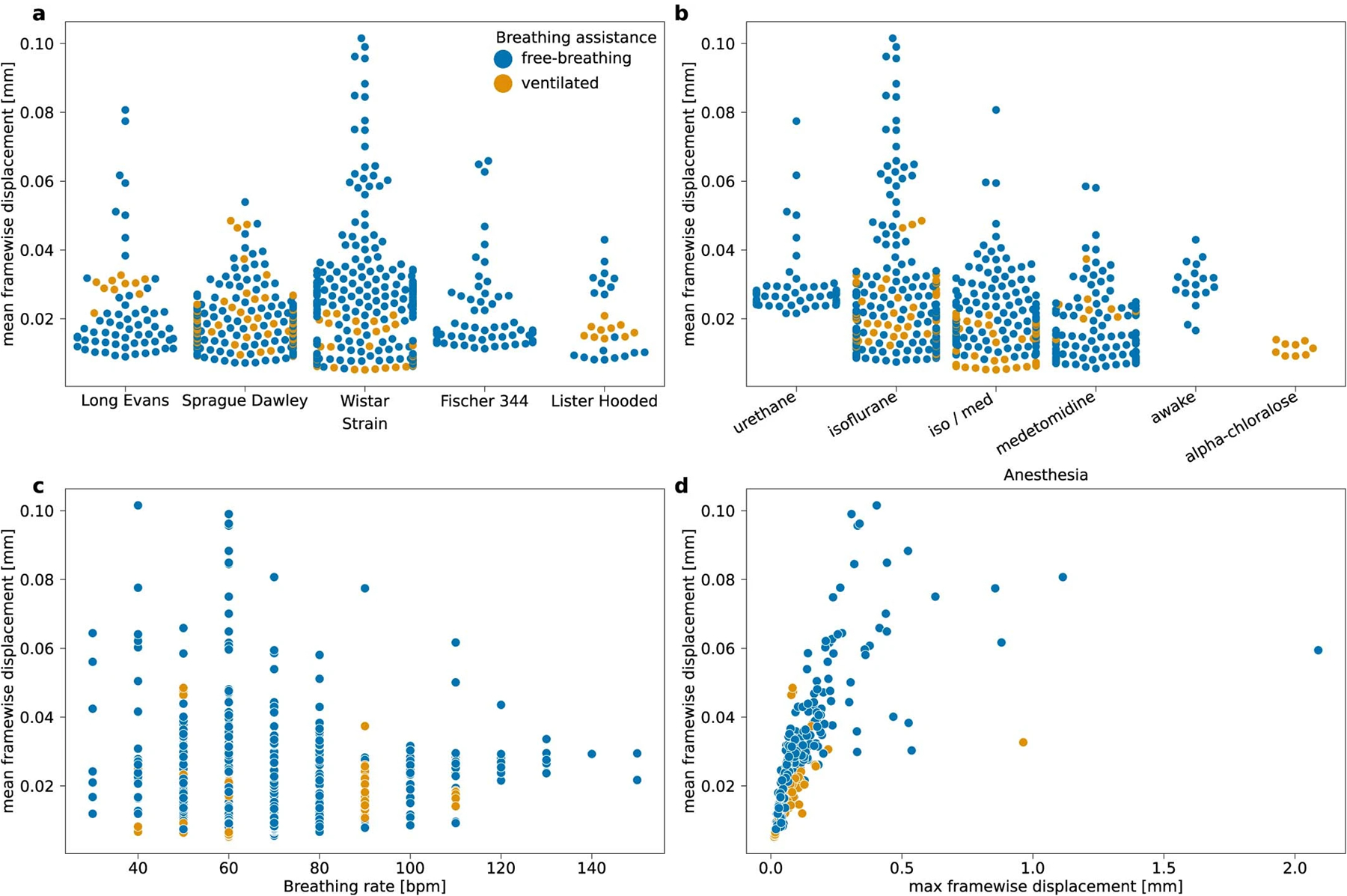
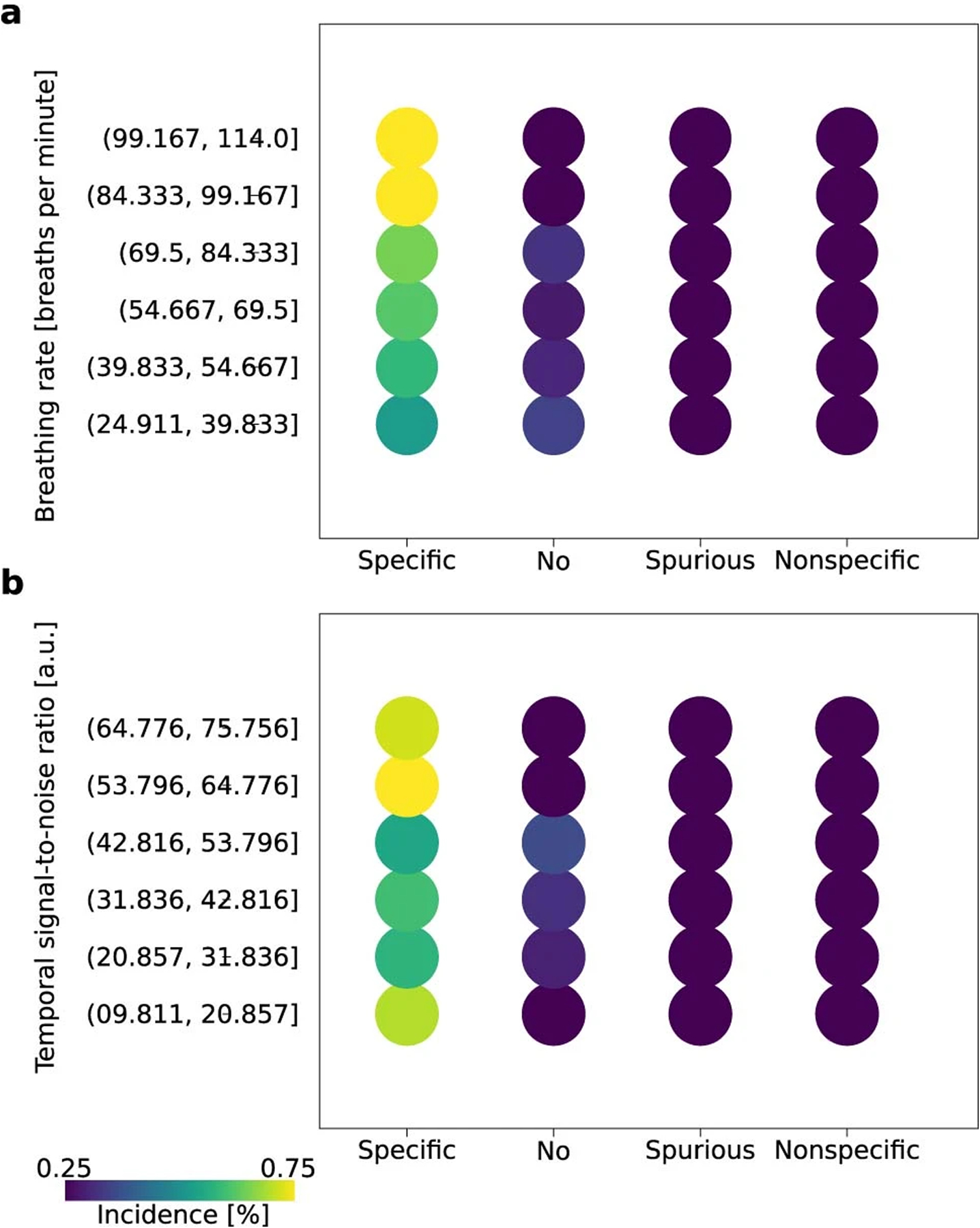
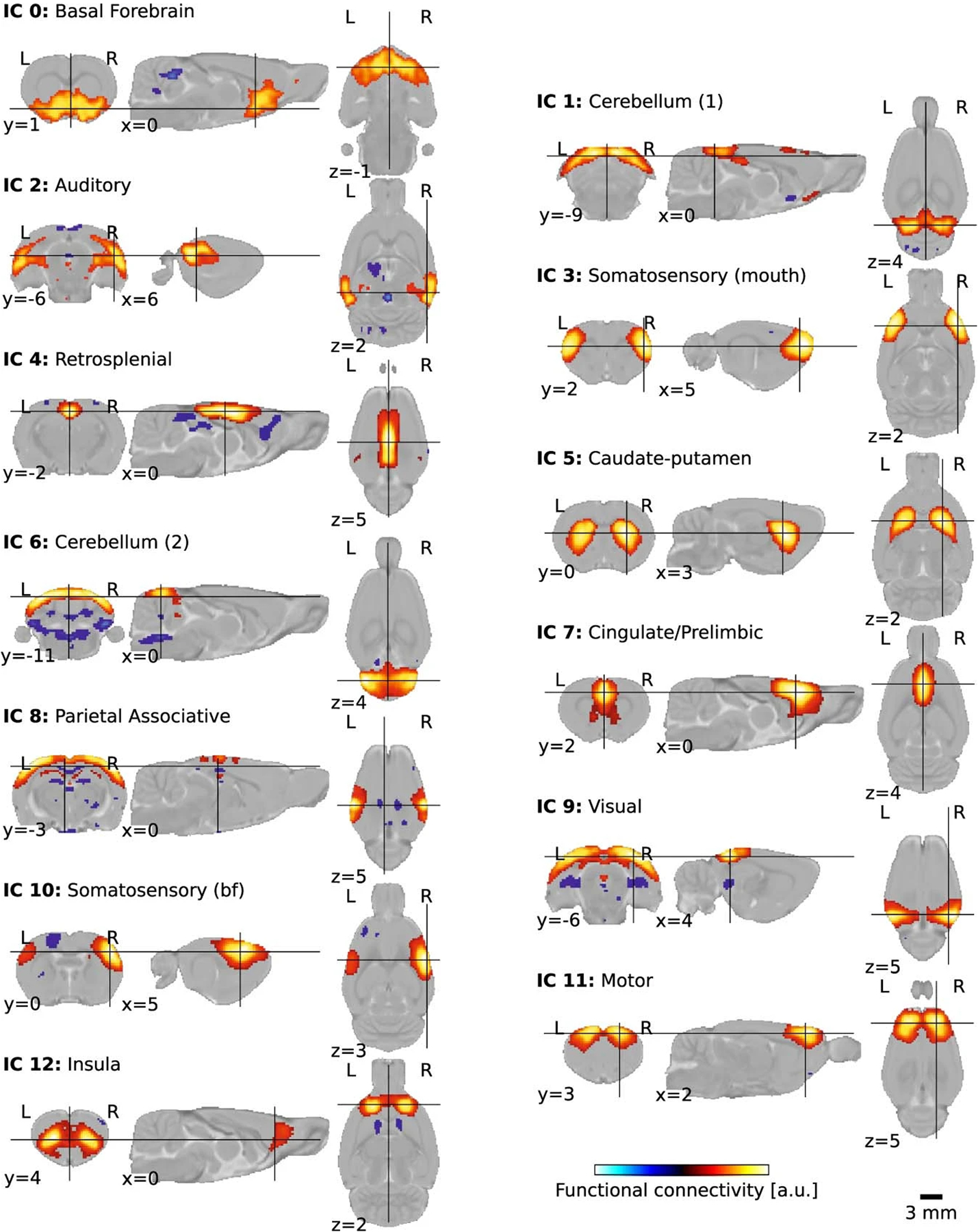
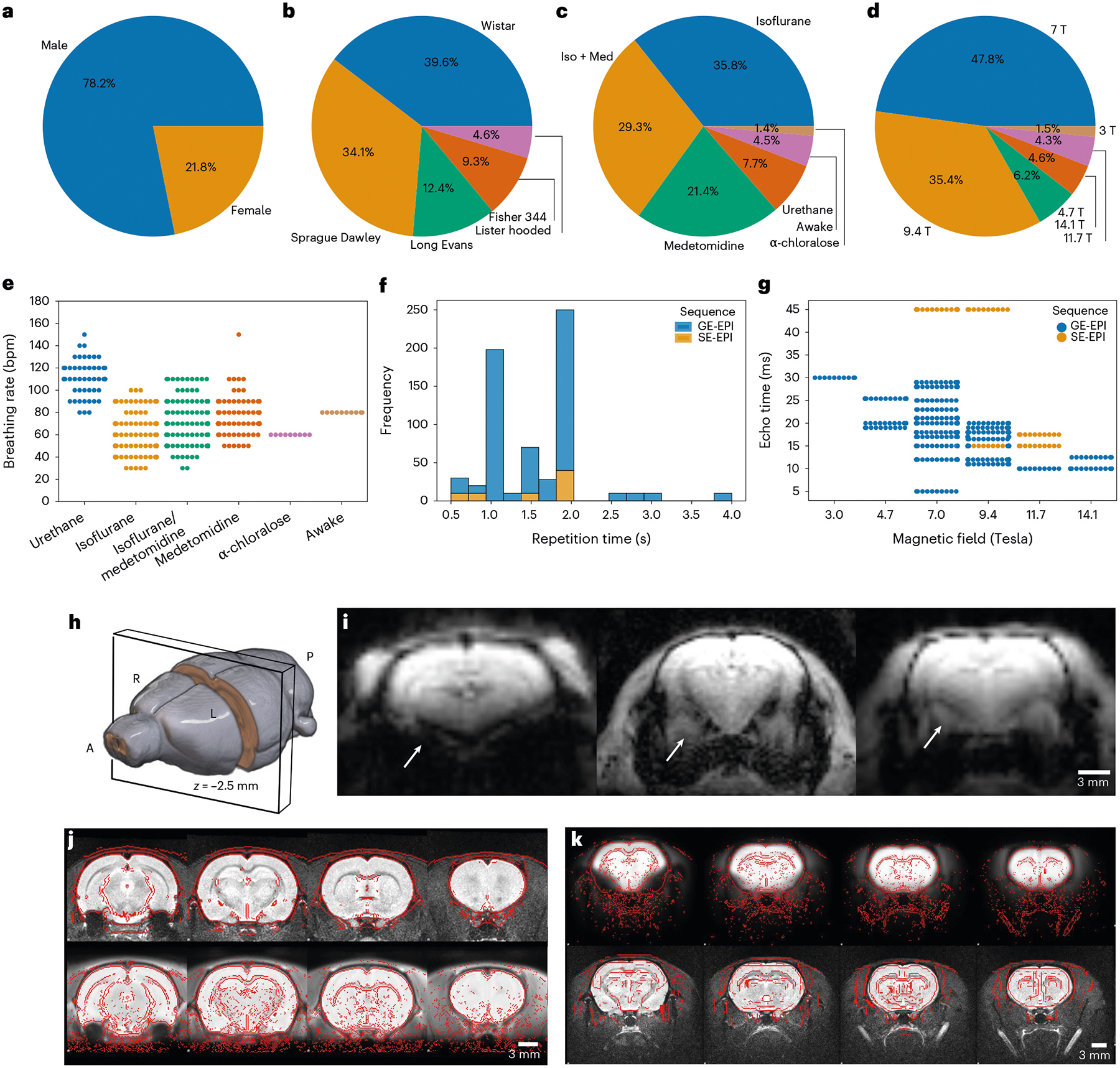
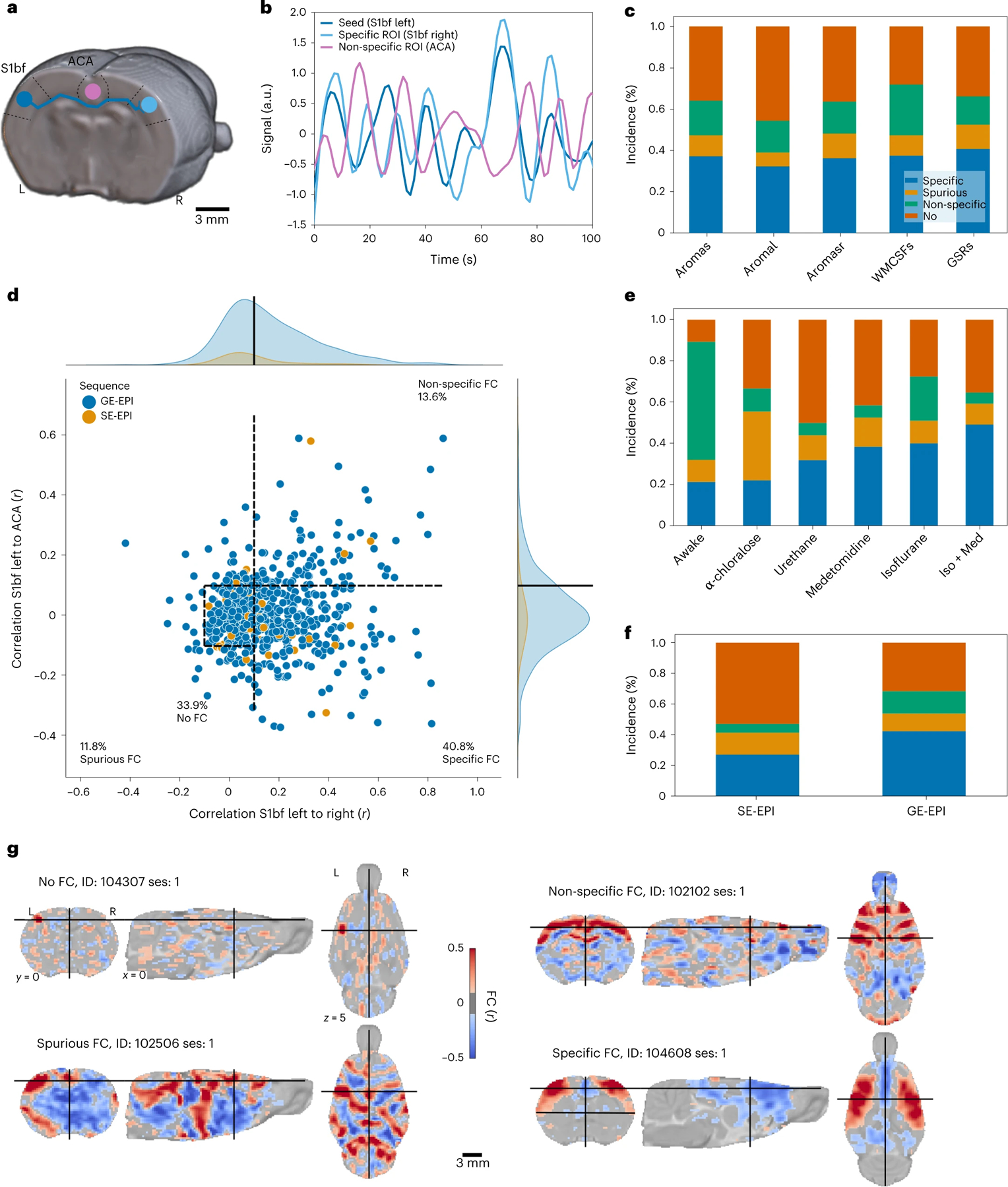
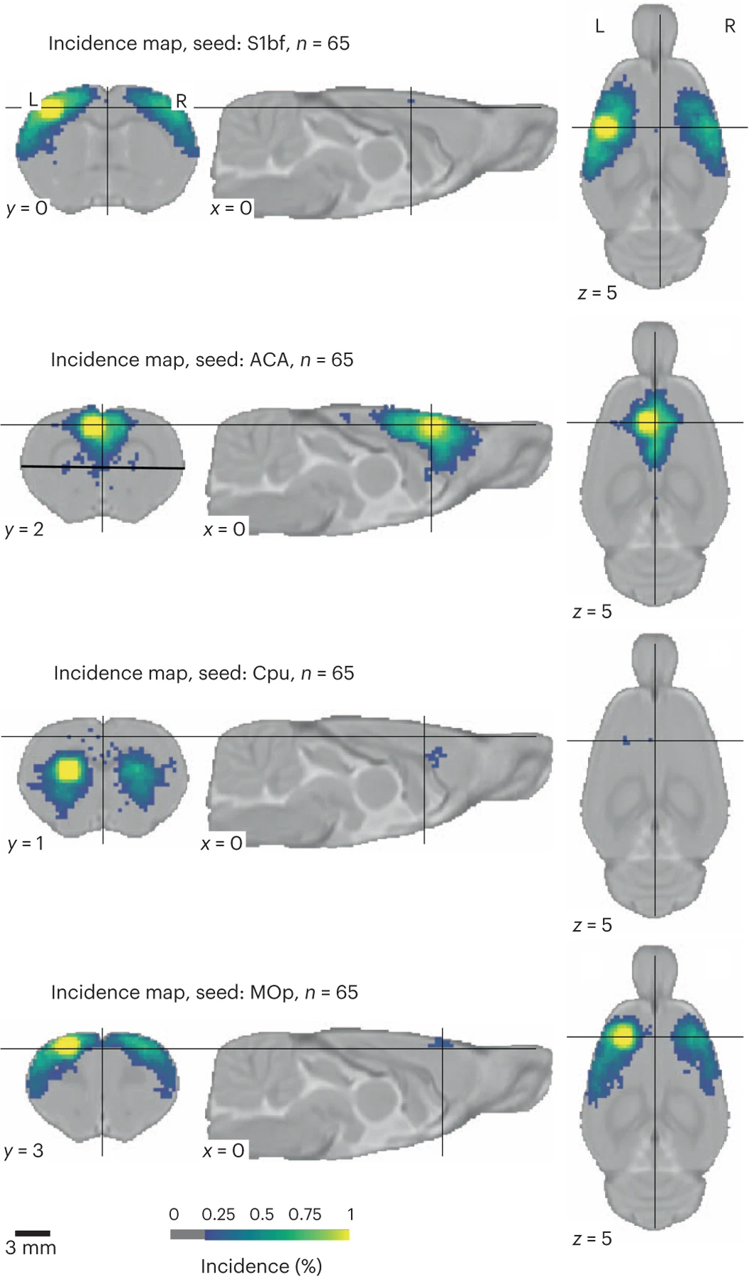
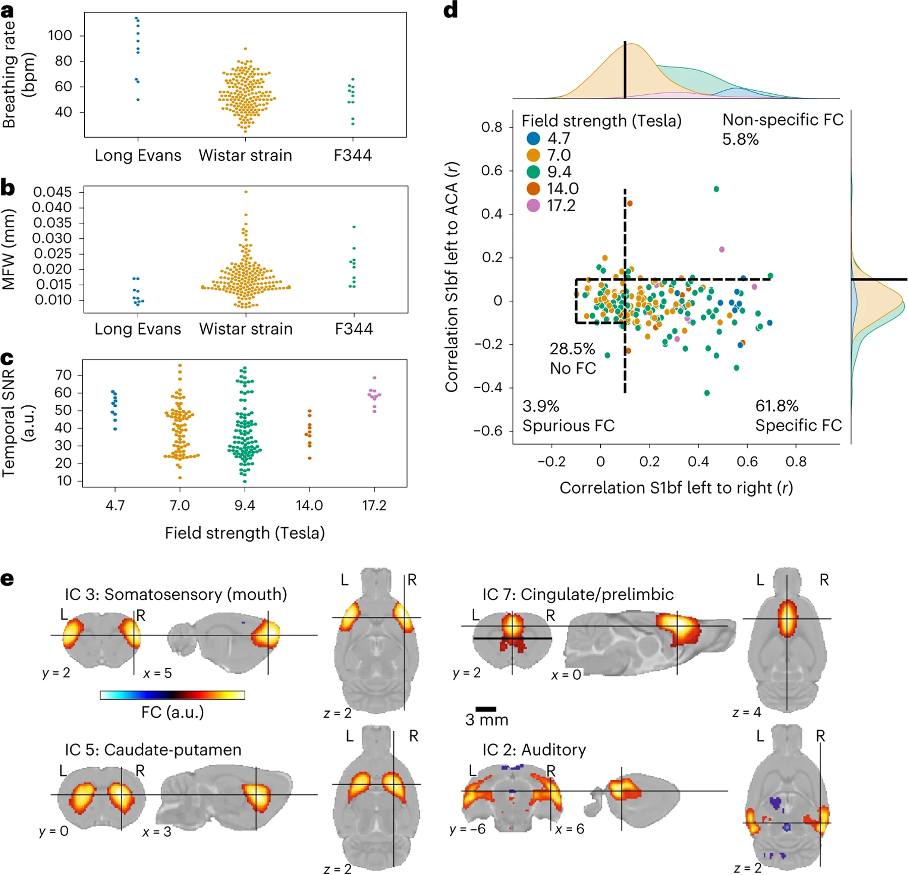
Task-free functional connectivity in animal models provides an experimental framework to examine connectivity phenomena under controlled conditions and allows for comparisons with data modalities collected under invasive or terminal procedures. Currently, animal acquisitions are performed with varying protocols and analyses that hamper result comparison and integration. Here we introduce StandardRat, a consensus rat functional magnetic resonance imaging acquisition protocol tested across 20 centers. To develop this protocol with optimized acquisition and processing parameters, we initially aggregated 65 functional imaging datasets acquired from rats across 46 centers. We developed a reproducible pipeline for analyzing rat data acquired with diverse protocols and determined experimental and processing parameters associated with the robust detection of functional connectivity across centers. We show that the standardized protocol enhances biologically plausible functional connectivity patterns relative to previous acquisitions. The protocol and processing pipeline described here is openly shared with the neuroimaging community to promote interoperability and cooperation toward tackling the most important challenges in neuroscience.


Conflict of interest statement

Competing interests

A.S. is an employee of Bruker, the manufacturer of preclinical MRI systems used for the acquisition of most of the datasets in this collection. E.L.B. is a consultant for Bruker. B.V. is an employee of Theranexus. S.Z., A.D. and N.B. are employees of Novartis Pharma AG. T.N. is founder and CEO of MRI.TOOLS GmbH. The other authors declare no competing interests.

Figures

Extended Data Fig. 1 |
Extended Data Fig. 1 |. Age and weight distributions.
Age (a) and weight (b) distribution for the rats in the MultiRat_rest collection.
Extended Data Fig. 2 |
Extended Data Fig. 2 |. Quality control examples.
Failed quality controls for anatomical to template registrations (a) and functional to anatomical registrations (b). The top rows are the moving objects, bottom rows are the reference objects. The red lines indicate the outlines of the other object. Four slices along the sagittal, axial, and coronal axis are shown for each case.
Extended Data Fig. 3 |
Extended Data Fig. 3 |. Temporal signal-to-noise ratio.
Temporal signal-to-noise ratio in the sensory cortex (tSNR S1) in the MultiRat_rest dataset collection as a function of (a) magnetic field strength, (b) repetition time, (c) echo time, (d) temporal signal-to-noise ratio in the striatum.
Extended Data Fig. 4 |
Extended Data Fig. 4 |. Framewise displacement.
MFW in the MultiRat_rest dataset collection as a function of (a) strain, (b) anesthesia, (c) breathing rate, (d) maximal framewise displacement.
Extended Data Fig. 5 |
Extended Data Fig. 5 |. FC in the default-mode network.
The reference seed is positioned in the anterior cingulate cortex (Fig. 2a), the specific region-of-interest is positioned 3.3 mm posterior in the cingulate cortex and the nonspecific region-of-interest is positioned in the S1bf.
Extended Data Fig. 6 |
Extended Data Fig. 6 |. StandardRat dataset description.
a. Strain. b. Sex. c. Field strength. d. Weight. e. Breathing rate as a function of MFW. f. FC specificity as a function of confound correction models.
Extended Data Fig. 7 |
Extended Data Fig. 7 |. FC incidence.
Incidence of FC at the group level in the StandardRat collection for four selected seeds (n = 21 datasets, n ~ 10 subjects per dataset). Connectivity incidence is improved in the StandardRat collection relative to MultiRat_rest (Fig. 3).
Extended Data Fig. 8 |
Extended Data Fig. 8 |. Between-datasets connectivity comparisons.
FC category comparison between MultiRat_rest and StandardRat (a) and between the awake datasets of MultiRat_rest and the awake dataset from Lui et al. 2020 (b).
Extended Data Fig. 9 |
Extended Data Fig. 9 |. Connectivity specificity as a function of breathing rate and signal-to-noise ratio.
FC specificity as a function of binned breathing rate (a) AND temporal signal-to-noise ratio (b) in the StandardRat collection. The percentage of each condition is size and color-coded. High levels of connectivity specificity were achieved in scans where the breathing rates were in the 84 to 114 bpm range. Similarly, higher connectivity specificity incidences were found when the cortical temporal signal-to-noise ratio was > 53. These observations support the notion of an optimal breathing rate when applying the StandardRat protocol, along with temporal signal-to-noise ratio and movement targets.
Extended Data Fig. 10 |
Extended Data Fig. 10 |. Group independent components analysis.
Plausible independent components overlapping with known rodent networks, obtained after group-level decomposition with n = 20 components. Labels are based on the SIGMA anatomical atlas.
Fig. 1 |
Fig. 1 |. MultiRat_rest dataset description.
a, Sex. b, Strain. c, Anesthesia. d, Magnetic field strength. e, Breathing rate as a function of anesthesia. f, Repetition time. g, Echo time as a function of magnetic field strength. h, Slice position for the examples. i, Example of representative raw functional images. Arrows indicate different susceptibility artifact-related geometric distortions in the amygdala. j, Successful anatomical (top) to standard (bottom) space registration. Red lines indicate the outlines of the standard image (top) and the anatomical (bottom). k, Successful functional (top) to anatomical (bottom) registration. Red lines indicate the outlines of the anatomical image (top) and the functional (bottom).
Fig. 2 |
Fig. 2 |. FC specificity.
a, Diagram illustrating the logic behind FC specificity. The sensory (barrel field, S1bf) area (blue) chiefly projects to the contralateral homotopic area (light blue) but not to the ACA area (purple). b, Example of temporal dynamics in the resting-state signal. Correlated signal between the ipsilateral and contralateral S1bf and anti-correlated signal from the ACA. c, Distribution of FC categories as a function of confound correction models. d, FC in left S1bf relative to specific (right S1bf) and non-specific (ACA) regions of interest using the global regression correction model. Dots represent scans (n = 638 rats); dotted lines indicate the thresholds used to delineate the categories. e, Distribution of connectivity categories as a function of anesthesia. Example of individual seed-based analysis maps for each connectivity category. f, Distribution of connectivity categories as a function of imaging sequence (EPI, echo-planar imaging; GE, gradient echo; SE, spin echo). Group-level FC incidence map (n = 65 datasets). g, Example of individual seed-based analysis maps for each connectivity category. a.u., arbitrary units; ROI, region of interest, WMCSFs, white-matter + cerebrospinal fluid.
Fig. 3 |
Fig. 3 |. Incidence of FC at the group level (n = 65 datasets of n ~ 10 subjects per dataset) for four seeds.
Cpu, caudate-putamen; MOp, primary motor area. MOp and CPu seeds were generally observed in 50–75% of the datasets.
Fig. 4 |
Fig. 4 |. StandardRat dataset description.
a, Breathing rate (bpm) as a function of strain. b, MFW as a function of strain. c, Temporal signal-to-noise ratio in the sensory cortex as a function of field strength. d, FC in left S1bf relative to specific (right S1bf) and non-specific (ACA) regions of interest using the global regression correction model. Dots represent scans (n = 207 rats); dotted lines indicate the thresholds used to delineate the categories. e, Representative independent components. a.u., arbitrary units.

References

    1. Elam JS et al. The Human Connectome Project: a retrospective. Neuroimage 244, 118543 (2021). - PMC - PubMed
    1. Mennes M, Biswal B, Castellanos FX & Milham MP Making data sharing work: the FCP/INDI experience. Neuroimage 82, 683 (2013). - PMC - PubMed
    1. Miller KL et al. Multimodal population brain imaging in the UK Biobank prospective epidemiological study. Nat. Neurosci 19, 1523–1536 (2016). - PMC - PubMed
    1. Smith SM et al. Resting-state fMRI in the Human Connectome Project. Neuroimage 80, 144–168 (2013). - PMC - PubMed
    1. Van Essen DC et al. The WU-Minn Human Connectome Project: an overview. Neuroimage 80, 62–79 (2013). - PMC - PubMed

Publication types