. 2023 Mar 27;18(1):248.
doi: 10.1186/s13018-023-03717-5.

Daurisoline attenuates H2O2-induced chondrocyte autophagy by activating the PI3K/Akt/mTOR signaling pathway

Affiliations

Daurisoline attenuates H2O2-induced chondrocyte autophagy by activating the PI3K/Akt/mTOR signaling pathway

Yang Zhang et al. J Orthop Surg Res. .

Erratum in

Abstract

Background: Osteoarthritis (OA) is a chronic degenerative joint disease characterized by cartilage degeneration and intra-articular inflammation. Daurisoline (DAS) is an isoquinoline alkaloid isolated from Rhizoma Menispermi, whose antitumor and anti-inflammatory pharmacological effects have been demonstrated, but the effects of DAS on OA have rarely been researched. In this study, we aimed to explore the potential role of DAS in OA and its partial mechanism.

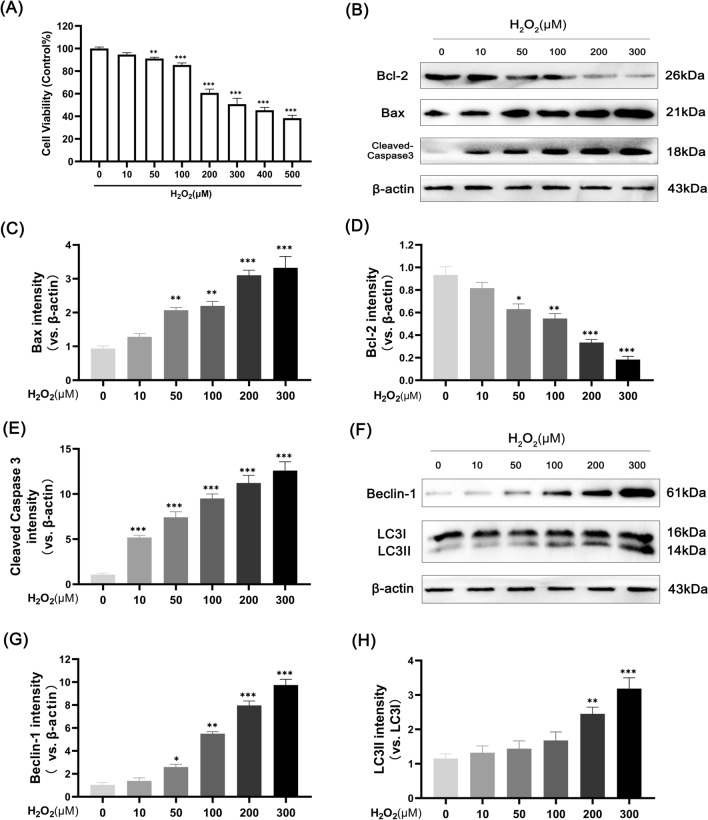
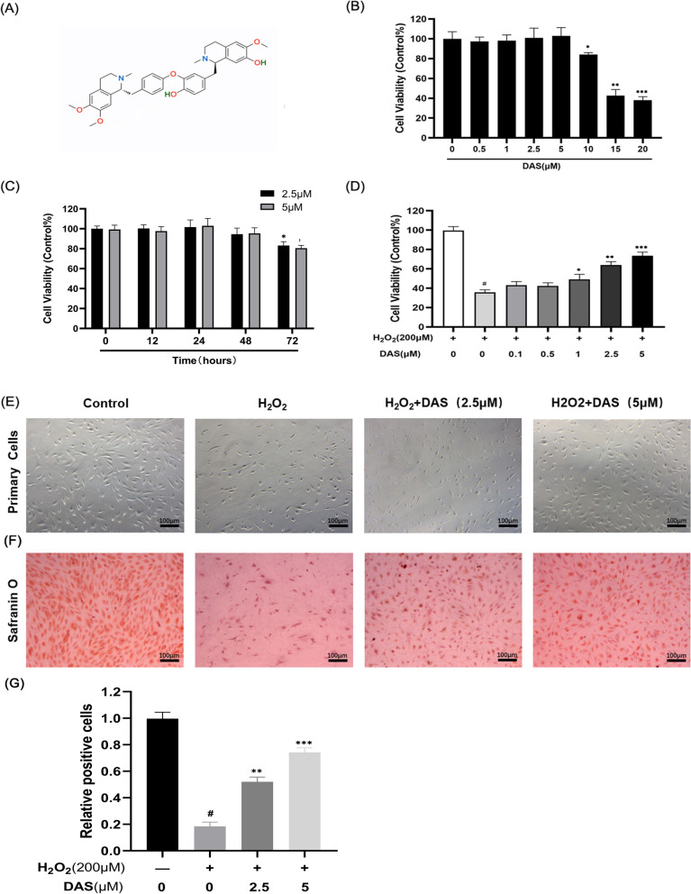
Materials and methods: The cytotoxicity of H2O2 and DAS toward chondrocytes was detected by the Cell Counting Kit-8 assay. Safranin O staining was used to detect chondrocyte phenotype changes. Cell apoptosis was measured by both flow cytometry and quantitative analysis of the protein levels of the apoptosis-related factors Bax, Bcl-2 and cleaved caspase-3 by western blot. Western blotting and immunofluorescence were used to assess the expression of the autophagy-related proteins LC3, Beclin-1 and p62. In addition, key signal pathway targets and matrix-degrading indicators were measured by western blot.

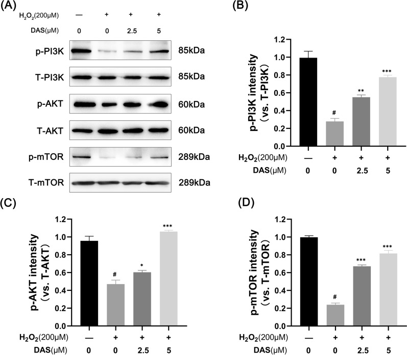
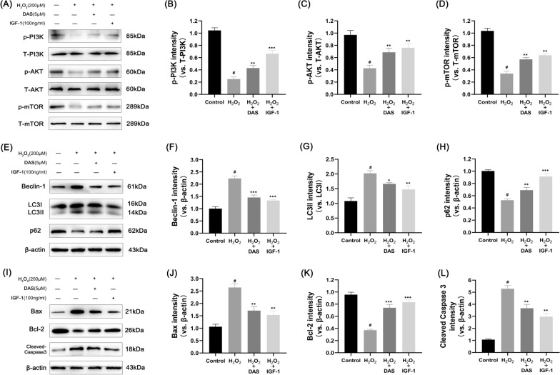
Results: Our results indicated that H2O2 induced human chondrocyte apoptosis and activated autophagy in a dose-dependent manner. DAS treatment dose-dependently reversed the expression of apoptosis-related proteins (Bax, Bcl-2 and cleaved caspase3) and the apoptosis rate induced by H2O2. Western blot and immunofluorescence analyses showed that DAS decreased the H2O2-induced upregulation of the autophagy marker Beclin-1 and the LC3 II/LC3 I ratio and upregulated the p62 protein level. Mechanistically, DAS inhibited autophagy through the activation of the classical PI3K/AKT/mTOR signaling pathway and protected chondrocytes from apoptosis. In addition, DAS alleviated the H2O2-induced degradation of type II collagen and the high expression of matrix metalloproteinase 3 (MMP3) and MMP13.

Conclusion: Our research demonstrated that DAS alleviated chondrocyte autophagy caused by H2O2 through activation of the PI3K/AKT/mTOR signaling pathway and protected chondrocytes from apoptosis and matrix degradation. In conclusion, these findings suggest that DAS may serve as a promising therapeutic strategy for OA.

Keywords: Apoptosis; Autophagy; Daurisoline; Osteoarthritis; PI3K/AKT/mTOR signaling pathway.


Conflict of interest statement

The authors declare that they have no known competing financial interests or personal relationships that could have appeared to influence the work reported in this paper.

Figures

Fig. 1
Fig. 1
H2O2 induces apoptosis and activates autophagy in human chondrocytes. A Cell cytotoxicity detected by CCK-8 assay. Chondrocytes were treated with 0, 10, 50, 100, 200, 300, 400 and 500 μM H2O2 for 4 h. BE western blot analysis and quantitative correlation analysis of Bax, cleaved caspase3 and Bcl-2 in chondrocytes. FH western blot analysis and correlation quantitative analysis of LC3 and Beclin-1 in cells. Values are mean ± SD. *p < 0.05, **p < 0.01, and ***p < 0.001 versus the control group
Fig. 2
Fig. 2
Protective effects of DAS on chondrocytes induced by H2O2. A The chemical structures of DAS. B Effects of DAS on chondrocyte activity. After being cultured with DAS (0.5, 1, 2.5, 5, 10, 15 and 20 μM) for 24 h, cell proliferation was determined by CCK8 assay. C Effects of DAS on chondrocyte activity. After being cultured with DAS (2.5 and 5 μM) for different treatment times (12, 24, 48 and 72 h), cell proliferation was determined by CCK8 assay. D Chondrocytes were pretreated with DAS (0.5, 1, 2.5, and 5 μM) for 24 h in the presence and absence of H2O2 (200 μM), and cell activity was measured by CCK8 assay. The values are mean ± SD. #p < 0.05 versus control group, *p < 0.05, **p < 0.01, and ***p < 0.001 versus control group. E Morphological analysis of chondrocytes after different treatments (magnification ×100, scale bar = 100 μm). F Chondrocyte phenotype, glycosaminoglycan production and matrix degradation were evaluated by Safranin O staining (magnification ×100, scale bar = 100 μm). G Statistical analysis of Safranin O staining
Fig. 3
Fig. 3
Daurisoline (DAS) protects chondrocytes from H2O2-induced apoptosis. AD Western blot was performed to quantitatively analyze the expression of Bax, Bcl-2 and cleaved caspase-3. The values are mean ± SD. #p < 0.05 versus the control group. *p < 0.05, **p < 0.01, and ***p < 0.001 versus the control group. E Apoptosis was quantified by flow cytometry. (F) Relevant quantitative analysis of flow cytometry
Fig. 4
Fig. 4
DAS inhibits H2O2-induced chondrocyte autophagy. AD Western blot and correlation quantitative analysis of Beclin-1, LC3 and p62 in chondrocytes. The values are mean ± SD. #p < 0.05 versus the control group. *p < 0.05, **p < 0.01, and ***p < 0.001 versus the control group
Fig. 5
Fig. 5
Beclin-1 expression as assayed by immunofluorescence. A Beclin-1 expression in chondrocytes was determined by immunofluorescence. Blue, DAPI; green, Beclin-1. Magnification ×400. Scale bar, 50 μm. B Fluorescence intensity quantification of Beclin1 expression
Fig. 6
Fig. 6
DAS activates the PI3K/AKT/mTOR signaling pathway. A The p-AKT, T-AKT, p-PI3K, T-PI3K, p-mTOR and T-mTOR expression levels in H2O2-stimulated chondrocytes with or without DAS were assayed by western blot. BD Relevant quantitative analysis of the blots shown in A. The data represent the mean ± SD. #p < 0.05 versus the control group. *p < 0.05, **p < 0.01, and ***p < 0.001 versus the control group
Fig. 7
Fig. 7
DAS inhibits autophagy markers and apoptosis-related factors through the PI3K/AKT/mTOR signaling pathway. AD Western blot analysis of the protein levels of p-AKT, T-AKT, p-PI3K, T-PI3K, p-mTOR and T-mTOR and the quantification of associated proteins in the blots shown. EH western blot and quantitative correlation analysis of Beclin-1, LC3 and p62 in chondrocytes. IL Western blot was performed to quantitatively analyze the expression of Bax, Bcl-2 and cleaved caspase-3. The values represent the mean ± SD. #p < 0.05 versus the control group. *p < 0.05, **p < 0.01, and ***p < 0.001 versus the control group
Fig. 8
Fig. 8
Effects of DAS on H2O2-induced matrix degradation of human chondrocytes. AD Western blot and quantitative correlation analysis of collagen II, MMP-3 and MMP-13 in chondrocytes. The values represent the mean ± SD. #p < 0.05 versus the control group. *p < 0.05, **p < 0.01, and ***p < 0.001 versus the control group

Similar articles

Cited by

References

    1. Bortoluzzi A, Furini F, Scirè CA. Osteoarthritis and its management—epidemiology, nutritional aspects and environmental factors. Autoimmun Rev. 2018;17(11):1097–104. - PubMed
    1. Coryell PR, Diekman BO, Loeser RF. Mechanisms and therapeutic implications of cellular senescence in osteoarthritis. Nat Rev Rheumatol. 2021;17(1):47–57. - PMC - PubMed
    1. Al Faqeh H, Nor Hamdan BM, Chen HC, Aminuddin BS, Ruszymah BH. The potential of intra-articular injection of chondrogenic-induced bone marrow stem cells to retard the progression of osteoarthritis in a sheep model. Exp Gerontol. 2012;47(6):458–64. - PubMed
    1. Guilak F, Nims RJ, Dicks A, Wu CL, Meulenbelt I. Osteoarthritis as a disease of the cartilage pericellular matrix. Matrix Biol. 2018;71–72:40–50. - PMC - PubMed
    1. Burr DB, Gallant MA. Bone remodelling in osteoarthritis. Nat Rev Rheumatol. 2012;8(11):665–73. - PubMed

MeSH terms

Grants and funding