. 2023 Apr 11;39(14):4863-4871.
doi: 10.1021/acs.langmuir.2c02859. Epub 2023 Mar 27.

Scalable Synthesis of Planar Macroscopic Lipid-Based Multi-Compartment Structures

Affiliations

Scalable Synthesis of Planar Macroscopic Lipid-Based Multi-Compartment Structures

Richard J Archer et al. Langmuir. .

Abstract

As life evolved, the path from simple single cell organisms to multicellular enabled increasingly complex functionalities. The spatial separation of reactions at the micron scale achieved by cellular structures allowed diverse and scalable implementation in biomolecular systems. Mimicking such spatially separated domains in a scalable approach could open a route to creating synthetic cell-like structured systems. Here, we report a facile and scalable method to create multicellular-like, multi-compartment (MC) structures. Aqueous droplet-based compartments ranging from 50 to 400 μm were stabilized and connected together by hydrophobic layers composed of phospholipids and an emulsifier. Planar centimeter-scale MC structures were formed by droplet deposition on a water interface. Further, the resulting macroscopic shapes were shown to be achieved by spatially controlled deposition. To demonstrate configurability and potential versatility, MC assemblies of both homogeneous and mixed compartment types were shown. Notably, magnetically heterogeneous systems were achieved by the inclusion of magnetic nanoparticles in defined sections. Such structures demonstrated actuated motion with structurally imparted directionality. These novel and functionalized structures exemplify a route toward future applications including compartmentally assembled "multicellular" molecular robots.


Conflict of interest statement

The authors declare no competing financial interest.

Figures

Figure 1
Figure 1
Schematic representing the basic procedure to achieve lipid-based MC assemblies. (a) SEM image of MG sponge, (b) schematic of MG sponge, (c) MG sponge is soaked with a chloroform based lipid solution (red), (d) the chloroform carrier of the lipid solution is then allowed to evaporate, (e) dried MG sponges with deposited lipids can then be soaked with an aqueous solution (green), (f) aqueous solution-soaked MG–lipid sponges can be placed into an oil phase (orange), which causes the aqueous phase to be spontaneously expelled as stabilized droplets as the MG sponge favorably absorbs the oil, (g) the expelled aqueous droplets can be pipetted out of the solution onto the surface of an external aqueous phase (blue), (h) the external oil is allowed to evaporate, and (h) the aqueous droplets then assemble together into a MC structure floating as a planar layer on an aqueous interface.
Figure 2
Figure 2
(a) Centimeter scale (∼2 × 4 cm) MC structure floating on top of water with a standard ruler for size comparison, image taken by a smartphone camera; (b) phase contrast image; (c) green fluorescence image showing the location of hydrophilic calcein dye; and (d) purple fluorescence image showing the location of hydrophobic Nile red dye in the lipid membrane. No significant difference was observed when MCs were made without internal dye (Figure S1). Images (b–d) scale bar 400 μm.
Figure 3
Figure 3
Microscopy phase contrast images of representative structures formed by various concentrations of DOPC and PGPR with fluorescence images of the same area at a DOPC concentration of 32.5 mM and PGPR concentrations of 2.5, 5.0, and 10.0 wt % (a–c), respectively. DOPC concentration of 3.25 mM and PGPR concentrations of 2.5, 5.0, and 10.0 wt % (d–f), respectively. A DOPC concentration of 0 mM and 2.5 wt % PGPR did not show any encapsulation (g) and a DOPC concentration of 0 mM and PGPR concentrations of 5.0 and 10.0 wt % shows amorphous structures (h,i, respectively). Scale bar 400 μm.
Figure 4
Figure 4
Aqueous droplets produced by calcein dye (100 μM) soaked lipid–MG sponge (lipid solution concentrations: DOPC 3.25 mM and PGPR 5 wt %) with (a) bright field and inset fluorescence images of droplets produced from placing MG sponge in hexane, (b) phase contrast and inset fluorescence images of droplets after hexane evaporation and droplet fusing, (c) 3D confocal images of post-hexane evaporation, with connected droplets showing tilted (upper) and side on (bottom) views, (d) bright field and inset fluorescence images of droplets produced from placing MG sponge in hexane and vortexing for 20 s after MG-sponge removal, (e) phase contrast and inset fluorescence images of vortexed droplets after hexane evaporation and droplet fusion, and (f) 3D confocal images of vortexed droplets post-hexane evaporation, showing tilted (upper) and side on (bottom) views. Scale bars represent 400 μm.
Figure 5
Figure 5
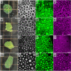
Images showing MC structures on macroscopic and microscopic scales with the addition of hexadecane oil of concentrations (a–d) 0 vol %, (e–h) 0.5 vol %, (i–l) 5.0 vol %, and (m–p) 100.0 vol %; (a,e,i,m) macroscopic scale images taken by a smartphone camera, where gridlines for the scale represent 1 cm2; (b,f,j,n) phase contrast imaging with the scale bar of 200 μm, (c,g,k,o) confocal images at 473 nm showing aqueous calcein green, and (d,h,l,p) confocal images at 543 nm showing hydrophobic Nile red, where the scale bar of all confocal images represents 400 μm.
Figure 6
Figure 6
Heterogeneous MC structures containing calcein green and Dextran Texas Red filled compartments, whose macroscopic images, phase contrast images, and confocal microscopy images are shown from left to right, respectively, for (a,b,c) random distribution and (d,e,f) separated distribution of compartments.
Figure 7
Figure 7
Magnetic MC structures showing (a) schematic for the location of oleic-modified SPIONs in the hydrophobic membrane of the MC structure and (b) magnetic MC structure (green) moving toward and over the neodymium magnet placed under the Petri dish. (c) Schematic of the heterogeneous magnetic MC structure with a magnetic head (purple) and a non-magnetic body (green) and (d) heterogeneous magnetic MC structure with a magnetic head (purple) and a non-magnetic body (green) turning in response to the neodymium magnet, where images (i–iv) are in a chronological sequence, black arrows point in the direction of the magnetic “head”, and white arrows indicate the direction the structure has turned.

References

    1. Kaiser D. Building a Multicellular Organism. Annu. Rev. Genet. 2001, 35, 103–123. 10.1146/annurev.genet.35.102401.090145. - DOI - PubMed
    1. Kin K.; Schaap P. Evolution of Multicellular Complexity in the Dictyostelid Social Amoebas. Genes 2021, 12, 487–502. 10.3390/genes12040487. - DOI - PMC - PubMed
    1. Furusawa C.; Kaneko K. Origin of Complexity in Multicellular Organisms. Phys. Rev. Lett. 2000, 84, 6130–6133. 10.1103/PhysRevLett.84.6130. - DOI - PubMed
    1. Driscoll W. W.; Travisano M. Synergistic Cooperation Promotes Multicellular Performance and Unicellular Free-Rider Persistence. Nat. Commun. 2017, 8, 1–10. 10.1038/ncomms15707. - DOI - PMC - PubMed
    1. Chen A. H.; Silver P. A. Designing Biological Compartmentalization. Trends Cell Biol. 2012, 22, 662–670. 10.1016/j.tcb.2012.07.002. - DOI - PubMed

Publication types