

Identification of AFG3L2 dominant optic atrophy following reanalysis of clinical exome sequencing
- PMID: 36974169
- PMCID: PMC10038781
- DOI: 10.1016/j.ajoc.2023.101825
Identification of AFG3L2 dominant optic atrophy following reanalysis of clinical exome sequencing
Abstract
Purpose: To highlight the importance of the utility of clinical exome sequencing, and show how it led to the diagnosis of nonsyndromic autosomal dominant optic atrophy arising from an autosomal dominant variant in AFG3L2.
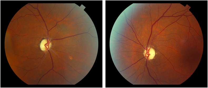
Observations: A healthy father and daughter of East African heritage experienced the onset of vision loss in the first decade of life due to optic atrophy. No additional neurologic or neuroimaging abnormalities were detected. Clinical exome sequencing was initially performed and provided a negative result. Reanalysis of the sequencing data revealed an autosomal dominant pathogenic variant in AFG3L2, c.1064C>T (p.Thr355Met), a gene that was recently identified to be associated with non-syndromic optic atrophy. This variant has previously been reported in a patient with optic atrophy, motor disturbances, and an abnormal brain MRI.
Conclusions: As the causes of dominant optic atrophy continue to expand, accurate genetic diagnosis is aided by an iterative reanalysis process for individuals and families when initial exome and genome testing does not provide an answer.
Keywords: AFG3L2 mutation; Dominant optic atrophy; SCA28.
© 2023 Published by Elsevier Inc.
Conflict of interest statement
None. Supported in part by a grant from the 10.13039/100001209Knights Templar Eye Foundation, Flower Mound, TX (Dr Brodsky). The supporter had no input for the conduct of research and preparation of the article, study design, collection, analysis or collection of data, writing the report, and the decision to submit article for publication. All authors attest that they meet the current ICMJE criteria for authorship.
Figures

References
-
- Brodsky M.C. Springer; NY: 2016. Pediatric Neuro-Ophthalmology; pp. 222–227.
-
- Kjer P. Hereditary infantile optic atrophy with dominant transmission (preliminary report) Dan Med Bull. 1956;3:135–141. - PubMed
-
- Kline L.B., Glaser J.S. Dominant optic atrophy: the clinical profile. Arch Ophthalmol. 1979;97:1680–1686. - PubMed
-
- Hoyt C.S. Autosomal dominant optic atrophy. A spectrum of disability. Ophthalmology. 1980;87:245. - PubMed
-
- Eliott D., Traboulsi E.I., Maumenee I.H. Visual prognosis in autosomal dominant optic atrophy. Am J Ophthalmol. 1993;115:360–367. - PubMed
Publication types
LinkOut - more resources
Full Text Sources
Research Materials
