

Collagen-based injectable and self-healing hydrogel with multifunction for regenerative repairment of infected wounds
- PMID: 36974203
- PMCID: PMC10039733
- DOI: 10.1093/rb/rbad018
Collagen-based injectable and self-healing hydrogel with multifunction for regenerative repairment of infected wounds
Abstract
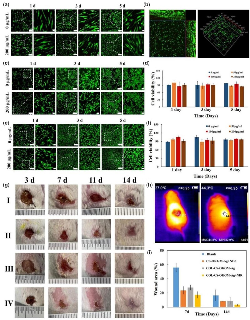
At present, the development trend of dressing materials is being multifunctional for convenient and long-term nursing care process of some complicated wounds. Here, basing on the theory of wound moist healing, an injectable and self-healing hydrogel comprising of collagen (COL), chitosan (CS) and oxidation modified Konjac glucomannan (OKGM), which acts as a macromolecular cross-linker to construct dynamic Schiff-base bonds was smartly designed. The strategy of introducing the silver nanoparticles (Ag NPs) into the COL-CS-OKGM hydrogel matrix achieved a markedly enhanced antibacterial activity derived from the synergistical effect between the Ag+ and the mild photothermal efficacy of Ag NPs, which also improved the local capillary blood circulation of the wound area to further facilitate wound healing process. The excellent syringeability and self-healing behaviors endowed the COL-CS-OKGM-Ag hydrogel with self-adapting ability for the wounds with irregular and large area needing frequent applying and changing without secondary injuries. In vitro and in vivo evaluations verified that so-designed COL-CS-OKGM-Ag hydrogel also with hemostatic performance is a promising multifunctional dressing for the treatments of infected wound with not only good biocompatibility and convenient use, but also with desired regenerative healing prognoses benefited from hydrogel moist environment and physiotherapy.
Keywords: collagen; hydrogel; injectable and self-healing; regenerative repair; wounds.
© The Author(s) 2023. Published by Oxford University Press.
Figures


Similar articles
-
A self-adapting hydrogel based on chitosan/oxidized konjac glucomannan/AgNPs for repairing irregular wounds.Biomater Sci. 2020 Mar 31;8(7):1910-1922. doi: 10.1039/c9bm01635j. Biomater Sci. 2020. PMID: 32026892
-
MMP-Responsive Nanoparticle-Loaded, Injectable, Adhesive, Self-Healing Hydrogel Wound Dressing Based on Dynamic Covalent Bonds.Biomacromolecules. 2023 Dec 11;24(12):5769-5779. doi: 10.1021/acs.biomac.3c00773. Epub 2023 Nov 11. Biomacromolecules. 2023. PMID: 37950669
-
An injectable adhesive antibacterial hydrogel wound dressing for infected skin wounds.Biomater Adv. 2022 Mar;134:112584. doi: 10.1016/j.msec.2021.112584. Epub 2021 Dec 2. Biomater Adv. 2022. PMID: 35525738
-
From hemostasis to proliferation: Accelerating the infected wound healing through a comprehensive repair strategy based on GA/OKGM hydrogel loaded with MXene@TiO2 nanosheets.Biomaterials. 2024 Jul;308:122548. doi: 10.1016/j.biomaterials.2024.122548. Epub 2024 Mar 27. Biomaterials. 2024. PMID: 38554642
-
Wound healing strategies based on nanoparticles incorporated in hydrogel wound patches.RSC Adv. 2023 Jul 17;13(31):21345-21364. doi: 10.1039/d3ra03477a. eCollection 2023 Jul 12. RSC Adv. 2023. PMID: 37465579 Free PMC article. Review.
Cited by
-
Recent progress in functional metal-organic frameworks for bio-medical application.Regen Biomater. 2023 Dec 23;11:rbad115. doi: 10.1093/rb/rbad115. eCollection 2024. Regen Biomater. 2023. PMID: 38313824 Free PMC article. Review.
-
Polydopamine-Coated Polycaprolactone Electrospun Nanofiber Membrane Loaded with Thrombin for Wound Hemostasis.Polymers (Basel). 2023 Jul 22;15(14):3122. doi: 10.3390/polym15143122. Polymers (Basel). 2023. PMID: 37514511 Free PMC article.
-
In vivo dynamic visualization and evaluation of collagen degradation utilizing NIR-II fluorescence imaging in mice models.Regen Biomater. 2025 Apr 11;12:rbaf025. doi: 10.1093/rb/rbaf025. eCollection 2025. Regen Biomater. 2025. PMID: 40405872 Free PMC article.
-
Porous PLLA microspheres dispersed in HA/collagen hydrogel as injectable facial fillers to enhance aesthetic effects.Regen Biomater. 2025 May 23;12:rbaf049. doi: 10.1093/rb/rbaf049. eCollection 2025. Regen Biomater. 2025. PMID: 40556787 Free PMC article.
-
Antibacterial and antioxidant bifunctional hydrogel based on hyaluronic acid complex MoS2-dithiothreitol nanozyme for treatment of infected wounds.Regen Biomater. 2024 Mar 9;11:rbae025. doi: 10.1093/rb/rbae025. eCollection 2024. Regen Biomater. 2024. PMID: 38605853 Free PMC article.
References
-
- Dainichi T, Kitoh A, Otsuka A, Nakajima S, Nomura T, Kaplan DH, Kabashima K.. The epithelial immune microenvironment (EIME) in atopic dermatitis and psoriasis. Nat Immunol 2018;19:1286–98. - PubMed
-
- Gan DL, Xu T, Xing WS, Ge X, Fang LM, Wang KF, Ren FZ, Lu X.. Mussel-inspired contact-active antibacterial hydrogel with high cell affinity, toughness, and recoverability. Adv Funct Mater 2019;29:1805964.
-
- Xiao L, Ni WQ, Zhao XH, Guo YC, Li X, Wang F, Luo GX, Zhan RX, Xu XS.. A moisture balanced antibacterial dressing loaded with lysozyme possesses antibacterial activity and promotes wound healing. Soft Matter 2021;17:3162–73. - PubMed
-
- Koehler J, Brandl FP, Goepferich AM.. Hydrogel wound dressings for bioactive treatment of acute and chronic wounds. Eur Polym J 2018;100:1–11.
LinkOut - more resources
Full Text Sources
