

Comparing Dermatoscopic Features With Slit Skin Smear and Histopathology in Diagnosis of Cutaneous Leishmaniasis
- PMID: 36974240
- PMCID: PMC10039316
- DOI: 10.7759/cureus.35336
Comparing Dermatoscopic Features With Slit Skin Smear and Histopathology in Diagnosis of Cutaneous Leishmaniasis
Abstract
Objective: To evaluate dermatoscopic features of cutaneous leishmaniasis and to compare its diagnostic accuracy against slit skin smear and skin histopathology.
Methods: This cross-sectional study was conducted at the Department of Dermatology, Pakistan Navy Station (PNS) Shifa Hospital, Karachi, Pakistan, from August 2021 to August 2022. A total of 200 lesions from 70 patients of cutaneous leishmaniasis diagnosed with slit skin smear for Leishmania-Donovan (LD) bodies and skin biopsy were included via non-probability consecutive sampling technique. Dermatoscopic evaluation was performed via a handheld dermatoscope (DELTA 20T; HEINE, Gilching, Germany) on 10x magnification. All dermatoscopic images were analyzed by two different observers who had command of dermatoscopy. Data analysis was done using Statistical Package for the Social Sciences SPSS version 27 (IBM Corp., Armonk, NY, USA).
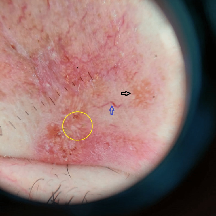
Results: Common dermatoscopic findings were erythema 200 (100%), hyperkeratosis 140 (70%), crusting 50 (25%), ulceration 42 (21%), milia-like structure 58 (29%), tear drop-like structure 46 (23%), yellow tears 70 (35%), and white starburst pattern 68 (34%). Less common findings were yellow hue 28 (14%), orange areas 26 (13%) and scar seven (3.5%). Vascular structures frequently observed were linear vessels 109 (54.5%), dotted vessels 80 (40%), and hairpin vessels 61 (30.5%); less common findings were comma-shaped vessels 52 (26%), arborizing vessels 20 (10%), crown vessels nine (4.5%). Comparison of dermatoscopic features was done with slit skin smear for LD bodies (p value = 0.003 ) and histopathology (p value = 0.001).
Conclusions: Dermatoscopy is a non-invasive technique that is helpful in diagnosing cutaneous leishmaniasis, saving time in making rapid diagnosis and saving the need to undergo extensive invasive investigation. Yield of dermatoscopy was comparable to slit smear for LD bodies and histopathology and was found to be effective in making rapid diagnosis with significant accuracy (p value <0.05).
Keywords: cutaneous leishmaniasis; dermatoscopy; histopathology; slit smear; star-burst pattern; yellow tears.
Copyright © 2023, Memon et al.
Conflict of interest statement
The authors have declared that no competing interests exist.
Figures

References
-
- Leishmaniasis in high-burden countries: an epidemiological update based on data reported in 2014. https://www.who.int/publications/i/item/who-wer9122. Wkly Epidemiol Rec. 2016;91(22):287–296. - PubMed
-
- A nested-PCR-based schizodeme method for identifying Leishmania kinetoplast minicircle classes directly from clinical samples and its application to the study of the epidemiology of Leishmania tropica in Pakistan. Noyes HA, Reyburn H, Bailey JW, Smith D. J Clin Microbiol. 1998;36:2877–2881. - PMC - PubMed
-
- New developments in diagnosis of leishmaniasis. Singh S. https://pubmed.ncbi.nlm.nih.gov/16778313/ Indian J Med Res. 2006;123:311–330. - PubMed
LinkOut - more resources
Full Text Sources
Research Materials