

Non-invasive detection of cardiac allograft rejection among heart transplant recipients using an electrocardiogram based deep learning model
- PMID: 36974261
- PMCID: PMC10039431
- DOI: 10.1093/ehjdh/ztad001
Non-invasive detection of cardiac allograft rejection among heart transplant recipients using an electrocardiogram based deep learning model
Abstract
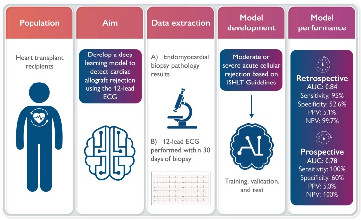
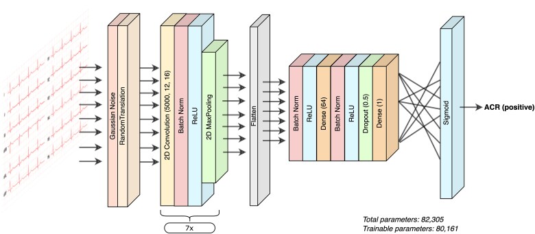
Aims: Current non-invasive screening methods for cardiac allograft rejection have shown limited discrimination and are yet to be broadly integrated into heart transplant care. Given electrocardiogram (ECG) changes have been reported with severe cardiac allograft rejection, this study aimed to develop a deep-learning model, a form of artificial intelligence, to detect allograft rejection using the 12-lead ECG (AI-ECG).
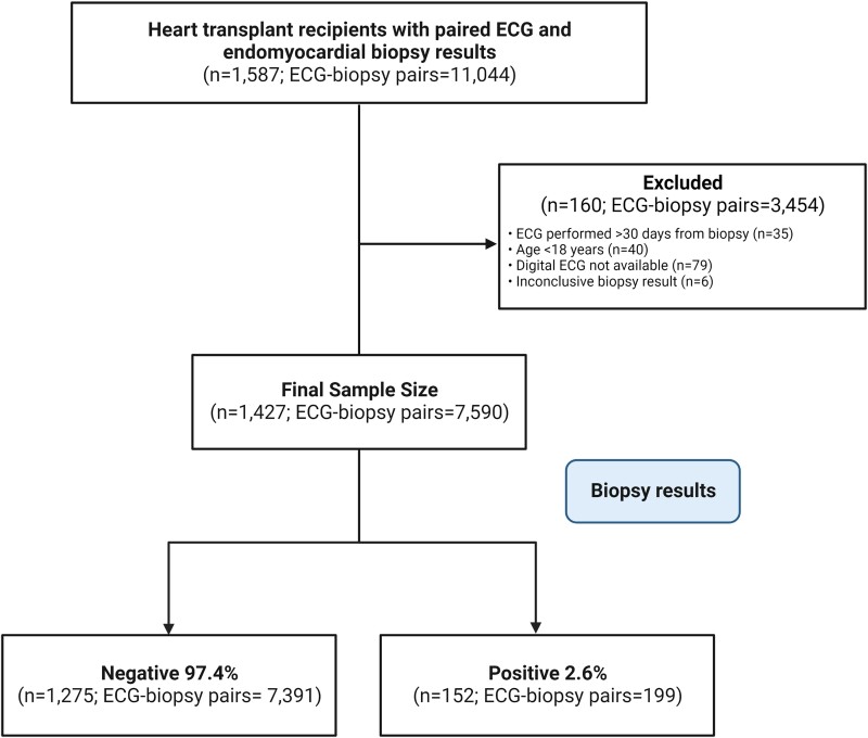
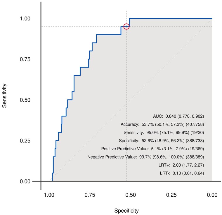
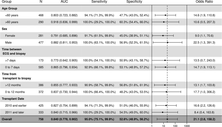
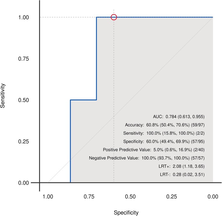
Methods and results: Heart transplant recipients were identified across three Mayo Clinic sites between 1998 and 2021. Twelve-lead digital ECG data and endomyocardial biopsy results were extracted from medical records. Allograft rejection was defined as moderate or severe acute cellular rejection (ACR) based on International Society for Heart and Lung Transplantation guidelines. The extracted data (7590 unique ECG-biopsy pairs, belonging to 1427 patients) was partitioned into training (80%), validation (10%), and test sets (10%) such that each patient was included in only one partition. Model performance metrics were based on the test set (n = 140 patients; 758 ECG-biopsy pairs). The AI-ECG detected ACR with an area under the receiver operating curve (AUC) of 0.84 [95% confidence interval (CI): 0.78-0.90] and 95% (19/20; 95% CI: 75-100%) sensitivity. A prospective proof-of-concept screening study (n = 56; 97 ECG-biopsy pairs) showed the AI-ECG detected ACR with AUC = 0.78 (95% CI: 0.61-0.96) and 100% (2/2; 95% CI: 16-100%) sensitivity.
Conclusion: An AI-ECG model is effective for detection of moderate-to-severe ACR in heart transplant recipients. Our findings could improve transplant care by providing a rapid, non-invasive, and potentially remote screening option for cardiac allograft function.
Keywords: Artificial intelligence; Cardiac allograft rejection; Deep learning; Electrocardiography; Heart transplantation.
© The Author(s) 2023. Published by Oxford University Press on behalf of the European Society of Cardiology.
Conflict of interest statement
Conflict of interest: None declared.
Figures
References
-
- Caves P, Billingham M, Stinson E, Shumway N. Serial transvenous biopsy of the transplanted human heart improved management of acute rejection episodes. Lancet 1974;303:821–826. - PubMed
-
- Costanzo MR, Dipchand A, Starling R, Anderson A, Chan M, Desai S, et al. The International Society of Heart and Lung Transplantation guidelines for the care of heart transplant recipients. J Heart Lung Transplant 2010;29:914–956. - PubMed
-
- Seferović PM, Tsutsui H, McNamara DM, Ristić AD, Basso C, Bozkurt B, et al. Heart Failure Association, Heart Failure Society of America, and Japanese Heart Failure Society position statement on endomyocardial biopsy. J Card Fail 2021;27:727–743. - PubMed
-
- Pham MX, Teuteberg JJ, Kfoury AG, Starling RC, Deng MC, Cappola TP, et al. Gene-expression profiling for rejection surveillance after cardiac transplantation. N Engl J Med 2010;362:1890–1900. - PubMed
Grants and funding
LinkOut - more resources
Full Text Sources