

Suture Fixation of Subacute Pediatric Seymour Fractures
- PMID: 36974281
- PMCID: PMC10039310
- DOI: 10.1016/j.jhsg.2022.10.013
Suture Fixation of Subacute Pediatric Seymour Fractures
Abstract
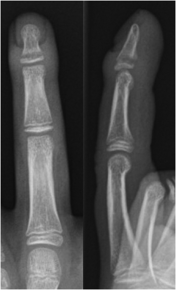
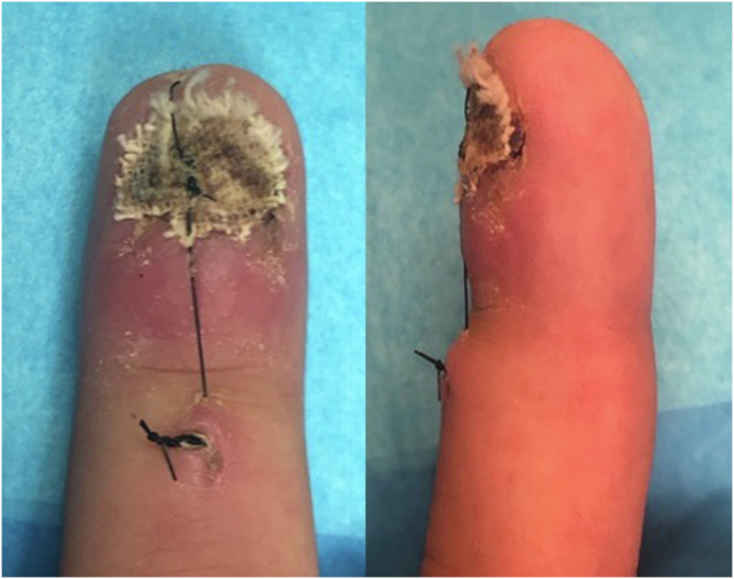
Seymour fractures are common injuries in the pediatric population. High rates of deep infection have been reported due to delayed presentation and subsequent treatment. This report describes the case of a 13-year-old male wrestler who presented 1 month after a finger injury that was later diagnosed as a subacute Seymour fracture with osteomyelitis. The patient underwent irrigation and debridement and fracture reduction stabilized with nonabsorbable suture fixation. After 6 weeks of intravenous antibiotics, the patient was recovering well, with radiographic evidence of fracture healing and clearance of infection. This case highlights the use of a single suture as a treatment option for fixation of unstable Seymour fractures with delayed presentation. The management of acute open distal phalangeal physeal fractures is well described in the literature; however, further investigations are warranted into the optimal management of chronically infected digits with unstable Seymour fractures.
Keywords: Infection; Nail trauma; Pediatric; Seymour fracture; Suture fixation.
© 2022 The Authors.
Figures


References
-
- Neustein T.M., Payne S.H., Jr., Seiler J.G., III Treatment of fingertip injuries. JBJS Rev. 2020;8(4) - PubMed
-
- Seymour N. Juxta-epiphysial fracture of the terminal phalanx of the finger. J Bone Joint Surg Br. 1966;48(2):347–349. - PubMed
-
- Abzug J.M., Kozin S.H. Seymour fractures. J Hand Surg Am. 2013;38(11):2267–2270. - PubMed
-
- Reyes B.A., Ho C.A. The high risk of infection with delayed treatment of open Seymour fractures: Salter-Harris I/II or juxta-epiphyseal fractures of the distal phalanx with associated nailbed laceration. J Pediatr Orthop. 2017;37(4):247–253. - PubMed
Publication types
LinkOut - more resources
Full Text Sources
