Case Reports
. 2022 Dec 8;5(2):242-245.
doi: 10.1016/j.jhsg.2022.11.005. eCollection 2023 Mar.

Cutis Calcinosis of the Hand in 2 Patients With Symbrachydactyly

Affiliations
Case Reports

Cutis Calcinosis of the Hand in 2 Patients With Symbrachydactyly

Sofia Bougioukli et al. J Hand Surg Glob Online. .

Abstract

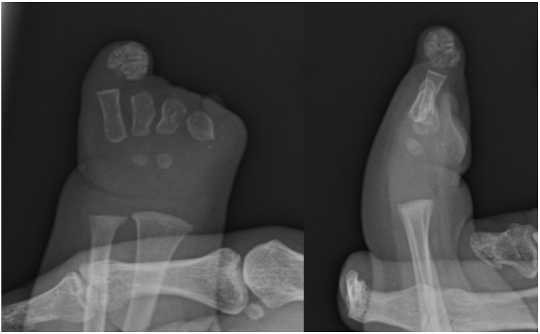
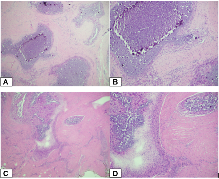
Cutis calcinosis of the hand in the setting of symbrachydactyly is presented in 2 unique patients. Both lesions were treated based on the standard of care protocols with resection of the calcified mass and hand reconstruction, as appropriate. The patients healed uneventfully without recurrence of the calcification at a the 1-year follow-up. The association between symbrachydactyly and calcinosis cutis is discussed along with a hypothesis on the pathophysiologic mechanism that could potentially explain this unusual occurrence and why it might be more common than previously identified.

Keywords: Congenital hand disorder; Cutis calcinosis; Symbrachydactyly.


Figures

Figure 1
Figure 1
Clinical and radiographic images of the first patient’s (19-month-old girl) left hand. A-C The fingers were comprised of digital skin tubes without phalanges. The thumb and ring fingers were the longest of the skin tubes, with very short underdeveloped skin tubes of the index, middle, and little fingers. The nail plate is absent in all “fingers,” with a hypoplastic nail bed at the dorsal distal tip of the thumb. Additionally, the left ring finger showed a soft tissue mass within the skin tube. Underdeveloped thenar and hypothenar eminences were also noted. D, E Radiographic imaging of the left hand demonstrates a small left hand containing 5 short metacarpals. The first, second, third, and fourth metacarpals are approximately equal in length. Synostosis of the proximal shafts of the second and third metacarpals is also noted. There are no phalanges in any of the skin tubes. A soft tissue mass with evidence of calcification is noted at the distal aspect of the ring finger stump.
Figure 2
Figure 2
Radiographic images of the second patient’s left hand at 13 months of age. The first metacarpal is absent. The rest of the metacarpals are present, with the second and third metacarpals hypoplastic. All phalanges are absent. A calcified heterogeneous soft tissue mass is noted at the distal aspect of the ring finger stump.
Figure 3
Figure 3
Hematoxylin and eosin (H&E) stained sections demonstrating soft tissue containing an intense inflammatory reaction composed primarily of aggregates of macrophages and multinucleated giant cells. The central portion of these aggregates contains calcific debris. The findings were consistent with tumoral calcinosis in both patients. A and B The first patient. C and D The second patient. (Hematoxylin and eosin stains; A and C, magnification × 40, B and D, magnification × 100).
Figure 4
Figure 4
Clinical (A) and radiographic (B) images of the first patient’s hand immediately after surgery. The patient underwent a release of her left thumb contracture using a standard Z-plasty, followed by the transfer of a nonvascularized middle phalanx from the left second and third toes into the left thumb and ring fingers to improve function and stabilize the skin tubes. Capsulodesis of the metacarpophalangeal joints of the left thumb and ring finger were also performed. Finally, the left ring finger mass was excised. C Radiographic image of the patient seen in the clinic 1 year later showing an excellent functional outcome and no recurrence of the cutis calcinosis in her hand.

References

    1. Oberg K.C., Feenstra J.M., Manske P.R., Tonkin M.A. Developmental biology and classification of congenital anomalies of the hand and upper extremity. J Hand Surg Am. 2010;35(12):2066–2076. - PubMed
    1. Bavinck J.N., Weaver D.D. Subclavian artery supply disruption sequence: hypothesis of a vascular etiology for Poland, Klippel-Feil, and Möbius anomalies. Am J Med Genet. 1986;23(4):903–918. - PubMed
    1. Reiter N., El-Shabrawi L., Leinweber B., Berghold A., Aberer E. Calcinosis cutis: part I. Diagnostic pathway. J Am Acad Dermatol. 2011;65(1):1–12. - PubMed
    1. White N., Chester D.L., Khanna A. Congenital bilateral calcinosis cutis of the hands. J Hand Surg Br. 2006;31(5):522–523. - PubMed
    1. Kim S.Y., Choi H.Y., Myung K.B., Choi Y.W. The expression of molecular mediators in the idiopathic cutaneous calcification and ossification. J Cutan Pathol. 2008;35(9):826–831. - PubMed

Publication types

LinkOut - more resources