. 2023 Jul;33(4):e13158.
doi: 10.1111/bpa.13158. Epub 2023 Mar 27.

Cortical iron accumulation in MAPT- and C9orf 72-associated frontotemporal lobar degeneration

Affiliations

Cortical iron accumulation in MAPT- and C9orf 72-associated frontotemporal lobar degeneration

Lucia A A Giannini et al. Brain Pathol. 2023 Jul.

Abstract

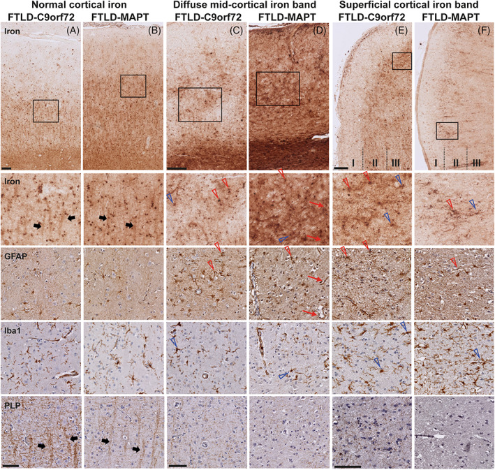
Neuroinflammation has been implicated in frontotemporal lobar degeneration (FTLD) pathophysiology, including in genetic forms with microtubule-associated protein tau (MAPT) mutations (FTLD-MAPT) or chromosome 9 open reading frame 72 (C9orf72) repeat expansions (FTLD-C9orf72). Iron accumulation as a marker of neuroinflammation has, however, been understudied in genetic FTLD to date. To investigate the occurrence of cortical iron accumulation in FTLD-MAPT and FTLD-C9orf72, iron histopathology was performed on the frontal and temporal cortex of 22 cases (11 FTLD-MAPT and 11 FTLD-C9orf72). We studied patterns of cortical iron accumulation and its colocalization with the corresponding underlying pathologies (tau and TDP-43), brain cells (microglia and astrocytes), and myelination. Further, with ultrahigh field ex vivo MRI on a subset (four FTLD-MAPT and two FTLD-C9orf72), we examined the sensitivity of T2*-weighted MRI for iron in FTLD. Histopathology showed that cortical iron accumulation occurs in both FTLD-MAPT and FTLD-C9orf72 in frontal and temporal cortices, characterized by a diffuse mid-cortical iron-rich band, and by a superficial cortical iron band in some cases. Cortical iron accumulation was associated with the severity of proteinopathy (tau or TDP-43) and neuronal degeneration, in part with clinical severity, and with the presence of activated microglia, reactive astrocytes and myelin loss. Ultra-high field T2*-weighted MRI showed a good correspondence between hypointense changes on MRI and cortical iron observed on histology. We conclude that iron accumulation is a feature of both FTLD-MAPT and FTLD-C9orf72 and is associated with pathological severity. Therefore, in vivo iron imaging using T2*-weighted MRI or quantitative susceptibility mapping may potentially be used as a noninvasive imaging marker to localize pathology in FTLD.

Keywords: C9orf72; MAPT; frontotemporal lobar degeneration; iron accumulation; neuroinflammation.


Figures

FIGURE 1
FIGURE 1
Schematic overview of the study design. Formalin‐fixed paraffin‐embedded tissue of the frontal cortex (i.e., middle frontal gyrus) and temporal cortex (i.e., either temporal pole [TP] or middle temporal gyrus [MTG]) of 11 FTLD‐MAPT and 11 FTLD‐C9orf72 cases was included for histological examination. Of these 22 cases, formalin‐fixed tissue of 4 FTLD‐MAPT and 2 FTLD‐C9orf72 cases was included for high‐resolution 7 T MRI. Anatomical images have been obtained and adapted from the Allen Reference Atlas (Adult Human). C9orf72, chromosome 9 open reading frame 72; FTLD, frontotemporal lobar degeneration; MAPT, microtubule‐associated protein tau
FIGURE 2
FIGURE 2
Normal and abnormal cortical iron distribution in nondemented controls and frontotemporal lobar degeneration (FTLD) cases. (A–D) Normal cortical iron was observed in nondemented controls (A,B) and a subset of FTLD (C,D) as iron concentrated in the myelin rich areas of the white matter and the lines of Baillarger, observable as a well‐defined thin line covering layer IV of the cortex (arrow heads in A2 and B2). (E–H) Abnormal cortical iron was observed as a diffuse mid‐cortical band‐shaped iron staining covering the middle layers of the cortex (III/IV), sometimes extending to layer V (arrows in E2 and F2), or as superficial cortical iron band in layer II of the cortex (arrow heads in G and H). The superficial cortical iron band occurred in some cases as the only abnormality present (G), or in other cases in conjunction with the diffuse mid‐cortical band (H). Scale bar in zoom: 2 mm.
FIGURE 3
FIGURE 3
Occurrence of a diffuse mid‐cortical iron band and a superficial cortical iron band in FTLD‐MAPT and FTLD‐C9orf72. (A) A diffuse mid‐cortical iron band was found in the frontal cortex in 72.7% of the FTLD‐MAPT cases and 45.5% of the FTLD‐C9orf72 cases. In the temporal cortex, relatively more FTLD‐MAPT cases showed a diffuse mid‐cortical iron band, that is, 81.8% in FTLD‐MAPT compared to 36.4% in FTLD‐C9orf72 (statistical trend, p = 0.083). (B) A superficial cortical iron band in the frontal cortex was found in 54.5% of the FTLD‐MAPT and 27.3% of the FTLD‐C9orf72 cases. Within the temporal cortex 36.4% of the FTLD‐MAPT and 27.3% of the FTLD‐C9orf72 cases showed this abnormal cortical iron pattern. (C) The cooccurrence of both bands was relatively more frequent in FTLD‐MAPT, in particular in the frontal cortex (p = 0.042). Numbers in plot depict the number of cases in the different categories used in these comparisons. C9orf72, chromosome 9 open reading frame 72; FTLD, frontotemporal lobar degeneration; MAPT, microtubule‐associated protein tau.
FIGURE 4
FIGURE 4
Association of the occurrence of the diffuse mid‐cortical iron band with proteinopathy severity in FTLD‐MAPT and FTLD‐C9orf72. (A) In FTLD‐MAPT, cases with moderate‐to‐severe tau pathology always had a diffuse mid‐cortical iron band in the frontal and temporal cortex. In FTLD‐C9orf72, the diffuse mid‐cortical iron band was only observed in cases with moderate‐to‐severe TDP‐43 pathology. Numbers in plot depict the number of cases in the different categories used in these comparisons. (B) Microscopic observation showed colocalization of relatively diffuse tau and TDP‐43 pathology in these cases and iron accumulation in the mid‐cortical layers corresponding to the laminar distribution of the diffuse mid‐cortical iron band. Scale bar in zoom = 100 μm. C9orf72, chromosome 9 open reading frame 72; FTLD, frontotemporal lobar degeneration; MAPT, microtubule‐associated protein tau.
FIGURE 5
FIGURE 5
Association of the occurrence of the diffuse mid‐cortical iron band with neuronal degeneration in FTLD‐MAPT and FTLD‐C9orf72. In FTLD‐MAPT, cases with moderate‐to‐(very) severe neuronal degeneration always had a diffuse mid‐cortical iron band in the frontal and temporal cortex. Similarly, in FTLD‐C9orf72, all cases with severe or very severe neuronal degeneration had a diffuse mid‐cortical iron band in the frontal cortex, while this association was less evident in the temporal cortex. Numbers in plot depict the number of cases in the different categories used in these comparisons. C9orf72, chromosome 9 open reading frame 72; FTLD, frontotemporal lobar degeneration; MAPT, microtubule‐associated protein tau.
FIGURE 6
FIGURE 6
Colocalization of iron with microglia, astrocytes, and myelin in cortex with normal and abnormal cortical iron. (A) FTLD‐C9orf72 and (B) FTLD‐MAPT cases with a normal cortical iron distribution showed iron in cells morphologically resembling glia cells (both microglia and astrocytes). Microscopically, the iron staining also showed iron reactivity within myelinated‐fiber bundles traversing the cortex (thick black arrows). In FTLD‐C9orf72 (C) and FTLD‐MAPT (D) cases with a diffuse mid‐cortical band‐shaped iron staining, an area of high iron load showed, next to diffuse iron present in the neuropil, iron within both active and dystrophic microglia (open blue arrows) and reactive astrocytes (open red arrows). Iron was also found within the vessel wall of small capillaries, probably caused by iron within processes of astrocytes surrounding these capillaries (thin red arrows). Within these areas with high iron load, cortical myelin was clearly reduced. In FTLD‐C9orf72 (E) and FTLD‐MAPT (F) cases with a superficial cortical iron band, layer II of the cortex was characterized by high amounts of activated and dystrophic microglia (open blue arrows), reactive astrocytes (open red arrows) and a reduction in myelin. Scale bars overview (A,B; C,D; E,F) = 200 μm; scale bars in zoom (A,B; C,D; E,F) = 100 μm. C9orf72, chromosome 9 open reading frame 72; FTLD, frontotemporal lobar degeneration; MAPT, microtubule‐associated protein tau.
FIGURE 7
FIGURE 7
Ex vivo 7 T T2*‐weighted MRI and iron histology in FTLD. (A) A normal distribution of iron was observed in the frontal cortex of the FTLD‐MAPT case, which was also reflected on MRI. The cortex was characterized by a homogeneous cortex containing two well‐defined layers separated by a thin layer (line of Baillarger, arrow heads) with lower signal intensity compared to the other layers. (B) The temporal cortex of this FTLD‐MAPT case showed a diffuse mid‐cortical iron band on histology, and the same pattern was observed on MRI (arrows). (C) The temporal cortex of this FTLD‐C9orf72 showed a superficial cortical iron band in layer II of the cortex, which was visible on both MRI and histology (red arrows), next to a diffuse mid‐cortical band in adjacent parts of the gyrus. C9orf72, chromosome 9 open reading frame 72; FTLD, frontotemporal lobar degeneration; MAPT, microtubule‐associated protein tau.

Similar articles

Cited by

References

    1. Onyike CU, Diehl‐Schmid J. The epidemiology of frontotemporal dementia. Int Rev Psychiatry. 2013;25:130–7. - PMC - PubMed
    1. Seelaar H, Rohrer JD, Pijnenburg YA, Fox NC, van Swieten JC. Clinical, genetic and pathological heterogeneity of frontotemporal dementia: a review. J Neurol Neurosurg Psychiatry. 2011;82:476–86. - PubMed
    1. Greaves CV, Rohrer JD. An update on genetic frontotemporal dementia. J Neurol. 2019;266:2075–86. - PMC - PubMed
    1. Rohrer JD, Guerreiro R, Vandrovcova J, Uphill J, Reiman D, Beck J, et al. The heritability and genetics of frontotemporal lobar degeneration. Neurology. 2009;73:1451–6. - PMC - PubMed
    1. Mackenzie IR, Neumann M, Bigio EH, Cairns NJ, Alafuzoff I, Kril J, et al. Nomenclature and nosology for neuropathologic subtypes of frontotemporal lobar degeneration: an update. Acta Neuropathol. 2010;119:1–4. - PMC - PubMed

Publication types

MeSH terms