Review
. 2023 Dec;55(1):1262-1286.
doi: 10.1080/07853890.2023.2191218.

The melatonin receptor 1B gene links circadian rhythms and type 2 diabetes mellitus: an evolutionary story

Affiliations
Review

The melatonin receptor 1B gene links circadian rhythms and type 2 diabetes mellitus: an evolutionary story

Hui Zhu et al. Ann Med. 2023 Dec.

Abstract

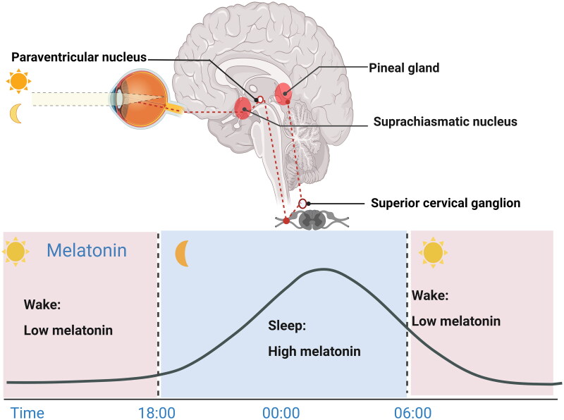
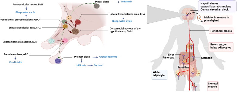
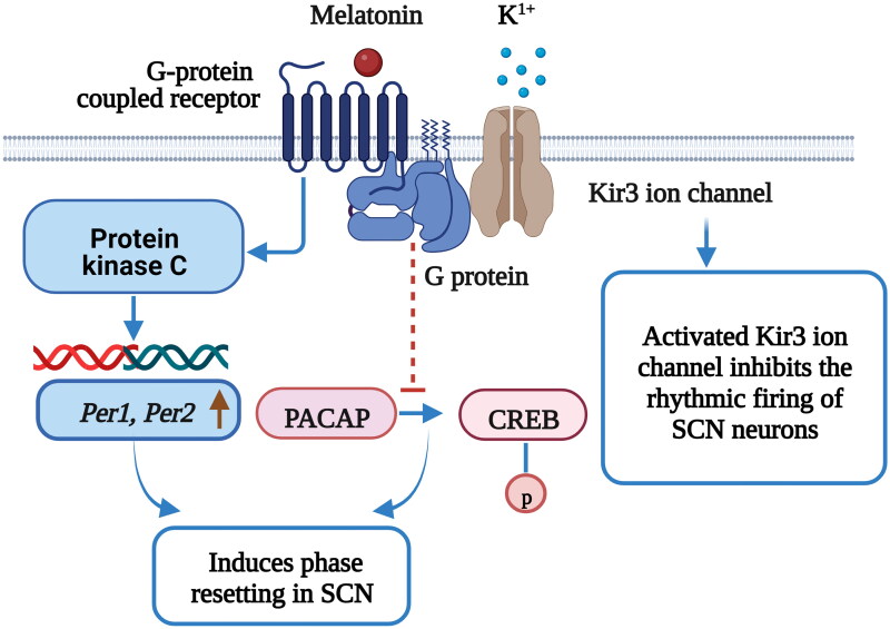
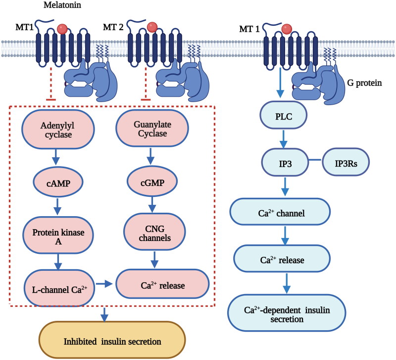
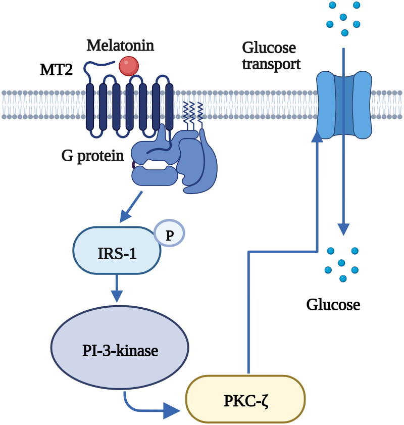
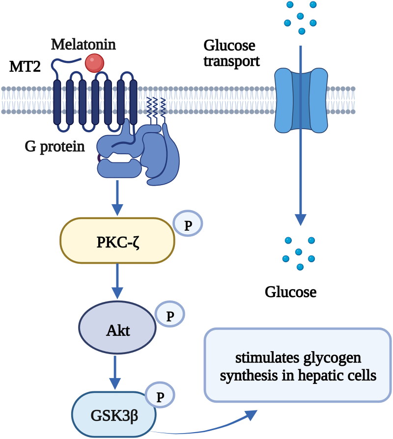
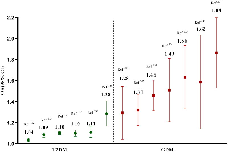
Disturbed circadian rhythms have been a risk factor for type 2 diabetes mellitus (T2DM). Melatonin is the major chronobiotic hormone regulating both circadian rhythm and glucose homeostasis. The rs10830963 (G allele) of the melatonin receptor 1B (MTNR1B) gene has the strongest genetic associations with T2DM according to several genome-wide association studies. The MTNR1B rs10830963 G allele is also associated with disturbed circadian phenotypes and altered melatonin secretion, both factors that can elevate the risk of diabetes. Furthermore, evolutionary studies implied the presence of selection pressure and ethnic diversity in MTNR1B, which was consistent with the "thrifty gene" hypothesis in T2DM. The rs10830963 G risk allele is associated with delayed melatonin secretion onset in dim-light and prolonged duration of peak melatonin. This delayed melatonin secretion may help human ancestors adapt to famine or food shortages during long nights and early mornings and avoid nocturnal hypoglycemia but confers susceptibility to T2DM due to adequate energy intake in modern society. We provide new insight into the role of MTNR1B variants in T2DM via disturbed circadian rhythms from the perspective of the "thrifty gene" hypothesis; these data indicate a novel target for the prevention and treatment of susceptible populations with the thrifty genotype.

Keywords: MTNR1B; Melatonin; Type 2 diabetes mellitus; thrifty gene.


Conflict of interest statement

The authors declare that they have no conflict of interest.

Figures

Figure 1.
Figure 1.
Melatonin levels fluctuate across the 24-h light-dark cycle [41,46,47]. Melatonin synthesis and release occur in dim light and are inhibited by daytime light. In the eyes, environmental light reaches intrinsic photosensitive retinal ganglion cells (ipRGCs) and is then transmitted to the SCN via the retinal hypothalamic tract. SCN signals are conveyed to the medial forebrain bundle by descending hypothalamic projections and then project to the spinal cord and superior cervical ganglia. Afterward, the sympathetic nerve from the superior cervical ganglion stimulates the pineal gland to secrete melatonin, thus entraining circadian rhythms to environmental light. Figure 1 was created with BioRender (https://biorender.com).
Figure 2.
Figure 2.
Circadian rhythms maintains glucose homeostasis. Circadian signals are conveyed from the SCN to the adjacent subparaventricular zone (SPZ), and the input is then integrated and amplified in the dorsomedial nucleus of the hypothalamus (DMH). Neurons in the DMH relay information to the ventrolateral preoptic nucleus (VLPO), the lateral hypothalamic area (LHA), orexin receptors and the paraventricular nucleus (PVN), which drive the circadian cycles of sleep, activity, feeding and corticosteroid secretion, respectively. The SCN could directly exerts excitatory-inhibitory effects on the neuronal response of the arcuate nucleus (ARC) to hypoglycemia and modulate food intake. The central circadian clock synchronizes peripheral clocks, and jointly regulate glucose metabolism. Figure 2 was created with BioRender (https://biorender.com).
Figure 3.
Figure 3.
Melatonin acts as a circadian pacemaker and advances the SCN phase [93–97]. The retinohypothalamic tract mediates cAMP responsive element binding protein (CREB) phosphorylation via pituitary adenylate cyclase-activating polypeptide (PACAP) release under light stimulation in SCN cells; PACAP release is responsible for light-induced phase shifts. The binding of melatonin to MT1 inhibits PACAP-induced CREB phosphorylation in the SCN. Melatonin receptors activate G-protein-coupled Kir3 ion channels, inhibit the rhythmic firing of SCN neurons and regulate circadian rhythms. Melatonin activates PKC in the SCN and induces phase resetting, and through this signaling, the expression of core clock genes, Period 1 (Per1) and Period 2 (Per2), increases within the SCN. Figure 2 was created with BioRender (https://biorender.com).
Figure 4.
Figure 4.
Melatonin regulates insulin secretion in pancreatic β-cells via the cAMP, cGMP and IP3 signaling pathways [98,236]. The binding of melatonin to MT1 inhibits cyclic adenosine monophosphate (cAMP) signaling and decreases insulin secretion in pancreatic β-cells. The binding of melatonin to MT2 inhibits cyclic guanosine monophosphate (cGMP) signaling and decreases insulin secretion in pancreatic β-cells. Melatonin stimulates IP3 release accompanied by a transient increase in Ca2+ concentrations and leads to Ca2+-dependent insulin secretion. Figure 3 was created with BioRender (https://biorender.com).
Figure 5.
Figure 5.
The binding of melatonin to MT2 stimulates glucose transport to skeletal muscle cells via the IRS-1/PI-3-kinase pathway [101]. Melatonin increases the phosphorylation level of insulin receptor substrate-1 (IRS-1) and the activity of phosphoinositide 3-kinase (PI-3-kinase), activates downstream protein kinase C (PKC)-ζ and stimulates glucose transport to muscle cells. Figure 4 was created with BioRender (https://biorender.com).
Figure 6.
Figure 6.
The binding of melatonin to MT1 stimulates glycogen synthesis in hepatic cells via the protein kinase Cζ (PKCζ)-Akt-glycogen synthase kinase 3B (GSK3β) pathway [102]. Melatonin increases the phosphorylation of PKCζ, Akt, and GSK3β and stimulates glycogen synthesis in hepatic cells. Figure 5 was created with BioRender (https://biorender.com).
Figure 7.
Figure 7.
The different genetic effect sizes of MTNR1B in T2DM and GDM.

References

    1. International Diabetes Federation (IDF) 2021. Available from: https://diabetesatlasorg/.
    1. Smushkin G, Vella A.. Genetics of type 2 diabetes. Curr Opin Clin Nutr Metab Care. 2010;13(4):471–477. - PMC - PubMed
    1. Panda S. Circadian physiology of metabolism. Science. 2016;354(6315):1008–1015. - PMC - PubMed
    1. Qian J, Scheer F.. Circadian system and glucose metabolism: implications for physiology and disease. Trends Endocrinol Metab. 2016;27(5):282–293. - PMC - PubMed
    1. Mason IC, Qian J, Adler GK, et al. Impact of circadian disruption on glucose metabolism: implications for type 2 diabetes. Diabetologia. 2020;63(3):462–472. - PMC - PubMed

Publication types