. 2023 Apr 28;132(9):1110-1126.
doi: 10.1161/CIRCRESAHA.122.322227. Epub 2023 Mar 28.

ZBP1 Protects Against mtDNA-Induced Myocardial Inflammation in Failing Hearts

Affiliations

ZBP1 Protects Against mtDNA-Induced Myocardial Inflammation in Failing Hearts

Nobuyuki Enzan et al. Circ Res. .

Abstract

Background: Mitochondrial DNA (mtDNA)-induced myocardial inflammation is intimately involved in cardiac remodeling. ZBP1 (Z-DNA binding protein 1) is a pattern recognition receptor positively regulating inflammation in response to mtDNA in inflammatory cells, fibroblasts, and endothelial cells. However, the role of ZBP1 in myocardial inflammation and cardiac remodeling remains unclear. The aim of this study was to elucidate the role of ZBP1 in mtDNA-induced inflammation in cardiomyocytes and failing hearts.

Methods: mtDNA was administrated into isolated cardiomyocytes. Myocardial infarctionwas conducted in wild type and ZBP1 knockout mice.

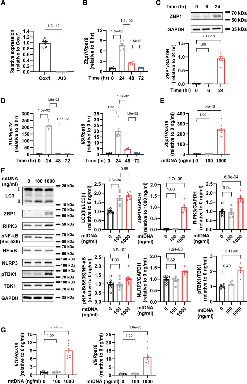
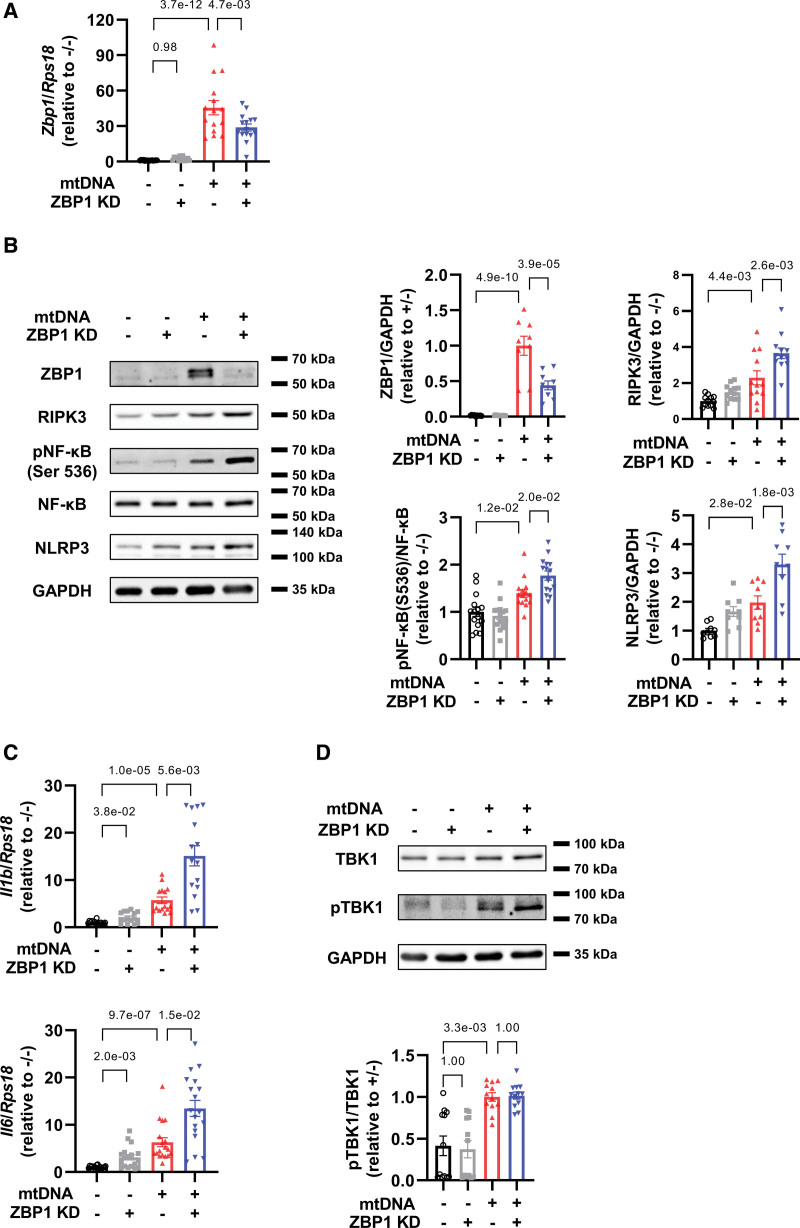
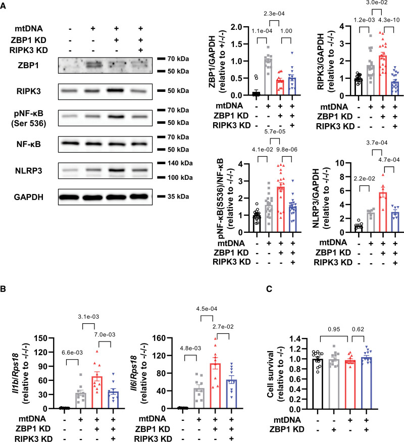
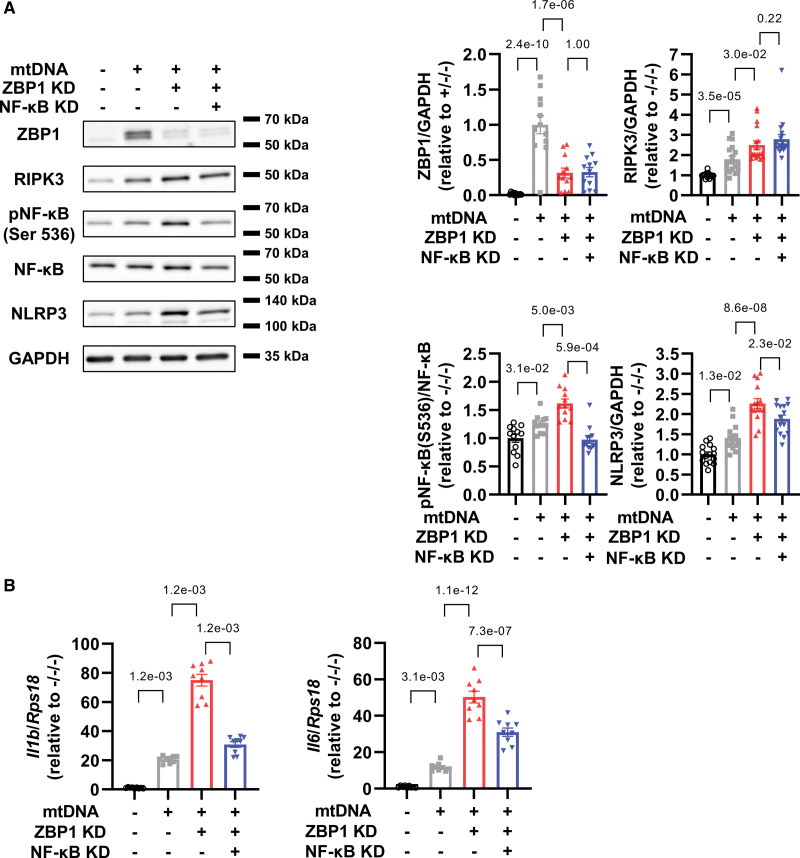
Results: We here found that, unlike in macrophages, ZBP1 knockdown unexpectedly exacerbated mtDNA-induced inflammation such as increases in IL (interleukin)-1β and IL-6, accompanied by increases in RIPK3 (receptor interacting protein kinase 3), phosphorylated NF-κB (nuclear factor-κB), and NLRP3 (nucleotide-binding domain and leucine-rich-repeat family pyrin domain containing 3) in cardiomyocytes. RIPK3 knockdown canceled further increases in phosphorylated NF-κB, NLRP3, IL-1β, and IL-6 by ZBP1 knockdown in cardiomyocytes in response to mtDNA. Furthermore, NF-κB knockdown suppressed such increases in NLRP3, IL-1β, and IL-6 by ZBP1 knockdown in response to mtDNA. CpG-oligodeoxynucleotide, a Toll-like receptor 9 stimulator, increased RIPK3, IL-1β, and IL-6 and ZBP1 knockdown exacerbated them. Dloop, a component of mtDNA, but not Tert and B2m, components of nuclear DNA, was increased in cytosolic fraction from noninfarcted region of mouse hearts after myocardial infarction compared with control hearts. Consistent with this change, ZBP1, RIPK3, phosphorylated NF-κB, NLRP3, IL-1β, and IL-6 were increased in failing hearts. ZBP1 knockout mice exacerbated left ventricular dilatation and dysfunction after myocardial infarction, accompanied by further increases in RIPK3, phosphorylated NF-κB, NLRP3, IL-1β, and IL-6. In histological analysis, ZBP1 knockout increased interstitial fibrosis and myocardial apoptosis in failing hearts.

Conclusions: Our study reveals unexpected protective roles of ZBP1 against cardiac remodeling as an endogenous suppressor of mtDNA-induced myocardial inflammation.

Keywords: cytokines; fibrosis; heart failure; inflammation; macrophages; nucleotides; receptors, pattern recognition.


Conflict of interest statement

H. Tsutsui reports personal fees from Merck Sharp and Dohme (MSD), Astellas, Pfizer, Bristol-Myers Squibb, Otsuka Pharmaceutical, Daiichi-Sankyo, Mitsubishi Tanabe Pharma, Nippon Boehringer Ingelheim, Takeda Pharmaceutical, Bayer Yakuhin, Novartis Pharma, Kowa Pharmaceutical, Teijin Pharma, Medical Review Co, and Japanese Journal of Clinical Medicine; nonfinancial support from Actelion Pharmaceuticals, Japan Tobacco Inc, Mitsubishi Tanabe Pharma, Nippon Boehringer Ingelheim, Daiichi-Sankyo, IQVIA Services Japan, and Omron Healthcare Co; grants from Astellas, Novartis Pharma, Daiichi-Sankyo, Takeda Pharmaceutical, Mitsubishi Tanabe Pharma, and Teijin Pharma, MSD, outside the submitted work. The other authors report no conflicts.

Figures

Figure 1.
Figure 1.
Mitochondrial DNA (mtDNA) increases ZBP1 (Z-DNA binding protein 1) and activates inflammatory signaling in cardiomyocytes. A, Cox1 and At3 DNA levels in mtDNA extracted from rat livers was quantified by real-time polymerase chain reaction (n=8). The experiment was conducted 2×. B, mRNA levels of Zbp1 in cultured neonatal rat ventricular myocytes (NRVMs) treated with 1000 ng/mL of mtDNA in indicated time point (n=6). The experiment was conducted 2×. C, Representative immunoblots of ZBP1 and GAPDH in NRVMs treated with 1000 ng/mL of mtDNA in indicated time point (n=6). The experiment was conducted 2×. D, Il1b and Il6 mRNA levels in NRVMs treated with 1000 ng/mL of mtDNA in indicated time point (n=6). The experiment was conducted 2×. E, mRNA levels of Zbp1 in NRVMs treated with indicated concentrations of mtDNA (n=8). The experiment was conducted 2×. F, Representative immunoblots of LC3 (light chain 3; n=6), ZBP1 (n=6), RIPK3 (n=6), phosphorylated NF-κB (nuclear factor-κB; Ser 536), NF-κB (n=9), NLRP3 (n=6), phosphorylated TBK1 (TANK-binding kinase 1), TBK1 (n=12), and GAPDH in NRVMs treated with indicated concentrations of mtDNA. The experiment was conducted 2× except for NF-κB (3×). G, mRNA levels of Il1b and Il6 in NRVMs treated with indicated concentrations of mtDNA (n=8). The experiment was conducted 2×. Error bars denote standard errors. Data were analyzed using the Student t test (A), Wilcoxon rank sum test (B and D; adjust=3. F NF-κB; adjust=2), and 2-way ANOVA followed by Tukey multiple comparisons test (C, E, F except for NF-κB and G).
Figure 2.
Figure 2.
ZBP1 (Z-DNA binding protein 1) knockdown exacerbates increases in inflammatory cytokines in cardiomyocytes treated with mitochondrial DNA (mtDNA). A, mRNA levels of Zbp1 in neonatal rat ventricular myocytes (NRVMs) treated with or without small interfering RNA (siRNA) for ZBP1 (10 nmol/L) in the presence or absence of mtDNA (1000 ng/mL) for 24 hours (n=15). The experiment was conducted 5×. B, Representative immunoblots of ZBP1 (n=9), RIPK3 (receptor interacting protein kinase; n=12), phosphorylated NF-κB (nuclear factor-κB; Ser 536), NF-κB (n=15), NLRP3 (nucleotide-binding domain and leucine-rich-repeat family pyrin domain containing 3; n=9), and GAPDH in NRVMs treated with or without small interfering RNA (siRNA) for ZBP1 (10 nmol/L) in the presence or absence of mtDNA (1000 ng/mL) for 24 hours. The experiment was conducted 3 (ZBP1 and NLRP3), 4 (RIPK3), and 5× (NF-κB). C, mRNA levels of Il1b (n=15) and Il6 (n=18) in NRVMs treated with or without siRNA for ZBP1 (10 nmol/L) in the presence or absence of mtDNA (1000 ng/mL) for 24 hours. The experiment was conducted 5 (Il1b) or 6 (Il6) ×. D, Representative immunoblots of TBK1, phosphorylated TBK1, and GAPDH in NRVMs treated with or without siRNA for ZBP1 (10 nmol/L) in the presence or absence of mtDNA (1000 ng/mL) for 24 hours (n=12). The experiment was conducted 4×. Error bars denote standard errors. Data were analyzed using the Wilcoxon rank sum test (C and E; adjust=3), and 2-way ANOVA followed by Tukey multiple comparisons test (A, B, and D).
Figure 3.
Figure 3.
RIPK3 (receptor interacting protein kinase) knockdown cancels increases in NF-κB (nuclear factor-κB)-NLRP3 (nucleotide-binding domain and leucine-rich-repeat family pyrin domain containing 3) axis by ZBP1 (Z-DNA binding protein 1) knockdown in cardiomyocytes treated with mitochondrial DNA (mtDNA). A, Representative immunoblots of ZBP1 (n=12; number of experiments, 4), RIPK3 (n=18; number of experiments, 6), phosphorylated NF-κB (Ser 536), NF-κB (n=18; number of experiments, 6), NLRP3 (n=6; number of experiments, 2), and GAPDH in neonatal rat ventricular myocytes (NRVMs) treated with or without small interfering RNA (siRNA) for ZBP1 (10 nmol/L) and RIPK3 (10 nmol/L) in the presence or absence of mtDNA (1000 ng/mL) for 24 hours. B, mRNA levels of Il1b and Il6 in NRVMs treated with or without siRNA for ZBP1 (10 nmol/L) and RIPK3 (10 nmol/L) in the presence or absence of mtDNA (1000 ng/mL) for 24 hours (n=9). The experiment was conducted 3×. C, Cell viability was assessed using Cell Titer Blue assays in NRVMs treated with or without small interfering RNA for ZBP1 (10 nmol/L) in the presence or absence of mtDNA (1000 ng/mL) for 24 hours (n=12). The experiment was conducted 2×. Error bars denote standard errors. Data were analyzed using the Wilcoxon rank sum test (A ZBP1; adjust, 3), and 2-way ANOVA followed by Tukey multiple comparisons test (A except for ZBP1, B, and C).
Figure 4.
Figure 4.
NF-κB (nuclear factor-κB) knockdown suppresses increases in NLRP3 (nucleotide-binding domain and leucine-rich-repeat family pyrin domain containing 3) axis by ZBP1 (Z-DNA binding protein 1) knockdown in cardiomyocytes treated with mitochondrial DNA (mtDNA). A, Representative immunoblots of ZBP1 (n=12; number of experiments, 4), RIPK3 (receptor interacting protein kinase; n=18; number of experiments, 6), phosphorylated NF-κB (Ser 536), NF-κB (n=12; number of experiments, 4), NLRP3 (n=15; number of experiments, 5), and GAPDH in neonatal rat ventricular myocytes (NRVMs) treated with or without small interfering RNA (siRNA) for ZBP1 (10 nmol/L) and NF-κB p65 subunit (10 nmol/L) in the presence or absence of mtDNA (1000 ng/mL) for 24 hours. B, mRNA levels of Il1b and Il6 in NRVMs treated with or without siRNA for ZBP1 (10 nmol/L) and NF-κB p65 subunit (10 nmol/L) in the presence or absence of mtDNA (1000 ng/mL) for 24 hours (n=9). The experiment was conducted 3×. Error bars denote standard errors. Data were analyzed using the Wilcoxon rank sum test (A, RIPK3 and NF-κB; B, Il1b; adjust=3), and 2-way ANOVA followed by Tukey multiple comparisons test (A, ZBP1 and NLRP3; B, Il6).
Figure 5.
Figure 5.
Interaction of ZBP1 (Z-DNA binding protein 1) and CpG-ODN (oligodeoxynucleotides) is involved in the anti-inflammatory effect of ZBP1 in CpG-ODN-treated cardiomyocytes. A, Representative images of neonatal rat ventricular myocytes (NRVMs) treated with adenovirus harboring mcherry-tagged ZBP1 in the presence of fluorescein isothiocyanate (FITC)-labeled CpG-oligodeoxynucleotide. Arrows indicated colocalized CpG-ODN and ZBP1. B, mRNA levels of Zbp1 in NRVMs treated with or without small interring (siRNA) for ZBP1 (10 nmol/L) in the presence or absence of CpG-ODN (1000 ng/mL) for 24 hours (n=6). The experiment was conducted 2×. C, Representative immunoblots of ZBP1 and GAPDH in NRVMs treated with or without siRNA for ZBP1 (10 nmol/L) in the presence or absence of CpG-ODN (1000 ng/mL) for 24 hours (n=9). D, mRNA levels of Il1b and Il6 in NRVMs treated with or without siRNA for ZBP1 (10 nmol/L) in the presence or absence of CpG-ODN (1000 ng/mL) for 24 hours (n=6). The experiment was conducted 2×. E, Representative immunoblots of RIPK3 (receptor interacting protein kinase) and GAPDH in NRVMs treated with CpG-ODN (1000 ng/mL) for 24 hours (n=6). The experiment was conducted 2×. F and G, mRNA levels of Tlr9 and Zbp1 in NRVMs treated with or without siRNA for TLR9 (Toll-like receptor 9; 10 nmol/L) in the presence or absence of mitochondrial DNA (mtDNA; 1000 ng/mL) for 24 hours (n=9). The experiment was conducted 3×. H, Representative immunoblots of ZBP1 and GAPDH in NRVMs treated with or without siRNA for TLR9 (10 nmol/L) in the presence or absence of mtDNA (1000 ng/mL) for 24 hours (n=6). The experiment was conducted 2×. I, Representative immunoblots of ZBP1 and GAPDH in NRVMs treated with or without siRNA for TLR9 (10 nmol/L) in the presence or absence of CpG-ODN (1000 ng/mL) for 24 hours (n=6). The experiment was conducted 2×. Error bars denote standard errors. Data were analyzed using the Student t test (E) and 2-way ANOVA followed by Tukey multiple comparisons test (B, C, D, F, G, H, and I).
Figure 6.
Figure 6.
ZBP1 (Z-DNA binding protein 1) knockout (KO) enhances myocardial inflammation in failing hearts via RIPK3 (receptor interacting protein kinase)-NF-κB (nuclear factor-κB)-NLRP3 (nucleotide-binding domain and leucine-rich-repeat family pyrin domain containing 3) pathway. A, Representative immunoblots of ZBP1 and GAPDH in C57B/6J mouse hearts 3 days after left anterior descending artery (LAD) ligation or sham operation (n=6). The experiment was conducted 2×. *P<0.05: Student t test. B, Representative immunoblots of COX4 (cytochrome c oxidase subunit 4), LC3 (light chain 3), and GAPDH in cytosolic fraction of C57B/6J mouse hearts 3 days after LAD ligation or sham operation. DNA levels in this cytosolic fraction was quantified by real-time polymerase chain reaction (n=12 in sham group, and n=17 in LAD ligation group). The experiment was conducted 4×. C, Representative immunoblots of ZBP1 (n=9; number of experiments, 3), RIPK3 (n=9; number of experiments, 3), phosphorylated NF-κB (Ser 536), NF-κB (n=21; number of experiments, 7), NLRP3 (n=12; number of experiments, 4), and GAPDH in wild type (WT) and ZBP1 KO mice hearts 3 days after LAD ligation or sham operation. D, mRNA levels of Il1b (n=13) and Il6 (n=16) in WT and ZBP1 KO mice hearts 3 days after LAD ligation or sham operation. The experiment was conducted 2 (Il1b) or 3 (Il6) ×. E, Secretion levels of IL (interleukin)-1β (n=12) and IL-6 (n=6) measured with ELISA in WT and ZBP1 KO mice hearts 3 days after LAD ligation or sham operation. The experiment was conducted 2 (Il-6) or 3 (Il-1β) ×. F, Representative immunoblots of TBK1 (TANK-binding kinase 1), phosphorylated TBK1, and GAPDH in WT and ZBP1 knockout mice hearts 3 days after left anterior descending artery ligation or sham operation (n=6). The experiment was conducted 2×. Error bars denote standard errors. Data were analyzed using the Student t test (B, Dloop), Wilcoxon rank sum test (A and B, Tert and B2m; no adjustment; C, except for RIPK3; D adjust, 3), and 2-way ANOVA followed by Tukey multiple comparisons test (C, RIPK3; E; and F). MI indicates myocardial infarction.
Figure 7.
Figure 7.
ZBP1 (Z-DNA binding protein 1) knockout (KO) exacerbates cardiac dysfunction and remodeling after myocardial infarction (MI). A, The representative echocardiographic images of ZBP1 KO and wild type (WT) mice hearts 28 days after left anterior descending artery (LAD) ligation (n=38) or sham operation (n=5). Long 2-way arrows and short 2-way arrows indicate left ventricular end-diastolic diameter (LVDd) and left ventricular end-systolic diameter (LVDs), respectively. B, Heart weight to tibial length (TL) ratio and left ventricle (LV) weight to TL ratio (n of sham, 8; n of LAD ligation, 41) in each group. C, Masson-trichrome-stained heart section in each group. Interstitial fibrosis was assessed by collagen volume in each group (n=5). Cardiomyocyte hypertrophy was assessed by cross-sectional area in each group (n=5). Error bars denote standard errors. Data were analyzed using the Wilcoxon rank sum test (A, LVDd; B and C, collagen volume; adjust, 3), and 2-way ANOVA followed by Tukey multiple comparisons test (A, except for LVDd and C, cross sectional area).
Figure 8.
Figure 8.
A schematic representation of the role of ZBP1 (Z-DNA binding protein 1) in myocardial inflammation and remodeling. Excessive amount of mitochondrial DNA (mtDNA) activates TLR9 (Toll-like receptor 9) and induces inflammation through RIPK3 (receptor interacting protein kinase)-NF-κB (nuclear factor-κB)-NLRP3 (nucleotide-binding domain and leucine-rich-repeat family pyrin domain containing 3) pathway. ZBP1 protects against adverse cardiac remodeling through suppressing this pathway.

References

    1. Ponikowski P, Anker SD, AlHabib KF, Cowie MR, Force TL, Hu S, Jaarsma T, Krum H, Rastogi V, Rohde LE, et al. . Heart failure: preventing disease and death worldwide. ESC Heart Fail. 2014;1:4–25. doi: 10.1002/ehf2.12005 - PubMed
    1. Deswal A, Petersen NJ, Feldman AM, Young JB, White BG, Mann DL. Cytokines and cytokine receptors in advanced heart failure: an analysis of the cytokine database from the Vesnarinone trial (VEST). Circulation. 2001;103:2055–2059. doi: 10.1161/01.cir.103.16.2055 - PubMed
    1. Suematsu N, Tsutsui H, Wen J, Kang D, Ikeuchi M, Ide T, Hayashidani S, Shiomi T, Kubota T, Hamasaki N, et al. . Oxidative stress mediates tumor necrosis factor-alpha-induced mitochondrial DNA damage and dysfunction in cardiac myocytes. Circulation. 2003;107:1418–1423. doi: 10.1161/01.cir.0000055318.09997 - PubMed
    1. Westermann D, Lindner D, Kasner M, Zietsch C, Savvatis K, Escher F, von Schlippenbach J, Skurk C, Steendijk P, Riad A, et al. . Cardiac inflammation contributes to changes in the extracellular matrix in patients with heart failure and normal ejection fraction. Circ Heart Fail. 2011;4:44–52. doi: 10.1161/CIRCHEARTFAILURE.109.931451 - PubMed
    1. Sobirin MA, Kinugawa S, Takahashi M, Fukushima A, Homma T, Ono T, Hirabayashi K, Suga T, Azalia P, Takada S, et al. . Activation of natural killer T cells ameliorates postinfarct cardiac remodeling and failure in mice. Circ Res. 2012;111:1037–1047. doi: 10.1161/CIRCRESAHA.112.270132 - PubMed

Publication types

MeSH terms