

Patterns and prognostic values of programmed cell death-ligand 1 expression and CD8 + T-cell infiltration in small cell carcinoma of the esophagus: a retrospective analysis of 34 years of National Cancer Center data in China
- PMID: 36974732
- PMCID: PMC11254267
- DOI: 10.1097/JS9.0000000000000064
Patterns and prognostic values of programmed cell death-ligand 1 expression and CD8 + T-cell infiltration in small cell carcinoma of the esophagus: a retrospective analysis of 34 years of National Cancer Center data in China
Abstract
Background: Small cell carcinoma of the esophagus (SCCE) is an extremely rare and highly aggressive neuroendocrine malignancy with a strikingly poor prognosis. Given the great clinical successes of checkpoint immunotherapies, we explored the expression profile and clinical significance of programmed cell death-ligand 1 (PD-L1) and CD8 + T cell in SCCE for the first time.
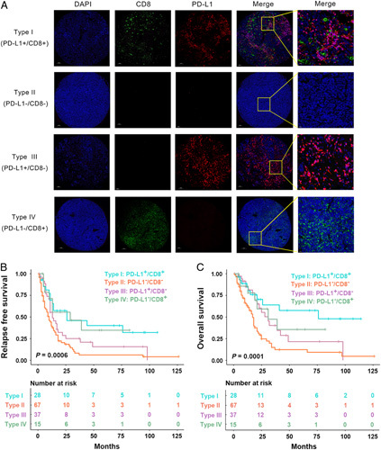
Materials and methods: Tumor-infiltrating immune cells (TIICs) and tumor cells in postoperative, whole tumor sections from 147 SCCE patients were stained for PD-LI expression. We also evaluated each patient's Combined Positive Score (CPS). Multiplex immunofluorescence staining (CD3, CD20, CD68, and PD-L1) was introduced to clarify the location of PD-L1. CD8 density was analyzed by digital imaging and analysis of entire slides. Clinical outcomes were tested for correlations with both PD-L1 expression and CD8 density.
Results: No patients had PD-L1 expressed in their tumor cells. PD-L1 + expression in TIICs was detected in 65 patients (44.2%) and 42 (28.6%) exhibited CPS positivity. Multiplex immunofluorescence staining demonstrated that most of the PD-L1 was expressed on the CD68 + monocytes/macrophages. PD-L1 expression in the TIICs and CPS was found to be correlated with paraffin block age, tumor length, macroscopic type, T stage, and increased overall survival (OS). Expression of PD-L1 in TIICs showed significantly prolonged relapse-free survival (RFS). Increasing CD8 densities were associated with increased PD-L1 expression ( Ptrend <0.0001). Multivariate regression confirmed that PD-L1 in TIICs and CD8 states were independent predictors of OS, and CD8 status were found to be independently predictive of RFS. A stratification based on PD-L1 and CD8 status was also significantly associated with both OS and RFS.
Conclusion: Expression of PD-L1 was only detected in TIICs from approximately half of the patients with SCCEs. In SCCEs, PD-L1 and CD8 status are novel prognostic biomarkers and may inform the implementation of risk-related therapeutic strategies. SCCEs with higher CD8 infiltration also had higher expression of PD-L1, suggesting the development of resistance against adaptive immunity. These findings support the assertion that PD-L1/programmed cell death 1 inhibitors should be investigated in this rare malignancy.
Copyright © 2024 The Author(s). Published by Wolters Kluwer Health, Inc.
Conflict of interest statement
The authors declare that they have no financial conflict of interest with regard to the content of this report.
Sponsorships or competing interests that may be relevant to content are disclosed at the end of this article.
Figures


References
-
- Ku GY, Minsky BD, Rusch VW, et al. Small-cell carcinoma of the esophagus and gastroesophageal junction: review of the Memorial Sloan-Kettering experience. Ann Oncol 2008;19:533–537. - PubMed
-
- Xu L, Li Y, Liu X, et al. Treatment strategies and prognostic factors of limited-stage primary small cell carcinoma of the esophagus. J Thorac Oncol 2017;12:1834–1844. - PubMed
MeSH terms
Substances
LinkOut - more resources
Full Text Sources
Medical
Research Materials
