. 2023 Apr 4;12(7):e027425.
doi: 10.1161/JAHA.122.027425. Epub 2023 Mar 28.

Circ_0022920 Maintains the Contractile Phenotype of Human Aortic Vascular Smooth Muscle Cells Via Sponging microRNA-650 and Promoting Transforming Growth Factor Beta Receptor 1 Expression in Angiotensin II-Induced Models for Aortic Dissection

Affiliations

Circ_0022920 Maintains the Contractile Phenotype of Human Aortic Vascular Smooth Muscle Cells Via Sponging microRNA-650 and Promoting Transforming Growth Factor Beta Receptor 1 Expression in Angiotensin II-Induced Models for Aortic Dissection

Mao-Xun Huang et al. J Am Heart Assoc. .

Abstract

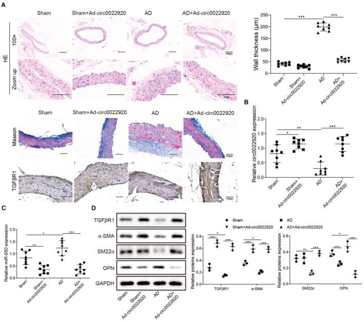
Background Abnormal regulation of vascular smooth muscle cells is regarded as the iconic pathological change of aortic dissection (AD). Herein, we aim to identify circ_0022920 as a crucial regulator in AD. Methods and Results Microarray analysis of circular RNAs, messenger RNAs, and micro RNAs in patients with AD was performed, and we identified that circ_0022920 was significantly downregulated in these patients. The Pearson correlation analysis uncovered the negative correlation between miR-650 and circ_0022920 or TGFβR1 (transforming growth factor beta receptor 1). Angiotensin II was used to treat human aortic vascular smooth muscle cells (HASMCs) and mice as models for AD. Hematoxylin and eosin and Masson's trichrome staining were used to analyze AD histopathology. Cell proliferation was analyzed with Cell Counting Kit-8 assay and EdU incorporation. Cell migration was assessed with transwell and wound healing assays. Enhanced circ_0022920 expression dramatically inhibited HASMC proliferation and migration and maintained contractile marker expression induced by angiotensin II, whereas miR-650 exerted opposite effects. MiR-650 was a target of circ_0022920. MiR-650 targeted IRF1 (interferon regulatory factor 1) and thus negatively regulated TGFβR1 expression to promote HASMC proliferation and migration and inhibit contractile marker expression. Circ_0022920 suppressed the progression of AD in vivo. Conclusions Circ_0022920 modulates the contractile phenotype of HASMCs via regulating the miR-650-IRF1-TGFβR1 axis in angiotensin II-induced models for AD, which provides potential therapeutic targets for AD.

Keywords: Circ_0022920; aortic dissection; contractile phenotype; miR‐650/IRF1/TGFβR1 axis.


Figures

Figure 1
Figure 1. Dysregulated expression of circ_0022920, miR‐650, and TGFβR1 in AD.
A through C, Differential expression analysis of circRNAs/miRNA/mRNA in AD patients with heatmap and volcano plot. D, Computationally constructed circRNA/miRNA/mRNA ceRNA network. Downregulated mRNAs (green circle), upregulated mRNAs (red circle), downregulated circRNAs (purple square), upregulated circRNAs (blue square), downregulated miRNAs (pink inverted triangle), and upregulated miRNAs (yellow inverted triangle). miRNA‐mRNA regulatory relationship (yellow arrow) and circRNA‐miRNA regulatory relationship (black T line). AD indicates aortic dissection; ceRNA, competing endogenous RNA; circRNA, circular RNA; mRNA, messenger RNA; miRNA, microRNA; and TGFβR1, transforming growth factor beta receptor 1.
Figure 2
Figure 2. Circ_002290 negatively correlates with TGFβR1 but positively correlates with miR‐650 in AD.
A, The relative expression of circ_0022920, miR‐650, and TGFβR1 in aortic tissues from controls and patients with AD (n=28). Circ_0022920 and TGFβR1 were normalized to GAPDH, and miR‐650 was normalized to U6 snRNA. B and C, The Pearson correlation analysis of circ_0022920/miR‐650 and circ_0022920/TGFβR1 in patients with AD and controls. All experiments were repeated 3 times independently. To examine statistical significance, t test, nonparametric Friedman test, and post hoc Dunn's multiple comparison test were used. *P<0.05, **P<0.01, ***P<0.001. AD indicates aortic dissection; miR‐650, microRNA‐650; and TGFβR1, transforming growth factor beta receptor 1.
Figure 3
Figure 3. Circ_0022920 inhibits proliferation and migration and maintains the expression of contractile phenotype markers in angiotensin II‐treated HASMCs.
A, The relative expression of circ_0022920 in control and angiotensin II‐treated HASMCs. B, The relative expression of circ_0022920 in angiotensin II‐treated HASMCs (Control), angiotensin II‐treated HASMCs transfected with Ad‐NC, Ad‐circ_0022920, sh‐NC, or sh‐circ_0022920, respectively. C, Proliferation and viability analysis of Control, angiotensin II‐treated HASMCs transfected with Ad‐NC, Ad‐circ_0022920, sh‐NC, or sh‐circ_0022920 with Cell Counting Kit 8 assay at 0, 24, 48, 72, and 96 hours. D, EdU incorporation of Control, ngiotensin II‐treated HASMCs transfected with Ad‐NC, Ad‐circ_0022920, sh‐NC, or sh‐circ_0022920. E and F, Migration analysis of Control, angiotensin II‐treated HASMCs transfected with Ad‐NC, Ad‐circ_0022920, sh‐NC, or sh‐circ_0022920 with transwell assay. G, Migration analysis of Control, angiotensin II‐treated HASMCs transfected with Ad‐NC, Ad‐circ_0022920, sh‐NC, or sh‐circ_0022920 with wound healing assay. H, Western blotting analysis of protein levels of α‐SMA, SM22α, osteopontin, and MYH11 in Control, angiotensin II‐treated HASMCs transfected with Ad‐NC, Ad‐circ_0022920, sh‐NC, or sh‐circ_0022920. U6 RNA was used as a normalization control in quantitative PCR analysis of miR‐650. GAPDH was used as a normalization control in Western blot analysis. N=3 for each group. All experiments were repeated 3 times independently. To examine statistical significance, t test, nonparametric Friedman test, and post hoc Dunn's multiple comparison test were used. *P<0.05, **P<0.01, ***P<0.001. α‐SMA indicates smooth muscle alpha actin; HASMC, human aortic vascular smooth muscle cell; MYH11, myosin heavy chain‐11; PCR, polymerase chain reaction; and SM22α, smooth muscle 22α.
Figure 4
Figure 4. Overexpression of miR‐650 promotes proliferation and migration and downregulates contractile phenotype markers in angiotensin II‐treated HASMCs.
A, The relative expression of miR‐650 in control and angiotensin II‐treated HASMCs. B, The relative expression of miR‐650 in angiotensin II‐treated HASMCs transfected with miR‐NC, miR‐650 mimic, anti‐miR‐NC, or anti‐miR‐650, respectively. C, Proliferation analysis of angiotensin II‐treated HASMCs transfected with miR‐NC, miR‐650 mimic, anti‐miR‐NC, or anit‐miR‐650 with Cell Counting Kit 8 assay at 0, 24, 48, 72, and 96 hours. D, EdU incorporation of angiotensin II‐treated HASMCs transfected with miR‐NC, miR‐650 mimic, anti‐miR‐NC, or anti‐miR‐650. E and F, Migration analysis of angiotensin II‐treated HASMCs transfected with miR‐NC, miR‐650 mimic, anti‐miR‐NC, or anti‐miR‐650 with transwell assay. G, Migration analysis of angiotensin II‐treated HASMCs transfected with miR‐NC, miR‐650 mimic, anti‐miR‐NC, or anti‐miR‐650 with wound healing assay. H, Western blotting analysis of protein levels of α‐SMA, SM22α, osteopontin, and MYH11 in angiotensin II‐treated HASMCs transfected with miR‐NC, miR‐650 mimic, anti‐miR‐NC, or anti‐miR‐650. U6 RNA was used as a normalization control in quantitative PCR analysis of miR‐650. GAPDH was used as a normalization control in Western blot analysis. N=3 for each group. All experiments were repeated 3 times independently. To examine statistical significance, t test, nonparametric Friedman test, and post hoc Dunn's multiple comparison test were used. *P<0.05, **P<0.01, ***P<0.001. α‐SMA indicates smooth muscle alpha actin; HASMC, human aortic vascular smooth muscle cell; miR‐650, microRNA‐650; MYH11, myosin heavy chain‐11; PCR, polymerase chain reaction; and SM22α, smooth muscle 22α.
Figure 5
Figure 5. Circ_0022920 binds directly to miR‐650 to regulate proliferation and migration in HASMCs.
A, The subcellular fractionation was employed to determine the location of circ_0022920. B, The enrichment of circ_0022920 in cell lysis input, cell lysates with circ_0022920 probe, or control probe. C, The enrichment of miR‐650 in cell lysis input, cell lysates with circ_0022920 probe, or control probe. D, Luciferase reporter assay was performed to verify the interaction of circ_0022920/miR‐650. E, MS2‐based RNA immunoprecipitation assay was used to verify the interaction of circ_0022920/miR‐650. F, Colocalization analysis of miR‐650 and circ_0022920 with fluorescence in situ hybridization assay; miR‐650 (Red), circ_0022920 (Green), and nuclear (Blue). G, The relative expression of miR‐650 in angiotensin II‐treated HASMCs transfected with Ad‐NC/miR‐NC, Ad‐circ_0022920/miR‐NC, or Ad‐circ_0022920/miR‐650 mimic, respectively. H, Proliferation analysis of angiotensin II‐treated HASMCs transfected with Ad‐NC/miR‐NC, Ad‐circ_0022920/miR‐NC, or Ad‐circ_0022920/miR‐650 mimic with Cell Counting Kit 8 assay at 0, 24, 48, 72, and 96 hours. I and J, Migration analysis of angiotensin II‐treated HASMCs transfected with Ad‐NC/miR‐NC, Ad‐circ_0022920/miR‐NC, or Ad‐circ_0022920/miR‐650 mimic with transwell assay. K and L, Migration analysis of angiotensin II‐treated HASMCs transfected with Ad‐NC/miR‐NC, Ad‐circ_0022920/miR‐NC, or Ad‐circ_0022920/miR‐650 mimic with wound healing assay. M, Western blotting analysis of protein levels of TGFβR1, α‐SMA, SM22α, osteopontin, and MYH11 in angiotensin II‐treated HASMCs transfected with Ad‐NC/miR‐NC, Ad‐circ_0022920/miR‐NC, or Ad‐circ_0022920/miR‐650 mimic. U6 RNA was used as a normalization control in quantitative PCR analysis of miR‐650. GAPDH was used as a normalization control in Western blot analysis. N=3 for each group. All experiments were repeated 3 times independently. To examine statistical significance, t test, nonparametric Friedman test, and post hoc Dunn's multiple comparison test were used. *P<0.05, **P<0.01, ***P<0.001. α‐SMA indicates smooth muscle alpha actin; HASMC, human aortic vascular smooth muscle cell; miR‐650, microRNA‐650; MYH11, myosin heavy chain‐11; PCR, polymerase chain reaction; SM22α, smooth muscle 22α; and TGFβR1, transforming growth factor beta receptor 1.
Figure 6
Figure 6. MiR‐650‐mediated regulation of HASMCs is dependent on TGFβR1.
A, The Pearson correlation analysis of miR‐650 and TGFβR1. B, The relative expression of TGFβR1 in angiotensin II‐treated HASMCs transfected with anti‐miR‐NC, anti‐miR‐650, miR‐NC, or miR‐650, respectively. C, Proliferation analysis of angiotensin II‐treated HASMCs transfected with anti‐miR‐NC/siNC, anti‐miR‐650/siNC, or anti‐miR‐650/siTGFβR1with Cell Counting Kit 8 assay at 0, 24, 48, 72, and 96 hours. D and E, Migration analysis of angiotensin II‐treated HASMCs transfected with anti‐miR‐NC/siNC, anti‐miR‐650/siNC, or anti‐miR‐650/siTGFβR1 with transwell assay. F and G, Migration analysis of angiotensin II‐treated HASMCs transfected with anti‐miR‐NC/siNC, anti‐miR‐650/siNC, or anti‐miR‐650/siTGFβR1 with wound healing assay. H, Western blot analysis of protein levels of α‐SMA, SM22α, osteopontin, and MYH11 in angiotensin II‐treated HASMCs transfected with anti‐miR‐NC/siNC, anti‐miR‐650/siNC, or anti‐miR‐650/siTGFβR1. GAPDH was used as a normalization control in quantitative PCR and Western blot analysis. N=3 for each group. All experiments were repeated 3 times independently. To examine statistical significance, t test, nonparametric Friedman test, and post hoc Dunn's multiple comparison test were used. *P<0.05, **P<0.01. α‐SMA indicates smooth muscle alpha actin; HASMC, human aortic vascular smooth muscle cell; miR‐650, microRNA‐650; MYH11, myosin heavy chain‐11; PCR, polymerase chain reaction; SM22α, smooth muscle 22α; and TGFβR1, transforming growth factor beta receptor 1.
Figure 7
Figure 7. Circ_0022920 inhibits the progression of AD in vivo.
A, Hematoxylin and eosin staining in murine aortic tissues from sham, sham+Ad‐circ_0022920, AD, and AD+Ad‐circ_0022920 groups. B and C, Masson's trichrome staining and immunohistochemistry (TGFβR1) from sham, sham+Ad‐circ_0022920, AD, and AD+Ad‐circ_0022920 groups. D, The relative expression of circ_0022920, miR‐650, and TGFβR1 in aortic tissues from sham, sham+Ad‐circ_0022920, AD, and AD+Ad‐circ_0022920 groups (n=8). E, Western blot analysis of protein levels of TGFβR1, α‐SMA, SM22α, osteopontin, and MYH11 in aortic tissues from sham, sham+Ad‐circ_0022920, AD, and AD+Ad‐circ_0022920 groups (n=3). U6 RNA was used as a normalization control in quantitative PCR analysis of miR‐650. GAPDH was used as a normalization control in quantitative PCR analysis of circ_0022920 and Western blot analysis. All experiments were repeated 3 times independently. To examine statistical significance, t test, nonparametric Friedman test, and post hoc Dunn's multiple comparison test were used. *P<0.05, **P<0.01, ***P<0.001. α‐SMA indicates smooth muscle alpha actin; AD, aortic dissection; HASMC, human aortic vascular smooth muscle cell; MYH11, myosin heavy chain‐11; PCR, polymerase chain reaction; SM22α, smooth muscle 22α; and TGFβR1, transforming growth factor beta receptor 1.
Figure 8
Figure 8. Molecular mechanism diagram. Circ_0022920 works as a miR‐650 sponge to reduce its expression and naturally promotes IRF1 expression.
Subsequently, IRF1 binds directly to TGFβR1 promoter and enhances its transcription and expression, thus inhibiting proliferation and migration and maintaining contractile marker expression in HASMCs, thus alleviating AD. AD indicates aortic dissection; HASMC, human aortic vascular smooth muscle cell; IRF1, interferon regulatory factor 1; miR, microRNA; TGFβR1, transforming growth factor beta receptor 1; and 3'UTR, three prime untranslated region.

Similar articles

Cited by

References

    1. Nienaber CA, Clough RE, Sakalihasan N, Suzuki T, Gibbs R, Mussa F, Jenkins MP, Thompson MM, Evangelista A, Yeh JS, et al. Aortic dissection. Nat Rev Dis Primers. 2016;2:16071. doi: 10.1038/nrdp.2016.71 - DOI - PubMed
    1. Juang D, Braverman AC, Eagle K. Cardiology patient pages. Aortic dissection. Circulation. 2008;118:e507–e510. - PubMed
    1. Daily PO, Trueblood HW, Stinson EB, Wuerflein RD, Shumway NE. Management of acute aortic dissections. Ann Thorac Surg. 1970;10:237–247. doi: 10.1016/S0003-4975(10)65594-4 - DOI - PubMed
    1. Khan IA, Nair CK. Clinical, diagnostic, and management perspectives of aortic dissection. Chest. 2002;122:311–328. doi: 10.1378/chest.122.1.311 - DOI - PubMed
    1. Nienaber CA, Fattori R, Mehta RH, Richartz BM, Evangelista A, Petzsch M, Cooper JV, Januzzi JL, Ince H, Sechtem U, et al. Gender‐related differences in acute aortic dissection. Circulation. 2004;109:3014–3021. doi: 10.1161/01.CIR.0000130644.78677.2C - DOI - PubMed

Publication types

LinkOut - more resources