. 2023 Feb 22;8(1):89.
doi: 10.3390/biomimetics8010089.

A Novel Peptide-Based Detection of SARS-CoV-2 Antibodies

Affiliations

A Novel Peptide-Based Detection of SARS-CoV-2 Antibodies

Aliye Bulut et al. Biomimetics (Basel). .

Abstract

The need for rapidly developed diagnostic tests has gained significant attention after the recent pandemic. Production of neutralizing antibodies for vaccine development or antibodies to be used in diagnostic tests usually require the usage of recombinant proteins representing the infectious agent. However, peptides that can mimic these recombinant proteins may be rapidly utilized, especially in emergencies such as the recent outbreak. Here, we report two peptides that mimic the receptor binding domain of the spike protein of severe acute respiratory syndrome coronavirus 2 (SARS-CoV-2) and investigate their binding behavior against the corresponding human immunoglobulin G and immunoglobulin M (IgG and IgM) antibodies in a clinical sample using a quartz crystal microbalance (QCM) sensor. These peptides were immobilized on a QCM sensor surface, and their binding behavior was studied against a clinical serum sample that was previously determined to be IgG and IgM-positive. It was determined that designed peptides bind to SARS-CoV-2 antibodies in a clinical sample. These peptides might be useful for the detection of SARS-CoV-2 antibodies using different methods such as enzyme-linked immunosorbent assay (ELISA) or lateral flow assays. A similar platform might prove to be useful for the detection and development of antibodies in other infections.

Keywords: SARS-CoV-2; antibody detection; biosensor; peptide mimetics; quartz crystal microbalance.


Conflict of interest statement

The authors declare no conflict of interest.

Figures

Figure 1
Figure 1
Schematic diagram of the quartz crystal microbalance (QCM) experiment steps. These steps are 3-mercaptopropyltrimethoxysilane (MPS) coating, peptide immobilization, serum treatment (Antibody (Ab) binding) and fluorescent reporter microspheres (FRMs) conjugated anti-human secondary antibody treatment.
Figure 2
Figure 2
Schematic diagram of the QCM experiment flow setup.
Figure 3
Figure 3
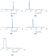
HPLC results of (a) pure TBT1 peptide, (b) pure TBT2 peptide and (c) MAL-PEG8-NHS linker molecules and HPLC results of the (d) TBT1 conjugates and (e) TBT2 conjugates collected after Sephadex-G25 column purification. Scanning was performed for 30 min between 5–80% B gradient at a flow rate of 0.4 mL/min.
Figure 4
Figure 4
Resonance frequency shifts (∆f) observed during TBT1 and TBT2 peptide immobilization on the MPS−coated QCM surface.
Figure 5
Figure 5
f versus time of COVID−19 Ab detection by human blood serum treatment for (a) TBT1 and (b) TBT2.
Figure 6
Figure 6
Resonance frequency shifts observed during confirmation experiments for (a) TBT1 and (b) TBT2. ∆f versus time of anti−human immunoglobulin G (anti−human IgG) and FRMs conjugated anti−human immunoglobulin M (anti−human IgM) detections for antibody-positive serum (Ab PS)−treated and negative/control serum (CS)−treated TBT1 are shown in a. ∆f of FRMs conjugated anti−human IgG detection for Ab PS−treated and CS−treated TBT2 are shown in (b).
Figure 7
Figure 7
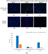
Blue, fluorescent images of the captured microspheres on the QCM surface after FRMs conjugated secondary antibody detection following Ab positive (ac) and control serum (df) treatments. (a,d) are the results of FRM/anti-human IgG treatments, (b,e) are the results of FRM/anti-human IgM treatments for TBT1. Also, (c,f) are the results of FRM/anti-human IgG treatments for TBT2. (g) shows the number of FRMs captured on the QCM surface after various treatments for TBT1 and TBT2. The numbers of the FRMs captured on the QCM surface are 1368, 140, 631, 419, 72 and 58, which are shown in (af), respectively.

References

    1. Afzal A., Mujahid A., Schirhagl R., Bajwa S.Z., Latif U., Feroz S. Gravimetric viral diagnostics: Qcm based biosensors for early detection of viruses. Chemosens. 2017;5:7. doi: 10.3390/chemosensors5010007. - DOI
    1. Rasmi Y., Li X., Khan J., Ozer T., Choi J.R. Emerging point-of-care biosensors for rapid diagnosis of COVID-19: Current progress, challenges, and future prospects. Anal. Bioanal. Chem. 2021;413:4137–4159. doi: 10.1007/s00216-021-03377-6. - DOI - PMC - PubMed
    1. Hwang Y.C., Lu R.M., Su S.C., Chiang P.Y., Ko S.H., Ke F.Y., Liang K.H., Hsieh T.Y., Wu H.C. Monoclonal antibodies for COVID-19 therapy and SARS-CoV-2 detection. J. Biomed. Sci. 2022;29:1. doi: 10.1186/s12929-021-00784-w. - DOI - PMC - PubMed
    1. Ali M.A., Hu C., Zhang F., Jahan S., Yuan B., Saleh M.S., Gao S.J., Panat R. N protein-based ultrasensitive SARS-CoV-2 antibody detection in seconds via 3d nanoprinted, microarchitected array electrodes. J. Med. Virol. 2022;94:2067–2078. doi: 10.1002/jmv.27591. - DOI - PMC - PubMed
    1. Carter L.J., Garner L.V., Smoot J.W., Li Y., Zhou Q., Saveson C.J., Sasso J.M., Gregg A.C., Soares D.J., Beskid T.R., et al. Assay techniques and test development for COVID-19 diagnosis. ACS Cent. Sci. 2020;6:591–605. doi: 10.1021/acscentsci.0c00501. - DOI - PMC - PubMed

LinkOut - more resources