. 2023 Mar 16;8(1):123.
doi: 10.3390/biomimetics8010123.

Influence of a Physiologically Formed Blood Clot on Pre-Osteoblastic Cells Grown on a BMP-7-Coated Nanoporous Titanium Surface

Affiliations

Influence of a Physiologically Formed Blood Clot on Pre-Osteoblastic Cells Grown on a BMP-7-Coated Nanoporous Titanium Surface

Leonardo Raphael Zuardi et al. Biomimetics (Basel). .

Abstract

Titanium (Ti) nanotopography modulates the osteogenic response to exogenous bone morphogenetic protein 7 (BMP-7) in vitro, supporting enhanced alkaline phosphatase mRNA expression and activity, as well as higher osteopontin (OPN) mRNA and protein levels. As the biological effects of OPN protein are modulated by its proteolytic cleavage by serum proteases, this in vitro study evaluated the effects on osteogenic cells in the presence of a physiological blood clot previously formed on a BMP-7-coated nanostructured Ti surface obtained by chemical etching (Nano-Ti). Pre-osteoblastic MC3T3-E1 cells were cultured during 5 days on recombinant mouse (rm) BMP-7-coated Nano-Ti after it was implanted in adult female C57BI/6 mouse dorsal dermal tissue for 18 h. Nano-Ti without blood clot or with blood clot at time 0 were used as the controls. The presence of blood clots tended to inhibit the expression of key osteoblast markers, except for Opn, and rmBMP-7 functionalization resulted in a tendency towards relatively greater osteoblastic differentiation, which was corroborated by runt-related transcription factor 2 (RUNX2) amounts. Undetectable levels of OPN and phosphorylated suppressor of mothers against decapentaplegic (SMAD) 1/5/9 were noted in these groups, and the cleaved form of OPN was only detected in the blood clot immediately prior to cell plating. In conclusion, the strategy to mimic in vitro the initial interfacial in vivo events by forming a blood clot on a Ti nanoporous surface resulted in the inhibition of pre-osteoblastic differentiation, which was minimally reverted with an rmBMP-7 coating.

Keywords: BMP-7; blood clot; nanotopography; osteoblast; titanium.


Conflict of interest statement

The authors declare no conflict of interest.

Figures

Figure 1
Figure 1
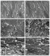
The epifluorescence of MC3T3-E1 cells on day 5 of culture (A,B), blood clot alone on time 0 (C,D), or the combination of both on day 5 of culture (E,F) on pristine Nano-Ti (A,C,E) or Nano-Ti functionalized with rmBMP-7 (B,D,F). Red fluorescence (Alexa Fluor 594 phalloidin) indicates the actin cytoskeleton (AC,E,F), green fluorescence indicates the GFP-positive blood clot leukocytes (CF), and blue fluorescence indicates the cell nuclei (DAPI nuclear stain) (AF) and the fibrin network (D). Red fluorescence is deleted in the left halves of (AC,E,F) using Adobe Photoshop CS6 for a better visualization of the cell nuclei and GFP-positive cells (white arrows in (E,F)) in the same microscopic field. Objectives: 20× (AD); 40× (E,F).
Figure 2
Figure 2
Scanning electron microscopy (SEM) imaging of MC3T3-E1 cells on day 5 of culture (A,B), blood clots alone on time 0 (C,D), or the combination of both on day 5 of culture (E,F) on pristine Nano-Ti (A,C,E) or Nano-Ti functionalized with rmBMP-7 (B,D,F). Original magnification: 1000× (AF). MC3T3-E1 cells exhibit elongated shapes (white arrows in (A)) irrespective of the presence of a blood clot.
Figure 3
Figure 3
Runx2, Osx, Alp, Bsp, Oc, and Opn mRNA expression levels normalized to Gapdh in MC3T3-E1 cell cultures grown for 5 days and in blood clots on time 0 on Nano-Ti surfaces either functionalized with or without rmBMP-7. The bars represent one biological replicate (resulting from the pooling of 16 independent wells/experimental replicates for each group) run in three technical replicates (mean and SD). The mean value of the control MC3T3-E1 group is assigned a value of 1. Additional technical replicates obtained in a different moment using the same cDNA result in a similar expression pattern (Figure S5).
Figure 4
Figure 4
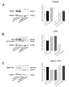
Detection and quantification of RUNX2 (A), OPN (B), and phosphorylated-SMAD 1/5/9 (C) by Western blotting (number of pixels of the protein bands normalized to GAPDH) in MC3T3-E1 cell cultures grown for 5 days and in blood clots on time 0 on Nano-Ti surfaces either functionalized with or without rmBMP-7. The bars represent one biological replicate (resulting from the pooling of 20 independent wells/experimental replicates for each group) and one technical replicate. The ratio of the control group (MC3T3-E1) is assigned a value of 1. Lower molecular weight bands in (B,C) are cropped to fit the figure.

References

    1. Holmes C., Tabrizian M. Surface Functionalization of Biomaterials. In: Vishwakarma A., Sharpe P., Shi S., Ramalingam M., editors. Stem Cell Biology and Tissue Engineering in Dental Sciences. Academic Press; Cambridge, MA, USA: 2015. pp. 187–206. - DOI
    1. Ferraris S., Cazzola M., Zuardi L.R., de Oliveira P.T. Metal nanoscale systems functionalized with organic compounds. In: Guarino V., Iafisco M., Spriano S., editors. Nanostructured Biomaterials for Regenerative Medicine. Woodhead Publishing; Sawston, UK: 2020. pp. 407–436. (Woodhead Publishing Series in Biomaterials). - DOI
    1. Jablonská E., Horkavcová D., Rohanová D., Brauer D.S. A review of in vitro cell culture testing methods for bioactive glasses and other biomaterials for hard tissue regeneration. J. Mater. Chem. B. 2020;8:10941–10953. doi: 10.1039/D0TB01493A. - DOI - PubMed
    1. Anderson J.M. Future challenges in the in vitro and in vivo evaluation of biomaterial biocompatibility. Regen. Biomater. 2016;3:73–77. doi: 10.1093/rb/rbw001. - DOI - PMC - PubMed
    1. Jablonská E., Kubásek J., Vojtěch D., Ruml T., Lipov J. Test conditions can significantly affect the results of in vitro cytotoxicity testing of degradable metallic biomaterials. Sci. Rep. 2021;11:6628. doi: 10.1038/s41598-021-85019-6. - DOI - PMC - PubMed