. 2023 Mar 13;14(3):282.
doi: 10.3390/insects14030282.

The Structure of the Female Genital System of the Diving Beetle Scarodytes halensis (Fabricius, 1787) (Hydroporinae, Dytiscidae), and the Organization of the Spermatheca and the Spermathecal Gland Complex

Affiliations

The Structure of the Female Genital System of the Diving Beetle Scarodytes halensis (Fabricius, 1787) (Hydroporinae, Dytiscidae), and the Organization of the Spermatheca and the Spermathecal Gland Complex

Romano Dallai et al. Insects. .

Abstract

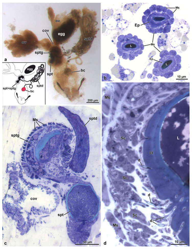
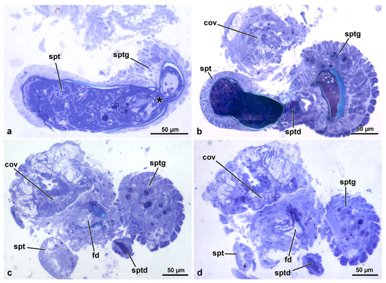
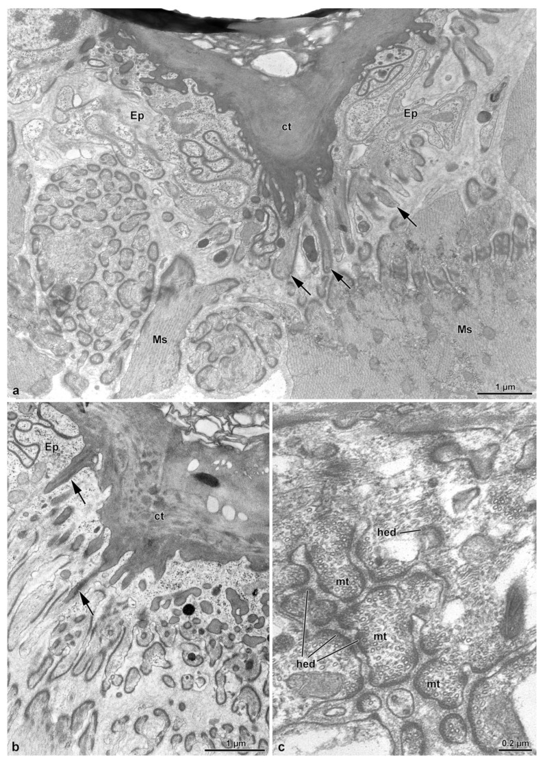
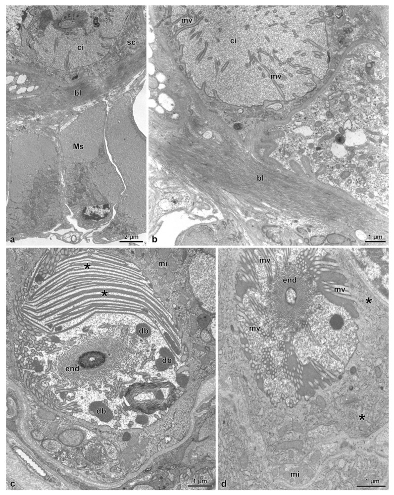
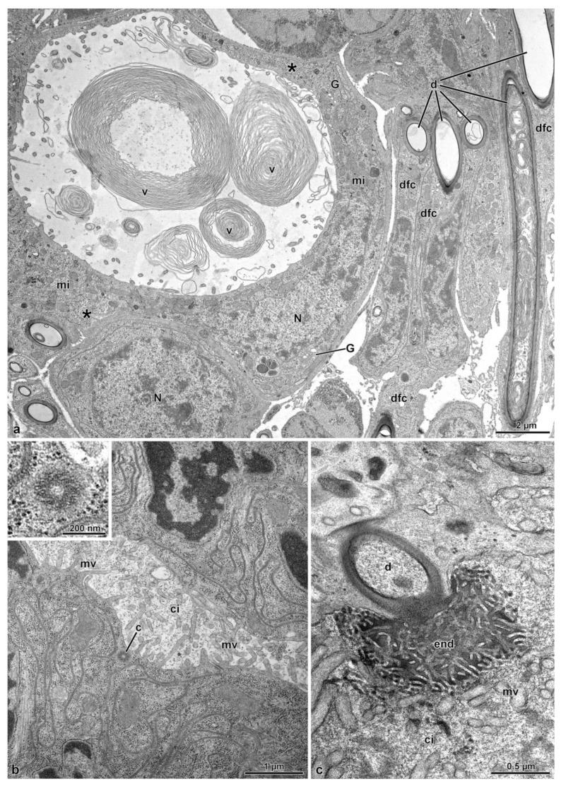
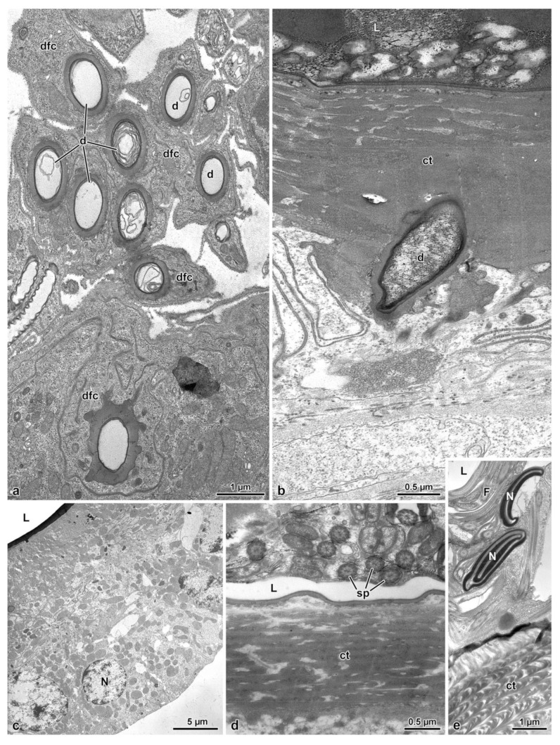
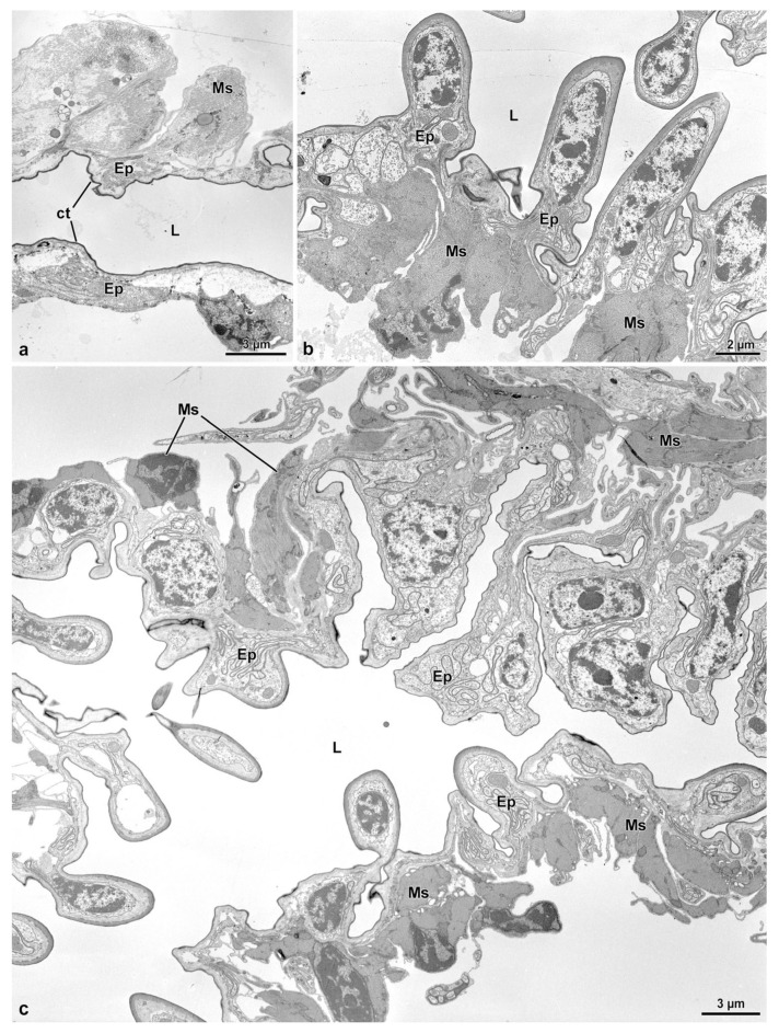
The fine structure of the female reproductive organs of the diving beetle Scarodytes halensis has been described, with particular attention to the complex organization of the spermatheca and the spermathecal gland. These organs are fused in a single structure whose epithelium is involved in a quite different activity. The secretory cells of the spermathecal gland have a large extracellular cistern with secretions; duct-forming cells, by their efferent duct, transport the secretions up to the apical cell region where they are discharged into the gland lumen. On the contrary, the spermatheca, filled with sperm, has a quite simple epithelium, apparently not involved in secretory activity. The ultrastructure of the spermatheca is almost identical to that described in a closely related species Stictonectes optatus. Sc. halensis has a long spermathecal duct connecting the bursa copulatrix to the spermatheca-spermathecal gland complex. This duct has a thick outer layer of muscle cells. Through muscle contractions, sperm can be pushed forwarding up to the complex of the two organs. A short fertilization duct allows sperm to reach the common oviduct where eggs will be fertilized. The different organization of the genital systems of Sc. halensis and S. optatus might be related to a different reproductive strategy of the two species.

Keywords: diving beetles; female reproductive system; insect ultrastructure.


Conflict of interest statement

The authors declare no conflict of interest.

Figures

Figure 1
Figure 1
(a). Light microscopic view of the female reproductive organs. bc, bursa copulatrix; cov, common oviduct; ov, oviduct; spt, spermatheca; sptd, spermathecal duct; sptg, spermathecal gland. Egg at the end of the oviduct. (b). Semi-thin section through the spermathecal duct showing the thick layer of muscle cells (Ms) and the epithelium (Ep). L, duct lumen. (c). Semi-thin section of the complex spermatheca (spt)–spermathecal gland (sptg). Note the spermathecal duct (sptd) and the common oviduct (cov). (d). Semi-thin section of the spermathecal gland epithelia (Ep) showing the outer muscle layer (Ms), the secretory cells (Sc), the ducts (d) of the duct-forming cells, the thick cuticle (ct) and the gland lumen (L).
Figure 2
Figure 2
(a). Semi-thin section of the spermatheca (spt) and the narrow connection (asterisk) to the spermathecal gland (sptg). Note the thin wall of the spermatheca compared to that of the spermathecal gland. (b). Semi-thin section of the complex spermatheca (spt)–spermathecal gland (sptg). Note the quite evident different thickness of the wall of the two districts. Note also the opening the spermathecal duct (sptd). cov, common oviduct. (c,d). Consecutive semi-thin sections showing the position of the fertilization duct (fd) beneath the complex spermatheca (spt)–spermathecal gland (sptg). cov, common oviduct; sptd, spermathecal duct.
Figure 3
Figure 3
(ac). Cross sections of the spermathecal duct. The thick cuticle (ct) lining the thin epithelium (Ep) shows long tubular prolongments (arrows) directed towards the thin cytoplasm. Around these structures, numerous longitudinal microtubules (mt) are visible reaching the basal lamina to form hemidesmosomes (hed). Beneath the epithelial cells, a thick layer of muscle cells (Ms) is present.
Figure 4
Figure 4
(a). Cross section through the basal epithelial region of the spermathecal gland. Note the secretory cell (sc) with the cistern (ci). Beneath the epithelium, a fibrous basal lamina (bl) and a thick layer of muscle cells (Ms) are visible. (b). Cross section of a secretory cell showing a large cistern (ci) lined by long microvilli (mv). (c). Cross section through a cistern of a secretory cell with the lumen filled with numerous laminar structures (asterisks) and dense bodies (db). In the center of the cistern, the “end apparatus” (end) of a duct of the duct-forming cell is visible. In the cytoplasm, mitochondria (mi) and few cisterns of rough endoplasmic reticulum are present. (d). Cross section through a cistern of a secretory cell. Note the long microvilli (mv) and the “end apparatus” (end). Note the mitochondria (mi) and the few cisterns of rough endoplasmic reticulum (asterisks).
Figure 5
Figure 5
(a). Cross section of a large secretory cell with a cistern rich of membrane vortices (v). In the cytoplasm, an elliptical nucleus (N) is visible, and numerous mitochondria (mi), some cisterns of rough endoplasmic reticulum (asterisks) and Golgi apparatuses (G) are present. A neighboring cell shows a spherical nucleus (N). On the right side some duct-forming cells (dfc) with their ducts (d) are visible. (b). Cross section of a secretory cell with the cistern still to be developed. Short microvilli (mv) are visible lining the cistern. Just beneath this region, a centriole (c) is visible. In the inset, a detail of the centriole. (c). Cross section of the “end apparatus” (end) and of the beginning of the duct (d) of the duct-forming cell. ci, cistern; mv, microvilli.
Figure 6
Figure 6
(a). Cross section of a complex of duct-forming cells (dfc) with their ducts (d). (b). Cross section of the apical region of the spermathecal gland epithelium lined by a thick cuticle (ct) that is crossed by a duct (d) of a duct-forming cell. L, gland lumen. (c). Cross section through the spermatheca epithelial cells. L, spermathecal lumen; N, nucleus. (d,e). Cross sections through the apical region of the spermatheca to show the lumen (L) filled with sperm cells (sp) sectioned at the flagellar (F) and nuclear (N) levels. ct, cuticle.
Figure 7
Figure 7
(a). Cross section of the fertilization duct showing a cuticular (ct) thin epithelium (Ep) with scattered muscle (Ms). L, duct lumen. (b,c). Cross sections through the characteristic epithelium (Ep) of the common oviduct, with epithelial cells often protruding into the lumen (L). Ms, Muscle cells.

References

    1. Miller G.T., Pitnick S. Sperm-female coevolution in Drosophila. Science. 2002;298:1230–1233. doi: 10.1126/science.1076968. - DOI - PubMed
    1. Miller G.T., Pitnick S. Functional significance of seminal receptacle length in Drosophila melanogaster. J. Evol. Biol. 2003;16:114–126. doi: 10.1046/j.1420-9101.2003.00476.x. - DOI - PubMed
    1. Pattarini J.M., Starmer W.T., Bjork A., Pitnick S. Mechanisms underlying the sperm quality advantage in Drosophila melanogaster. Evolution. 2006;60:2064–2080. - PubMed
    1. Higginson D.M., Miller K.B., Segraves K.A., Pitnick S. Convergence, recurrence and diversification of complex sperm traits in diving beetles (Dytiscidae) Evolution. 2012;66:1650–1661. doi: 10.1111/j.1558-5646.2011.01532.x. - DOI - PMC - PubMed
    1. Lüpold S., Manier M.K., Berben K.S., Smith B.D., Daley S.H., Buckley S.H., Belote J.M., Pitnick S. How multivariate ejaculate traits determine competitive fertilization success in Drosophila melanogaster. Curr. Biol. 2012;22:1667–1672. doi: 10.1016/j.cub.2012.06.059. - DOI - PubMed

LinkOut - more resources