. 2023 Feb 27;14(3):133.
doi: 10.3390/jfb14030133.

Functionalization of a Cortical Membrane with a Photodynamic Protocol

Affiliations

Functionalization of a Cortical Membrane with a Photodynamic Protocol

Tania Vanessa Pierfelice et al. J Funct Biomater. .

Abstract

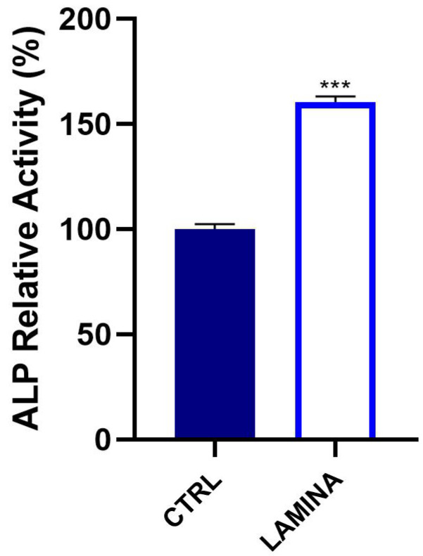
Guided bone regeneration (GBR) comprehends the application of membranes to drive bone healing and to exclude non-osteogenic tissues from interfering with bone regeneration. However, the membranes may be exposed to bacterial attack, with the risk of failure of the GBR. Recently, an antibacterial photodynamic protocol (ALAD-PDT) based on a gel with 5% 5-aminolevulinic acid incubated for 45 min and irradiated for 7 min by a LED light at 630 nm, also showed a pro-proliferative effect on human fibroblasts and osteoblasts. The present study hypothesized that the functionalization of a porcine cortical membrane (soft-curved lamina, OsteoBiol) with ALAD-PDT might promote its osteoconductive properties. TEST 1 aimed to verify the response of osteoblasts seeded on lamina with respect to the plate surface (CTRL). TEST 2 aimed to investigate the effects of ALAD-PDT on the osteoblasts cultured on the lamina. SEM analyses were performed to study the topographical characteristics of the membrane surface, the adhesion, and the morphology of cells at 3 days. The viability was assessed at 3 days, the ALP activity at 7 days, and calcium deposition at 14 days. Results showed the porous surface of the lamina and the increase in cell attachment of osteoblasts with respect to controls. The proliferation, the ALP, and bone mineralization activity of osteoblasts seeded on lamina resulted in being significantly higher (p < 0.0001) than controls. Results also showed an additional significative enhancement (p < 0.0001) in the proliferative rate in ALP and calcium deposition after applying ALAD-PDT. In conclusion, the functionalization of the cortical membranes cultured with osteoblasts with the ALAD-PDT improved their osteoconductive properties.

Keywords: 5-aminolevulinic acid; ALP; GBR; LED; calcium deposition; lamina cortical membrane; osteoblasts; photodynamic therapy.


Conflict of interest statement

The authors declare no conflict of interest.

Figures

Figure 1
Figure 1
Cortical bone of heterologous origin OsteoBiol® lamina.
Figure 2
Figure 2
ALAD-PDT protocol consists of a gel containing 5% of delta-aminolevulinic acid distributed in a disposable syringe (A) and a LED device TL-01 as red-light source (B).
Figure 3
Figure 3
SEM observation of the lamina as manufactured. Mag = 1000×.
Figure 4
Figure 4
Viability of osteoblasts cultured on the lamina at 3 days. Statistically significant differences were expressed with respect to cells seeded on the well/plate as CTRL (*** p < 0.0001).
Figure 5
Figure 5
SEM images of oral osteoblasts (HOBs) cultured on the plate (A,C) and on the lamina cortical membrane (B,D) at 3 days of culture. Mag = 1000× (A,B), Mag = 3000× (C,D).
Figure 6
Figure 6
ALP levels of HOBs seeded on the lamina were analyzed at 7 days (*** p < 0.0001).
Figure 7
Figure 7
Mineralization of HOBs at 14 days of culture on the lamina (A). Qualitative evaluation by Alizarin red staining. (B) Quantitative evaluation by CPC (B). (** p < 0.001).
Figure 8
Figure 8
Viability of osteoblasts cultured on the lamina at 3 days and subjected to ALAD-PDT protocol (*** p < 0.0001).
Figure 9
Figure 9
SEM images of oral osteoblasts (HOBs) cultured on lamina cortical membrane and exposed to ALAD-PDT treatment at 3 days of culture. Mag = 1000× (AD). Mag = 3000× (EH).
Figure 10
Figure 10
ALP levels of HOBs seeded on the lamina and subjected to ALAD-PDT protocol were analyzed at 7 days (* p < 0.05, *** p < 0.001).
Figure 11
Figure 11
Mineralization of HOBs at 14 days of culture on the lamina and after ALAD-PDT. (A) Qualitative evaluation by Alizarin red staining. (B) Quantitative evaluation by CPC (B). (*** p < 0.0001).

References

    1. Lan M., Zhao S., Liu W., Lee C.S., Zhang W., Wang P. Photosensitizers for Photodynamic Therapy. Adv. Healthc. Mater. 2019;8:1900132. doi: 10.1002/adhm.201900132. - DOI - PubMed
    1. Kwiatkowski S., Knap B., Przystupski D., Saczko J., Kędzierska E., Knap-Czop K., Kotlińska J., Michel O., Kotowski K., Kulbacka J. Photodynamic Therapy—Mechanisms, Photosensitizers and Combinations. Biomed. Pharmacother. 2018;106:1098–1107. doi: 10.1016/j.biopha.2018.07.049. - DOI - PubMed
    1. Kou J., Dou D., Yang L. Porphyrin Photosensitizers in Photodynamic Therapy and Its Applications. Oncotarget. 2017;8:81591–81603. doi: 10.18632/oncotarget.20189. - DOI - PMC - PubMed
    1. Stájer A., Kajári S., Gajdács M., Musah-Eroje A., Baráth Z. Utility of Photodynamic Therapy in Dentistry: Current Concepts. Dent. J. 2020;8:43. doi: 10.3390/dj8020043. - DOI - PMC - PubMed
    1. Qian Y., Wang J., Bu W., Zhu X., Zhang P., Zhu Y., Fan X., Wang C. Targeted Implementation Strategies of Precise Photodynamic Therapy Based on Clinical and Technical Demands. Biomater. Sci. 2022;11:704–718. doi: 10.1039/D2BM01384C. - DOI - PubMed

LinkOut - more resources