. 2023 Aug;261(8):2265-2280.
doi: 10.1007/s00417-023-06014-3. Epub 2023 Mar 28.

A rabbit model for outer retinal atrophy caused by surgical RPE removal

Affiliations

A rabbit model for outer retinal atrophy caused by surgical RPE removal

Sami Al-Nawaiseh et al. Graefes Arch Clin Exp Ophthalmol. 2023 Aug.

Abstract

Purpose: We aimed to establish a rabbit model with retinal atrophy induced by an iatrogenic retinal pigment epithelium (RPE) removal, for future testing of the efficacy and safety of cell therapy strategies.

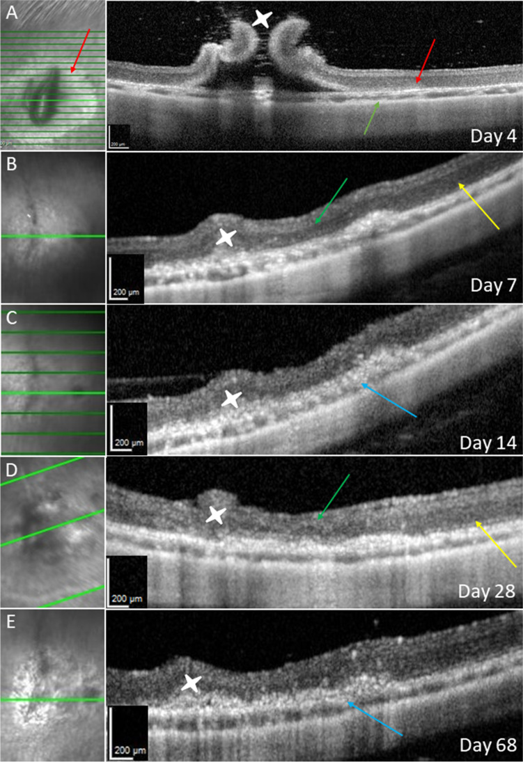
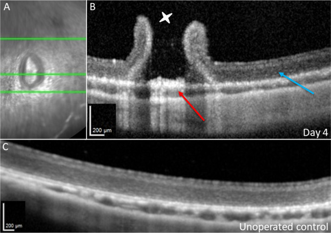
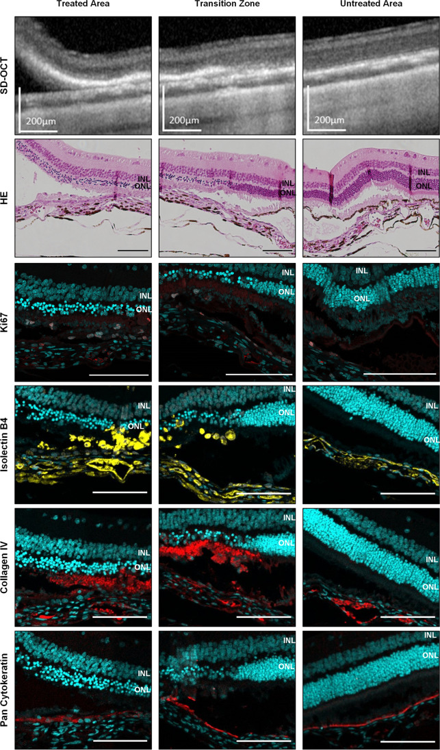
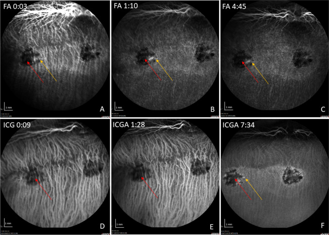
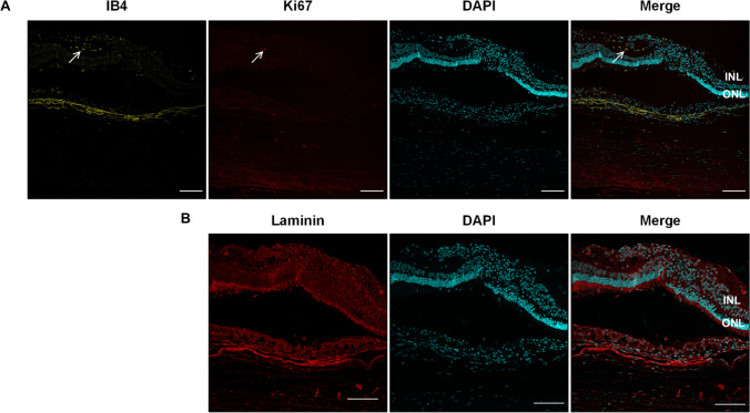
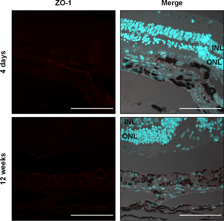
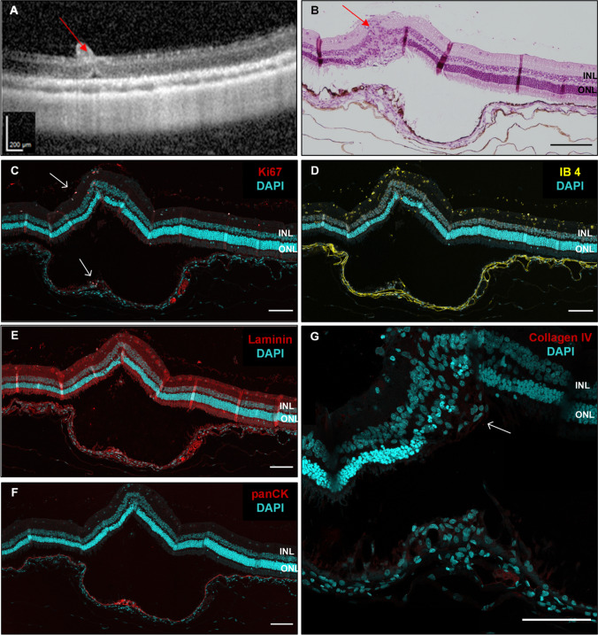
Methods: A localized detachment of the retina from the RPE/choroid layer was created in 18 pigmented rabbits. The RPE was removed by scraping with a custom-made extendable loop instrument. The resulting RPE wound was observed over a time course of 12 weeks with optical coherence tomography and angiography. After 4 days (group 1) and 12 weeks (group 2), histology was done and staining with hematoxylin and eosin, as well as immunofluorescence performed to further investigate the effects of debridement on the RPE and the overlying retina.

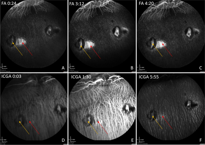
Results: Already after 4 days, we observed a closure of the RPE wound by proliferating RPE and microglia/macrophage cells forming a multilayered clump. This pattern continued over the observation time course of 12 weeks, whereby the inner and outer nuclear layer of the retina became atrophic. No neovascularization was observed in the angiograms or histology. The observed changes were limited to the site of the former RPE wound.

Conclusions: Localized surgical RPE removal induced an adjacent progressive retinal atrophy. Altering the natural course of this model may serve as a basis to test RPE cell therapeutics.

Keywords: Animal model; Cell therapy; Mechanical debridement; RPE degeneration; Rabbit; Subretinal surgery.


Conflict of interest statement

Geuder (F, C, P), Zeiss (F), MedOne Surgical Inc. (F). All other authors declare that they have no competing interests.

Figures

Fig. 1
Fig. 1
Intraoperative imaging of key steps in surgical procedure. (A) 25G vitrectomy over the optic nerve to disrupt the Alae canalis Cloqueti [39]. (B) Triamcinolone-assisted posterior vitreous detachment. (C) Formation of circumscribed bleb retinal detachment. (D) Performing the retinotomy. (E) Bleb during subretinal scraping. (F) Bleb after scraping, note the removed RPE still sticking to scraping instrument at tip of pink miOCT scan line
Fig. 2
Fig. 2
OCT after RPE scraping. Day 4 (A): bRD with RPE wound, red arrows show RPE defect and corresponding outer retinal change. The orange arrow shows the choroid with almost normal thickness, without iatrogenic bleeding. Follow-up on day 7 (B), 14 (C), 28 (D), and 68 (E): green arrows showing retinal atrophy above the RPE wound, and blue arrows showing RPE hypertrophy. White stars show the retinotomy with initially high standing retinal margins. The yellow arrows show the surrounding tissue without RPE manipulation
Fig. 3
Fig. 3
SD-OCT of bRD and retinotomy without RPE manipulation at 4 days post-operatively. (A) Infrared reflection image of the SLO. The green line marks the displayed cross-section in the corresponding SD-OCT image. (B) The red arrow shows central pigment epithelial hypertrophy at the retinotomy (white star) surrounded by raised retinal margins. Otherwise, near-normal retinal/choroidal OCT reflection bands can be discerned (blue arrow). In comparison, (C) shows a preoperative OCT, showing nearly the same reflection bands as in (B)
Fig. 4
Fig. 4
Representative FA/ICG with acquisition time (min:sec) at 1 week post-operatively. In the FA, a hyperfluorescence in the area of the induced RPE wound (red arrow) without leakage is shown over time (AC). In the late phase of the ICGA, the RPE wounds appear hyperfluorescent with signal blockage corresponding to that seen in FA, suggesting it originates from hyperpigmentation of the formerly created RPE defect (DF). The yellow arrow marks the retinotomy site
Fig. 5
Fig. 5
Visualization of the treated area, the transition zone, and untreated area as direct comparison by SD-OCT and histology at post-operative day 4. Hematoxylin and eosin (HE) staining already reveals atrophy of the photoreceptor outer segments and the outer nuclear layer (ONL) limited to the scraping site. The inner nuclear layer (INL) is not affected. Immunohistochemistry staining of the rabbit retina shows proliferating cells detected via Ki67 staining (red) which colocalize in part with subretinal microglia/macrophages shown by isolectin B4 staining (yellow). A strong deposition of collagen IV (red) is found around the scraping site beneath the ONL. Pan cytokeratin staining (red) for RPE already appears at the scraping site. Cell nuclei are stained with DAPI (cyan). Scale bar for SD-OCT = 200 µm, for histological images = 100 µm
Fig. 6
Fig. 6
Representative FA/ICG with acquisition time (min:sec) at 12 weeks post-operatively. In the FA, a small staining in the area of the induced RPE wound appears at the edge of a hypofluorescent lesion (yellow arrow), likely resulting from fluorescein blockage due to RPE hyperpigmentation (red arrow) (AC). In the ICG, choroidal perfusion remains intact under the blocked fluorescence of the pigmented lesion (DF). The subtle ICG hyperfluorescence (yellow arrow) at the edges of the RPE lesion at mid late phase may suggest perfused choriocapillaris (F)
Fig. 7
Fig. 7
SD-OCT (A) and corresponding histology at 12 weeks post-operatively. HE staining (B) shows atrophic and disorganized outer and inner retinal layers overlying the region of the original RPE wound. RPE hypertrophy is visible in both the HE and immunofluorescence staining (C). Note the normal RPE morphology adjacent to the RPE wound. INL, inner nuclear layer; ONL, outer nuclear layer. Scale bar = 100 µm
Fig. 8
Fig. 8
(A) 12 weeks post-operatively immunofluorescence staining of isolectin B4 (IB4, yellow) for endothelial cells, microglia and macrophages, and Ki67 to visualize proliferating cells (upper panel, red). Arrows point to proliferating IB4-positive cell. (B) Laminin is used to show integrity of Bruch’s membrane (lower panel, red). Cell nuclei are stained with DAPI (cyan). INL, inner nuclear layer; ONL, outer nuclear layer. Scale bar = 100 µm
Fig. 9
Fig. 9
Immunofluorescence images of ZO-1 staining of retinas 4 days and 12 weeks after RPE debridement surgery. The images show the transition zone, which contains the site of the RPE wound and the neighboring untouched RPE. ZO-1 (red) is used as epithelial polarity marker and DAPI (cyan) to visualize nuclei. The merged image contains also the transmission light microscopic picture. INL, inner nuclear layer; ONL, outer nuclear layer. Scale bar = 100 µm
Fig. 10
Fig. 10
SD-OCT (A) and corresponding histology of a retina 12 weeks after generation of a bRD without scraping the RPE. HE staining, scale bar = 150 µm (B) and immunohistochemistry (CG). Red arrows show subretinal injection site. White arrows indicate areas with ongoing cell proliferation (Ki67 staining, C). Without a generated RPE wound, there are only few infiltrating microglia/macrophages (IB4 staining, D). Laminin was used to visualize Bruch’s membrane (E) and panCK staining revealed a small spot of hypertrophic RPE beneath the bRD (F). Collagen IV is only found as small deposit in the outer nuclear layer (G); scale bar C-G = 100 µm

References

    1. Schmidt-Erfurth UM, Pruente C. Management of neovascular age-related macular degeneration. Prog Retin Eye Res. 2007;26:437–451. doi: 10.1016/j.preteyeres.2007.03.002. - DOI - PubMed
    1. Zarbin M, Sugino I, Townes-Anderson E. Concise review: update on retinal pigment epithelium transplantation for age-related macular degeneration. Stem Cells Transl Med. 2019;8:466–477. doi: 10.1002/sctm.18-0282. - DOI - PMC - PubMed
    1. Binder S, Stanzel BV, Krebs I, Glittenberg C. Transplantation of the RPE in AMD. Prog Retin Eye Res. 2007;26:516–554. doi: 10.1016/j.preteyeres.2007.02.002. - DOI - PubMed
    1. da Cruz L, Chen FK, Ahmado A, Greenwood J, Coffey P. RPE transplantation and its role in retinal disease. Prog Retin Eye Res. 2007;26:598–635. doi: 10.1016/j.preteyeres.2007.07.001. - DOI - PubMed
    1. Jin ZB, Gao ML, Deng WL, Wu KC, Sugita S, Mandai M, Takahashi M. Stemming retinal regeneration with pluripotent stem cells. Prog Retin Eye Res. 2019;69:38–56. doi: 10.1016/j.preteyeres.2018.11.003. - DOI - PubMed

LinkOut - more resources