. 2023 Sep;30(9):2780-2793.
doi: 10.1007/s43032-023-01218-8. Epub 2023 Mar 28.

Loss of the Maternal Effect Gene Nlrp2 Alters the Transcriptome of Ovulated Mouse Oocytes and Impacts Expression of Histone Demethylase KDM1B

Affiliations

Loss of the Maternal Effect Gene Nlrp2 Alters the Transcriptome of Ovulated Mouse Oocytes and Impacts Expression of Histone Demethylase KDM1B

Zahra Anvar et al. Reprod Sci. 2023 Sep.

Abstract

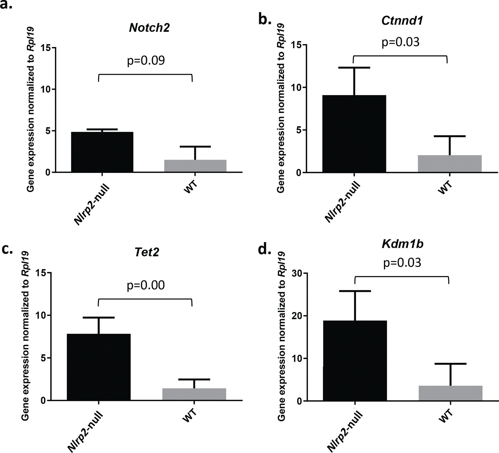
The subcortical maternal complex (SCMC) is a multiprotein complex in oocytes and preimplantation embryos that is encoded by maternal effect genes. The SCMC is essential for zygote-to-embryo transition, early embryogenesis, and critical zygotic cellular processes, including spindle positioning and symmetric division. Maternal deletion of Nlrp2, which encodes an SCMC protein, results in increased early embryonic loss and abnormal DNA methylation in embryos. We performed RNA sequencing on pools of meiosis II (MII) oocytes from wild-type and Nlrp2-null female mice that were isolated from cumulus-oocyte complexes (COCs) after ovarian stimulation. Using a mouse reference genome-based analysis, we found 231 differentially expressed genes (DEGs) in Nlrp2-null compared to WT oocytes (123 up- and 108 downregulated; adjusted p < 0.05). The upregulated genes include Kdm1b, a H3K4 histone demethylase required during oocyte development for the establishment of DNA methylation marks at CpG islands, including those at imprinted genes. The identified DEGs are enriched for processes involved in neurogenesis, gland morphogenesis, and protein metabolism and for post-translationally methylated proteins. When we compared our RNA sequencing data to an oocyte-specific reference transcriptome that contains many previously unannotated transcripts, we found 228 DEGs, including genes not identified with the first analysis. Interestingly, 68% and 56% of DEGs from the first and second analyses, respectively, overlap with oocyte-specific hyper- and hypomethylated domains. This study shows that there are substantial changes in the transcriptome of mouse MII oocytes from female mice with loss of function of Nlrp2, a maternal effect gene that encodes a member of the SCMC.

Keywords: NLRP2; Oocyte; RNA sequencing; Subcortical maternal complex.


Conflict of interest statement

Author Declarations: The authors declare no conflicts or competing financial interests.

Figures

Fig. 1
Fig. 1. Experimental design
Four-week-old WT and Nlrp2-null females were superovulated. The next day cumulus oocyte complexes (COCs) were collected from the ampulla of the oviduct. Total RNA sequencing was performed on RNA from pooled denuded oocytes. Left route: first strategy in which reads are mapped to the mouse reference genome. Right route: second strategy, in which reads are mapped to the mouse oocyte-specific transcriptome.
Fig. 2
Fig. 2. Principal component analysis and MA plot
a. Principal component analysis of normalized transcript counts from WT oocyte pools (red) and Nlrp2-null oocyte pools (blue) cluster separately. b. MA plot representing the Log2 Fold Change (Y-axis) versus normalized mean expression (X-axis) between WT and Nlrp2-null. Unchanged genes are in grey; differentially expressed genes are in red, adjusted p-value < 0.05.
Fig. 3
Fig. 3. qRT-PCR confirmation of selected upregulated genes
a. Notch2; b. Ctnnd1; c. Tet2; d. Kdm1b. Gene expression was normalized to oocyte housekeeping gene Rpl19. p-values are indicated for each gene.
Fig. 4
Fig. 4. Gene Network analysis
a. STRING protein-protein interaction and pathway enrichment analysis of upregulated differentially expressed genes result in one “Annotated Keyword” from the UniProt database, methylation (KW-0488, gene count=16, FDR=0.026). b. Direct interaction between the genes implicated in oocyte growth and embryonic development (selected for qRT-PCR) and methylation (KW-0488).
Fig. 5
Fig. 5. Principal component analysis
a. Principal component analysis of normalized transcript counts from WT oocyte pools (red) and Nlrp2-null oocyte pools (blue). b. PCA after removal of sample 2820. c. MA plot representing the Log2 Fold Change (Y-axis) versus normalized mean expression (X-axis) between WT and Nlrp2-null samples compared to reference oocyte transcriptome [28]. Unchanged genes are in grey; differentially expressed genes are in red, adjusted p-value < 0.05.
Fig. 6
Fig. 6. Overlap of differentially expressed genes with HyperDs and HypoDs
a. Overlap of differentially expressed genes from RNA-Seq data that were aligned to the mouse reference genome. b. Overlap of differentially expressed genes from RNA-Seq data were aligned to the reference oocyte transcriptome. The top Venn diagrams for each panel represents the overlap of overexpressed differentially expressed genes with HyperDs and HypoDs and the bottom panel represents the overlap of underexpressed differentially expressed genes with HyperDs and HypoDs; hypergeometric test; p < 0.05.
Fig. 7
Fig. 7. Re-distribution of HyperDs and HypoDs in absence of Nlrp2
a. Distribution of HyperDs and HypoDs within the WT oocytes genome. Black and white lollipops represent the presence and absence of DNA methylation, respectively. Gray blocks represent regions not identified as HyperDs/HypoDs. b, c. Transition and distribution of HyperDs and HypoDs within the Nlrp2-null oocytes genome. Red X shows the underexpressed differentially expressed genes within HyperDs and triple black arrow shows overexpressed differentially expressed genes within HypoDs. Altered transcription may cause change in the methylation status and consequently re-distribution of HyperDs/HypoDs (c).

References

    1. Wolf JB, Wade MJ. What are maternal effects (and what are they not)? Philos Trans R Soc Lond B Biol Sci. 2009; 10.1098/rstb.2008.0238 - DOI - PMC - PubMed
    1. Li L, Baibakov B, Dean J. A subcortical maternal complex essential for preimplantation mouse embryogenesis. Dev Cell. 2008; 10.1016/j.devcel.2008.07.010 - DOI - PMC - PubMed
    1. Lu X, Gao Z, Qin D, Li L. A Maternal Functional Module in the Mammalian Oocyte-To-Embryo Transition. Trends Mol Med. 2017; 10.1016/j.molmed.2017.09.004 - DOI - PubMed
    1. Qin D, Gao Z, Xiao Y, Zhang X, Ma H, Yu X, Nie X, Fan N, Wang X, Ouyang Y, Sun QY, Yi Z, et al. The subcortical maternal complex protein Nlrp4f is involved in cytoplasmic lattice formation and organelle distribution. Development. 2019; 10.1242/dev.183616 - DOI - PubMed
    1. Peng H, Chang B, Lu C, Su J, Wu Y, Lv P, Wang Y, Liu J, Zhang B, Quan F, Guo Z, Zhang Y. Nlrp2, a maternal effect gene required for early embryonic development in the mouse. PLoS One. 2012; 10.1371/journal.pone.0030344 - DOI - PMC - PubMed

Publication types

LinkOut - more resources