Observational Study
. 2023 May 15;133(10):e167957.
doi: 10.1172/JCI167957.

Pharmacologic improvement of CFTR function rapidly decreases sputum pathogen density, but lung infections generally persist

Affiliations
Observational Study

Pharmacologic improvement of CFTR function rapidly decreases sputum pathogen density, but lung infections generally persist

David P Nichols et al. J Clin Invest. .

Abstract

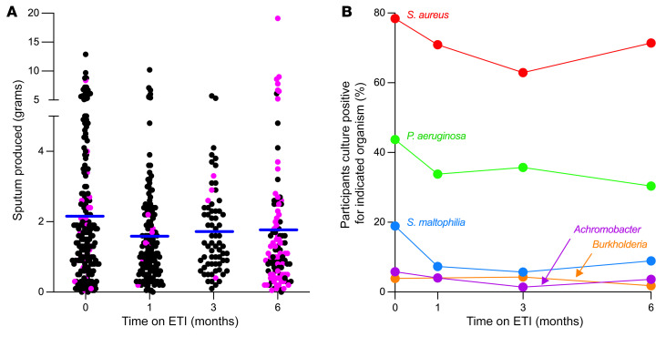
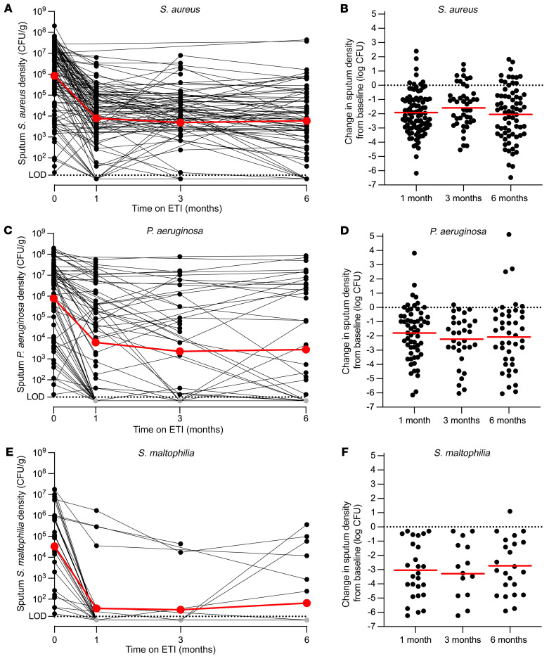
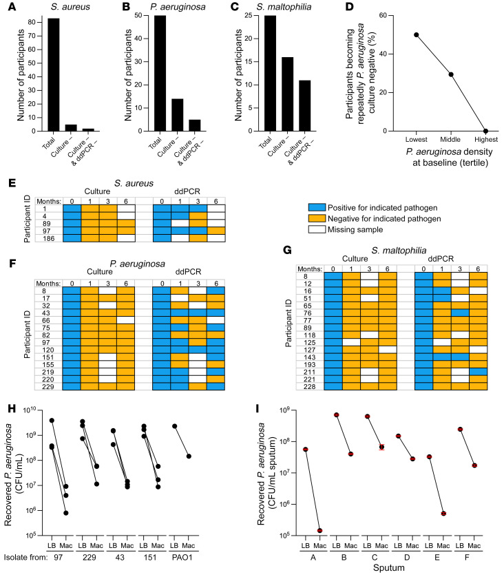
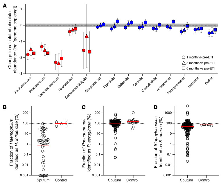
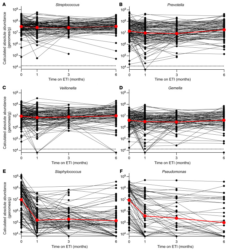
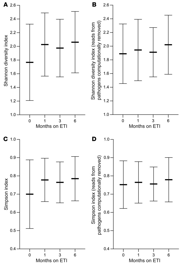
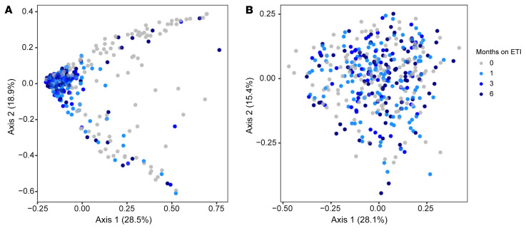
BackgroundLung infections are among the most consequential manifestations of cystic fibrosis (CF) and are associated with reduced lung function and shortened survival. Drugs called CF transmembrane conductance regulator (CFTR) modulators improve activity of dysfunctional CFTR channels, which is the physiological defect causing CF. However, it is unclear how improved CFTR activity affects CF lung infections.MethodsWe performed a prospective, multicenter, observational study to measure the effect of the newest and most effective CFTR modulator, elexacaftor/tezacaftor/ivacaftor (ETI), on CF lung infections. We studied sputum from 236 people with CF during their first 6 months of ETI using bacterial cultures, PCR, and sequencing.ResultsMean sputum densities of Staphylococcus aureus, Pseudomonas aeruginosa, Stenotrophomonas maltophilia, Achromobacter spp., and Burkholderia spp. decreased by 2-3 log10 CFU/mL after 1 month of ETI. However, most participants remained culture positive for the pathogens cultured from their sputum before starting ETI. In those becoming culture negative after ETI, the pathogens present before treatment were often still detectable by PCR months after sputum converted to culture negative. Sequence-based analyses confirmed large reductions in CF pathogen genera, but other bacteria detected in sputum were largely unchanged. ETI treatment increased average sputum bacterial diversity and produced consistent shifts in sputum bacterial composition. However, these changes were caused by ETI-mediated decreases in CF pathogen abundance rather than changes in other bacteria.ConclusionsTreatment with the most effective CFTR modulator currently available produced large and rapid reductions in traditional CF pathogens in sputum, but most participants remain infected with the pathogens present before modulator treatment.Trial RegistrationClinicalTrials.gov NCT04038047.FundingThe Cystic Fibrosis Foundation and the NIH.

Keywords: Bacterial infections; Microbiology; Pulmonology.


Conflict of interest statement

Conflict of interest: PKS has received unrestricted investigator-initiated research grants from Gilead Sciences and Vertex Pharmaceuticals.

Figures

Figure 1
Figure 1. Sputum production and sputum culture positivity after ETI.
(A) Grams of sputum produced after saline inhalation (black points) and spontaneously (pink points). Participants not producing sputum (due to inability or missed visit) are not shown. Blue lines indicate mean from all participants producing sputum at that visit. (B) Proportion of participants providing sputum samples that were culture positive for the indicated organism. Graph includes all participants, including those missing data at 1 or more study visit. Participants not providing sputum at a visit are not included in analysis of that visit. See Supplemental Figure 1 for complete case analyses. The number of participants studied at each time point is reported in Supplemental Table 3.
Figure 2
Figure 2. Sputum density of CF pathogens declines after ETI.
(A, C, and E) Density of indicated pathogens measured by quantitative culture for participants who were culture positive for the indicated pathogen at the baseline visit. Data from individual participants are indicated in black, averages are indicated in red. The limit of detection was 20 CFU/mL (dotted line). (B, D, and F) By-participant changes in sputum density of indicated pathogens measured by quantitative culture for participants who were culture positive for the indicated pathogen at the baseline visit. Data from individual participants are indicated in black, averages are indicated in red. Changes from baseline to 1 month were significant by repeated measures analysis for each organism (P < 0.0001). See Supplemental Table 4 for average values, the number of participants studied per time point, and statistical analysis; and Supplemental Figure 2 for data on Burkholderia and Achromobacter spp.
Figure 3
Figure 3. Some participants become repeatedly culture negative for CF pathogens after ETI.
(AC) The number of participants with sputum cultures positive for Staphylococcus aureus (A), Pseudomonas aeruginosa (B), or Stenotrophomonas maltophilia (C) at the baseline visit who provided at least 2 post-ETI samples are indicated as “Total”. The number of participants whose sputum became repeatedly culture negative (i.e., all samples after the baseline visit were culture negative) for the indicted pathogen are indicated as “Culture –”. The number of participants whose sputum became repeatedly culture and ddPCR negative (i.e., all samples after the baseline visit were culture and ddPCR negative) are indicated as “Culture – & ddPCR –”. (D) Inverse relationship between P. aeruginosa sputum culture density at the baseline visit and transition to repeatedly P. aeruginosa culture–negative status after ETI. Baseline P. aeruginosa culture density was categorized by tertile; the highest tertile exceeded 1.08 × 10⁷, middle was between 1.08 × 10⁷ and 4.21 × 10⁴, and lowest was less than or equal to 4.21 × 10⁴ P. aeruginosa CFU/gm of sputum. Significance of differences was significant by Fisher’s exact test, P = 0.005. See Supplemental Table 8 for the number of participants analyzed and statistical analysis. (EG) ddPCR assays detect pathogens in some culture-negative sputum samples. Participants becoming repeatedly culture negative for S. aureus (E), P. aeruginosa (F), or S. maltophilia (G) are indicated in rows and results of pathogen detection by culture and ddPCR are indicated by study time point in columns. Positive culture or ddPCR results are indicated in blue; negative culture or ddPCR results are indicated in yellow; missing data are indicated by white. See Supplemental Figure 7 for results of ddPCR assays in participants culture positive for P. aeruginosa and S. aureus. (H and I) Selective media used for P. aeruginosa culture reduced recovery of P. aeruginosa. Three isolates each from 4 PROMISE participants and the reference strain PAO1 (H) grown to late stationary phase, and sputum from 6 people with CF (I) were cultured on nonselective LB agar (LB) or the selective MacConkey agar (Mac) used for sputum P. aeruginosa culture in this study and by clinical labs generally, and viable P. aeruginosa counts measured (see Methods). CFU recovered from LB was higher than CFU recovered from MacConkey (P < 0.05 for all samples) by multiple unpaired, 2-tailed t tests on log-transformed values.
Figure 4
Figure 4. Sequence-based analyses find marked declines in the sputum density of traditional CF pathogens, but little change in other organisms.
(A) By-participant change in calculated absolute abundance after 1 (circles), 3 (triangles), and 6 (squares) months of ETI for genera detected at an average of 1% or greater in baseline sputum samples. Genera containing a traditional CF pathogen are indicated with red symbols, other bacterial genera are indicated in blue symbols. Mean and CIs as determined by mixed-model repeated measures analysis are shown. Both groups are ordered by the average cohort-wide relative abundance at baseline (high to low). Gray shading indicates technical variation in measurements of control samples (see Supplemental Figure 10C). See Supplemental Table 9 for number participants studied per time point and statistical analysis. (BD) Proportion of Haemophilus (B), Pseudomonas (C), and Staphylococcus (D) genera abundance attributable to the corresponding CF pathogen species. The proportion of Haemophilus, Pseudomonas, and Staphylococcus genera genomes that are H. influenzae, P. aeruginosa, or S. aureus species, respectively, was calculated by dividing H. influenzae, P. aeruginosa, or S. aureus species genome abundance measured by species-specific ddPCR by the corresponding genera absolute abundance. Sputum from participants with prior evidence of H. influenzae, P. aeruginosa, or S. aureus (from patient registry data) were studied. “Controls” were replicate cultures of laboratory strains of the indicated species. Values for controls sometimes exceed 100% due to variations in measurements of genera absolute abundance (using the product of total 16S rRNA and genera relative abundance) and species absolute abundance (using species specific ddPCR data). The difference between Haemophilus genera and H. influenzae abundance was significant (P = 0.0014); differences between Pseudomonas and P. aeruginosa abundance (P = 0.09) and Staphylococcus and S. aureus abundance (P = 0.38) were not different by 2-tailed t test on log-transformed data.
Figure 5
Figure 5. Sequence-based analyses show that other organisms in sputum exhibit little change, whereas traditional CF pathogens decrease.
Change in calculated absolute abundance of Streptococcus (A), Prevotella (B), Veillonella (C), Gemella (D), Staphylococcus (E), and Pseudomonas (F) genera in participants’ sputum. Data from individual participants are indicated in black, averages are indicated in red. Participants for which the indicated genera was not detected in baseline sputum samples were not included. See Supplemental Table 9 for the number of samples analyzed per time point and statistical analysis.
Figure 6
Figure 6. Sputum microbial diversity increases after ETI due to decreases in CF pathogen abundance.
(A and C) Sputum genera relative abundance data were used to calculate the Shannon (A) and Simpson (C) indices at each time point. (B and D) To examine the effect CF pathogen abundance changes on the observed increase in diversity, sequencing reads from traditional CF pathogen genera were computationally removed (see text) and the Shannon (B) and Simpson (D) indices were recalculated. Mean and SD for each time point are shown. There was a significant increase (P < 0.05 by 1-way ANOVA) in diversity after ETI compared with before ETI) only when all genera were included; this was true for all post-ETI visits. One hundred forty-five participants were studied at the pre-ETI visit, 123 at 1 month, 56 at 3 months, and 89 at 6 months.
Figure 7
Figure 7. Sputum microbial composition changes after ETI are driven by decreases in traditional CF pathogens.
Multidimensional scaling (MDS) representation of the Bray-Curtis index measurements of pair-wise dissimilarity in the abundances of genera in each sputum sample relative to all other samples. Gray indicates samples collected before ETI, light blue 1 month after ETI, blue 3 months after ETI, and dark blue 6 months after ETI. (A) Bray-Curtis analysis was performed on all taxonomic read counts from each participant. Pairwise differences between pre-ETI (0 months) and the 1-, 3-, and 6-month visits all P < 0.001 by PERMANOVA. (B) To examine the effect CF pathogen abundance changes on the shifts in sputum microbial composition, sequencing read counts from traditional CF pathogen genera were computationally removed (see text) and Bray-Curtis pair-wise dissimilarities between samples were recalculated. Pairwise differences between pre-ETI (0 months) and the 1-, 3-, and 6-month visits all not significant by PERMANOVA. One hundred forty-five participants were studied at the pre-ETI time point, 123 at 1 month, 56 at 3 months, and 89 at 6 months.

References

    1. Barry PJ, et al. Triple therapy for cystic fibrosis Phe508del-gating and -residual function genotypes. N Engl J Med. 2021;385(9):815–825. doi: 10.1056/NEJMoa2100665. - DOI - PMC - PubMed
    1. Nichols DP, et al. Clinical effectiveness of elexacaftor/tezacaftor/ivacaftor in people with cystic fibrosis: a clinical trial. Am J Respir Crit Care Med. 2022;205(5):529–539. doi: 10.1164/rccm.202108-1986OC. - DOI - PMC - PubMed
    1. Heijerman HGM, et al. Efficacy and safety of the elexacaftor plus tezacaftor plus ivacaftor combination regimen in people with cystic fibrosis homozygous for the F508del mutation: a double-blind, randomised, phase 3 trial. Lancet. 2019;394(10212):1940–1948. doi: 10.1016/S0140-6736(19)32597-8. - DOI - PMC - PubMed
    1. Middleton PG, et al. Elexacaftor-tezacaftor-ivacaftor for cystic fibrosis with a single Phe508del allele. N Engl J Med. 2019;381(19):1809–1819. doi: 10.1056/NEJMoa1908639. - DOI - PMC - PubMed
    1. Rowe SM, et al. Clinical mechanism of the cystic fibrosis transmembrane conductance regulator potentiator ivacaftor in G551D-mediated cystic fibrosis. Am J Respir Crit Care Med. 2014;190(2):175–184. doi: 10.1164/rccm.201404-0703OC. - DOI - PMC - PubMed

Publication types

Substances

Associated data