. 2023 May 4;31(5):584-594.e5.
doi: 10.1016/j.str.2023.03.003. Epub 2023 Mar 27.

Structural features of the Notch ankyrin domain-Deltex WWE2 domain heterodimer determined by NMR spectroscopy and functional implications

Affiliations

Structural features of the Notch ankyrin domain-Deltex WWE2 domain heterodimer determined by NMR spectroscopy and functional implications

Andrea A Carter et al. Structure. .

Abstract

The Notch signaling pathway, an important cell fate determination pathway, is modulated by the ubiquitin ligase Deltex. Here, we investigate the structural basis for Deltex-Notch interaction. We used nuclear magnetic resonance (NMR) spectroscopy to assign the backbone of the Drosophila Deltex WWE2 domain and mapped the binding site of the Notch ankyrin (ANK) domain to the N-terminal WWEA motif. Using cultured Drosophila S2R+ cells, we find that point substitutions within the ANK-binding surface of Deltex disrupt Deltex-mediated enhancement of Notch transcriptional activation and disrupt ANK binding in cells and in vitro. Likewise, ANK substitutions that disrupt Notch-Deltex heterodimer formation in vitro block disrupt Deltex-mediated stimulation of Notch transcription activation and diminish interaction with full-length Deltex in cells. Surprisingly, the Deltex-Notch intracellular domain (NICD) interaction is not disrupted by deletion of the Deltex WWE2 domain, suggesting a secondary Notch-Deltex interaction. These results show the importance of the WWEA:ANK interaction in enhancing Notch signaling.

Keywords: Deltex; NMR; Notch; Notch signaling; RING domain; WWE(2) motifs; ankyrin repeats; protein-protein interactions; ubiquitin ligase.


Conflict of interest statement

Declaration of interests The authors declare no competing interests.

Figures
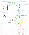
Figure 1.
Figure 1.. Mechanisms of Notch signaling and regulation by Deltex.
The left side of the figure shows canonical Notch signaling, in which a DSL-type ligand on an adjacent cell binds to the extracellular domain of the Notch receptor, stimulating S2 cleavage by an ADAM protease. This cleavage liberates the Notch extracellular domain (NECD) and leaves a transmembrane-bound fragment that contains the intracellular domain (NEXT). NEXT is a substrate for S3 cleavage by γ-secretase either at the plasma membrane or in an endocytic vesicle, which liberates the Notch intracellular domain (NICD). Cytoplasmic NICD then enters the nucleus, where it activates transcription through the transcription factor CSL. The right-side of the figure shows non-canonical Notch signaling, which is mediated by Deltex. In the absence of extracellular ligands, Deltex can stimulate full-length Notch receptor to enter endocytic vesicles in a Rab5-dependent process. These vesicles can either be recycled or can fuse with early endosomes (EE) and then late endosomes (LE). At this stage, the intact Notch receptor can either be down-regulated by internalization to a multivesicular body that is destined for degradation in the lysosome (Lys), or can be activated through a second Deltex-mediated process, again forming NICD. Down-regulation through internalization is promoted by Kurtz and Suppressor of Deltex (SuDx). Depending on the balance between the SuDx and Deltex activity at the late endosomal stage, Deltex can either promote Notch signaling by generating NICD or can inhibit Notch signaling by promoting internalization followed by degradation.
Figure 2.
Figure 2.. Primary structure of the Drosophila Deltex and Notch proteins, and constructs used in this study.
The top schematic shows the Notch intracellular domain, which extends from the γ-secretase cleavage site to the C-terminus. The seven ankyrin repeats are shown in blue, and the TWFP sequence that binds tightly to CSL is shown in green. The second schematic gives the boundaries of the Notch ankyrin domain used in this study. The third schematic shows the full-length Deltex protein. The N-terminal WWE2 tandem spans residues 35 to 201. Two polyglutamine segments span from residues 261 to 302 and from residues 488 to 513. The C-terminal region contains a RING and a DTC domain. The bottom schematic gives the boundaries of the Deltex WWE2 construct used in this study.
Figure 3.
Figure 3.. NMR spectra of the Drosophila Deltex WWE2 domain and its interaction with the Notch Ankyrin domain.
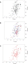
(A) 1H-15N-TROSY spectrum of a 2H-, 15N-labelled fragment of Drosophila Deltex spanning residues 26–261 (Dx1A*), showing high chemical shift dispersion. Using triple-resonance techniques, assignments were made for the 169 of the 217 backbone amides (excluding prolines). (B) Titration of 100 μM 2H-, 15N-labelled Dx1A* from (A) with the R2007A variant of unlabeled Notch ANK. Colors correspond to molar ratios of 1:0 (black), 1:1 (blue), 1:2 (green), and 1:4 (red) Deltex to ANK R2007A. (C) Spectrum of a 1:4 molar ratio of the Dx1A* (100 μM) and an unlabeled Notch ANK R2007A/R2027A (red; black spectrum shows Dx1A* on its own). Conditions: 300 mM NaCl, 25 mM NaPO4, pH 7.0, 25 °C (A) and 35°C (B, C).
Figure 4.
Figure 4.. Representative behavior of Deltex WWE2 cross peaks titrated with unlabeled Notch ANK domain.
Molar ratios of Deltex WWE2 domain to Notch ANK are 1:0, black; 1:1, blue; 1:2, green; 1:4, red. (A) T77 disappears at a molar ratio of 1:2 (category 1). (B) W133 disappears at a molar ratio of 1:4 (category 2). (C) S47 shifts slightly and disappears at a molar ratio of 1:4 (category 2). (D) G115 shifts completely from one location to another in a traceable manner at a molar ratio of 1:4 (category 3). (E) Y60 shifts slightly (category 4). (F) V153 loses intensity, but it does not disappear completely (category 4). (G) I156 both shifts slightly and loses intensity (category 4). (H) K56 broadens (category 4). (I) S53 exhibits no change (category 5). Conditions: 300 mM NaCl, 25 mM NaPO4, pH 7.0, 35 °C, 800 MHz.
Figure 5.
Figure 5.. Locations on the Deltex WWE2 domain of chemical shift perturbations resulting from binding of the Notch ANK domain.
(A) Ribbon diagram of the crystal structure of Dx1A* (2A90.pdb, Zweifel et al., 2005) showing the WWEA and WWEB motifs (orange and purple) and the C-terminal linker (dark grey) in different orientations. (B) Ribbon diagram showing each secondary structure element in the WWEA motif. Analogous secondary structure elements in the WWEB motifs have the same color (see Figure S1). (C) Ribbon diagram showing chemical shift perturbations from binding of Notch ANK. Resonances that disappear above 1:1 molar ratio of ANK to WWE2 (class 1) are colored black, residues that disappear above a molar ratio of 1:2 (class 2) are colored red, resonances that shift upon binding (class 3) are colored brown. Resonances that show modest shifts or increases in linewidth upon binding (class 4) are colored beige. Resonances that are unaffected by binding are colored light grey. Based on the intersection of conservation (see panel F) and chemical shift perturbations, we identified a set of five residues (sticks) to substitute to attempt to disrupt binding of the Notch ANK domain. Two of these (K74A and Y91A) destabilized the Dx1A*: Notch R2007A ANK complex (Table S3). (D) Surface representation with the same color scheme as (C). (E) Surface representation showing unassigned non-proline (yellow) and proline (green) residues. (F) Conservation based on CONSURF analysis. Residues are colored by conservation score, in the following order: gray (least conserved), light pink, light orange, pale yellow, pale green, cyan, blue, purple, black (most conserved). In each panel, the center-right image is an end-on view of WWEA, and the right image is an end-on view of WWEB.
Figure 6.
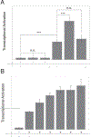
Figure 6.. Co-transfection with Deltex stimulates Notch transcriptional activation.
(A) Transfection of S2R+ cells with full-length Deltex alone or Deltex lacking the WWE2 domain alone does not activate transcription from a luciferase reporter gene under the control of a Notch-responsive promoter. Transfection with full-length Notch receptor (10 ng DNA) results in an increase in transcriptional activation. Co-transfection of Notch with full-length Deltex (40 ng DNA) enhances transcriptional activation by full-length Notch; this enhancement is eliminated when the Deltex WWE2 domain is deleted. The statistical significance (p) of differences in transcriptional activation was quantified using a two-sided t-test; *** corresponds to p < 0.001, n.s. indicates not significant (p>0.05). (B) Dose-dependence of Notch-mediated transcription by full-length Deltex.
Figure 7.
Figure 7.. Variants that disrupt ANK:WWE2 binding decrease Deltex-mediated transcriptional activation by full-length Notch.
Co-transfection of S2R+ cells with wild-type Notch (left), Notch R1985A (middle), or Notch R2027A variants (right) and either wild-type Deltex (grey bars) or Deltex K74A, Y91A, or K74A/Y91A variants (stippled bars). Luciferase activities were normalized to that of wild-type Notch in the absence of Deltex (white bars). Both Deltex single-residue variants enhance transcription activation by wild-type Notch, though to a lower level than wild-type Deltex. However, when combined into a double mutant construct, Deltex-mediated enhancement is no longer detected. In contrast, the Notch R1985A and R2027A variants are refractory to activation by Deltex, though both Notch variants do activate transcription over background. Error bars represent +/− the standard deviations of three replicates each from two independent experiments. Statistical significance from a two-sided t-test is indicated at p < 0.001 (***); n.s. indicates not statistically significant. Cells were transfected with 10ng Notch and 30ng Deltex as indicated.
Figure 8.
Figure 8.. Co-immunoprecipitation from S2R+ cells shows decreased interaction of NICD with Deltex variants.
(A) Lysates from S2R+ cells co-transfected with C-terminally FLAG-tagged Notch variants and C-terminally V5-tagged Deltex variants. Transfected cells were probed for NICD-FLAG (top), Deltex-V5 (middle) and control protein α-tubulin (bottom). (B) Co-immunoprecipitation of wild-type and variant full-length Deltex proteins with wild-type and variant NICD proteins. Co-IP elutions from anti-FLAG (Notch) beads were probed for NICD-FLAG (top) and Deltex-V5 (bottom). Red lines indicate the position of a 75kDa marker protein, illustrating the size difference between the WT (82kDa) ΔWWE (64kDa) Deltex constructs.
Figure 9.
Figure 9.. Potential roles of the Deltex RING domain in WWE2-mediated Notch activation and inhibition.
(A) Structural comparison of the Deltex WWE2 domain with the WWE and RING domains of RNF146. The two structures were superposed using the WWEB motif of Deltex (purple; 2A90.pdb) and the WWE motif of RNF146 (yellow; 4QPL.pdb). The RING finger of RNF146 (blue), which makes extensive interactions with the RNF146 WWE motif, localizes to a position near to but not overlapping with the Deltex WWEA motif (orange). If the Deltex RING finger were to interact similarly with the Deltex WWEB motif, it would be positioned adjacent to the binding site for the Notch ANK domain. (B) The Deltex WWEB motif may bind the RING domain as in panel (A); interaction of the Notch ANK domain with Deltex WWEA motif leads to Deltex-mediated stimulation of transcription from the late endosome (Figure 1). Disruption of the WWEA-ANK interaction by point-substitution at the ANK or WWEA binding site prevents this activation (left). Dissociation of RING domain from WWEB, which can be mimicked by deletion of the WWE2 domain, facilitates formation of the secondary interaction between the Deltex RING domain and an alternative site on NICD. This interaction fails to stimulate Notch-mediated transcription, and may lead to inhibition of Notch signaling through MBV/lysosomal degradation (Figure 1, lower right).

References

    1. Kopan R (2012). Notch Signaling. Cold Spring Harb. Perspect. Biol. 4, a011213. 10.1101/cshperspect.a011213. - DOI - PMC - PubMed
    1. Hori K, Sen A, and Artavanis-Tsakonas S (2013). Notch signaling at a glance. J. Cell Sci. 126, 2135–2140. 10.1242/jcs.127308. - DOI - PMC - PubMed
    1. Bigas A, and Porcheri C (2018). Notch and Stem Cells. Adv. Exp. Med. Biol. 1066, 235–263. 10.1007/978-3-319-89512-3_12. - DOI - PubMed
    1. Kushwah R, Guezguez B, Lee JB, Hopkins CI, and Bhatia M (2014). Pleiotropic roles of Notch signaling in normal, malignant, and developmental hematopoiesis in the human. EMBO Rep. 15, 1128–1138. 10.15252/embr.201438842. - DOI - PMC - PubMed
    1. Aster JC, Pear WS, and Blacklow SC (2017). The Varied Roles of Notch in Cancer. Annu. Rev. Pathol. Mech. Dis. 12, 245–275. 10.1146/annurev-pathol-052016-100127. - DOI - PMC - PubMed

Publication types

LinkOut - more resources