. 2023 Jun 8;33(12):7702-7713.
doi: 10.1093/cercor/bhad073.

Functional network reconfiguration supporting memory-guided attention

Affiliations

Functional network reconfiguration supporting memory-guided attention

Kylie Isenburg et al. Cereb Cortex. .

Abstract

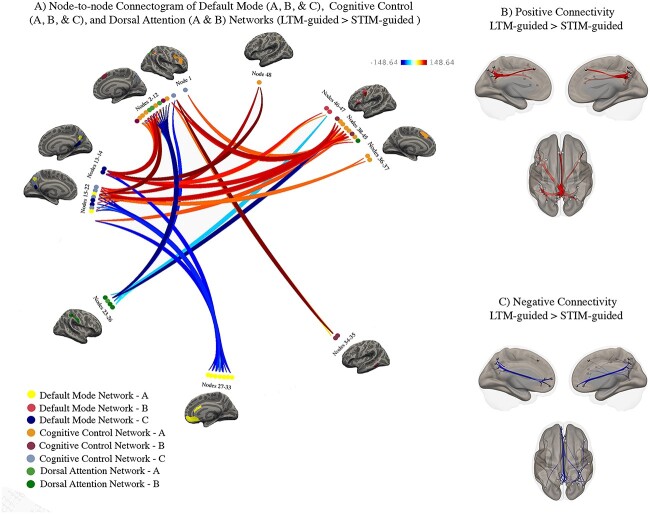
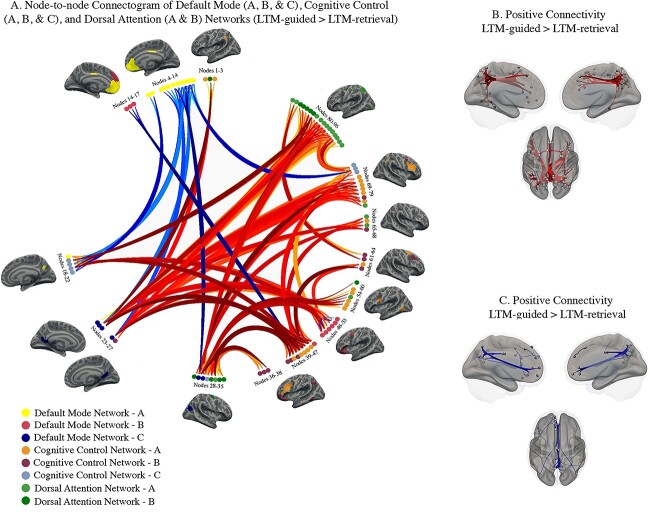
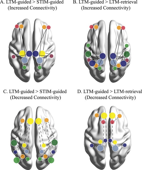
Studies have identified several brain regions whose activations facilitate attentional deployment via long-term memories. We analyzed task-based functional connectivity at the network and node-specific level to characterize large-scale communication between brain regions underlying long-term memory guided attention. We predicted default mode, cognitive control, and dorsal attention subnetworks would contribute differentially to long-term memory guided attention, such that network-level connectivity would shift based on attentional demands, requiring contribution of memory-specific nodes within default mode and cognitive control subnetworks. We expected that these nodes would increase connectivity with one another and with dorsal attention subnetworks during long-term memory guided attention. Additionally, we hypothesized connectivity between cognitive control and dorsal attention subnetworks facilitating external attentional demands. Our results identified both network-based and node-specific interactions that facilitate different components of LTM-guided attention, suggesting a crucial role across the posterior precuneus and restrosplenial cortex, acting independently from the divisions of default mode and cognitive control subnetworks. We found a gradient of precuneus connectivity, with dorsal precuneus connecting to cognitive control and dorsal attention regions, and ventral precuneus connecting across all subnetworks. Additionally, retrosplenial cortex showed increased connectivity across subnetworks. We suggest that connectivity from dorsal posterior midline regions is critical for the integration of external information with internal memory that facilitates long-term memory guided attention.

Keywords: attention; connectivity; fMRI; memory; precuneus.


Figures

Fig. 1
Fig. 1
Participants were trained and later scanned with three separate conditions: A) during STIM-guided attention, a red arrow prompted the participants to direct their attention and indicate whether the object in the location was a match to the named category, B) in the LTM-retrieval condition, the double-sided red arrows were uninformative, and participants had to indicate whether the central object was a paired-associate of the category word by memory, C) for LTM-guided attention, the double-sided arrows were again uninformative, however here participant were instructed to direct their attention to the paired location of the category word, and indicate whether the paired object was in the correct location. Original figure from Rosen et al. 2018, Cerebral Cortex.
Fig. 2
Fig. 2
Step by step method of gPPI analysis implemented in Conn toolbox. A) an inflated image of the 400 cortical nodes matched to the Yeo 17-network atlas (adapted with permission from https://github.com/ThomasYeoLab/CBIG/tree/master/stable_projects/brain_parcellation/Schaefer2018_LocalGlobal). B) A representation of the psychophysiological index regressors utilized in the gPPI analysis. For each of the three conditions, LTM-guided attention, STIM-guided attention, and LTM-retrieval, the block design was multiplied by the time-series of each of the 400 nodes separately to create individual PPI regressors. C) each PPI regressor is used to compute beta values for all possible pairwise comparisons amongst the 400 nodes. D) Beta values were extracted and sorted using hierarchical clustering available in Conn toolbox. This clustering method groups nodes together based on functional similarity and anatomical proximity. E) Threshold free cluster enhancement (TFCE) with P < 0.01 is run to correct each node-to-node connectivity pair for multiple comparisons. F) a node-to-node connectogram is constructed within Conn toolbox that displays clusters of connections between ROIs that pass TFCE correction.
Fig. 3
Fig. 3
Condition-modulated connectivity between default mode, cognitive control, and dorsal attention network nodes (LTM-guided attention > STIM-guided attention). A) Significant increases in node-to-node connectivity are shown in red/orange while significant decreases are shown in blue. Darker colors signify stronger task-modulated connectivity between nodes. Inflated brains beside clusters of nodes offer visualization of the nodes and the Yeo 17-network that they belong to. B) Visualization of node-specific locations and the significant positive connections from all clusters. C) Visualization of node location and the significant negative connections from all clusters.
Fig. 4
Fig. 4
Condition-modulated connectivity between default mode, cognitive control, and dorsal attention network nodes (LTM-guided attention > LTM-retrieval). A) Significant increases in node-to-node connectivity are shown in red/orange while significant decreases are shown in blue. Darker colors signify stronger task-modulated connectivity between nodes. Inflated brains beside clusters of nodes offer visualization of the nodes and the Yeo 17-network that they belong to. B) Visualization of node-specific locations and the significant positive connections from all clusters. C) Visualization of node location and the significant negative connections from all clusters.
Fig. 5
Fig. 5
Conceptual figure illustrating main network interactions resulting from each of the contrasts. Networks are color coded based on the Yeo 17 colors shown in Figs 3 and 4. Prominent nodes of the contrast are denoted by larger dots on the brain. Significant but less recurring nodes are denoted by smaller dots. Solid lines indicate increased connectivity between two nodes for a) LTM-guided > STIM-guided attention, and B) LTM-guided attention > LTM-retrieval. dotted lines indicate decreased connectivity between two nodes for C) LTM-guided > STIM-guided attention, and D) LTM-guided attention > LTM-retrieval. notably, LTM-guided > STIM-guided attention seems to employ connectivity from posterior nodes of the memory-attention network, while LTM-guided attention > LTM-retrieval highlights a role for brain regions involved in visuospatial and external processing.

References

    1. Alexander AS, Place R, Starrett MJ, Nitz DA, Chrastil ER. Rethinking retrosplenial cortex: perspectives and predictions. Neuron. 2022:111:176–189. - PMC - PubMed
    1. Bar-Joseph Z, Gifford DK, Jaakkola TS. Fast optimal leaf ordering for hierarchical clustering. Bioinformatics. 2001:17(Suppl 1):S22–S29. - PubMed
    1. Brodt S, Pöhlchen D, Flanagin VL, Glasauer S, Gais S, Schönauer M. Rapid and independent memory formation in the parietal cortex. PNAS. 2016:113(46):13251–13256. - PMC - PubMed
    1. Cavanaugh J, Alvarez BD, Hurtz W. Enhanced performance with brain stimulation:Attentional shift or visual cue. J Neurosci. 2006:26(44):11347–11358. - PMC - PubMed
    1. Chen D, Hutchinson JB. What is memory-guided attention? How past experiences shape selective visuospatial attention in the present. Curr Top Behav Neurosci. 2019:41:185–212. - PubMed

Publication types

MeSH terms

LinkOut - more resources