. 2023 Jun;128(11):2126-2139.
doi: 10.1038/s41416-023-02230-0. Epub 2023 Mar 28.

Immunogenic hypofractionated radiotherapy sensitising head and neck squamous cell carcinoma to anti-PD-L1 therapy in MDSC-dependent manner

Affiliations

Immunogenic hypofractionated radiotherapy sensitising head and neck squamous cell carcinoma to anti-PD-L1 therapy in MDSC-dependent manner

Liang Mao et al. Br J Cancer. 2023 Jun.

Abstract

Background: Enhancing the response rate of immunotherapy will aid in the success of cancer treatment. Here, we aimed to explore the combined effect of immunogenic radiotherapy with anti-PD-L1 treatment in immunotherapy-resistant HNSCC mouse models.

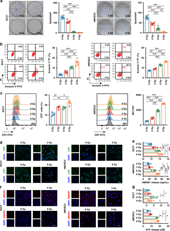
Methods: The SCC7 and 4MOSC2 cell lines were irradiated in vitro. SCC7-bearing mice were treated with hypofractionated or single-dose radiotherapy followed by anti-PD-L1 therapy. The myeloid-derived suppressive cells (MDSCs) were depleted using an anti-Gr-1 antibody. Human samples were collected to evaluate the immune cell populations and ICD markers.

Results: Irradiation increased the release of immunogenic cell death (ICD) markers (calreticulin, HMGB1 and ATP) in SCC7 and 4MOSC2 in a dose-dependent manner. The supernatant from irradiated cells upregulated the expression of PD-L1 in MDSCs. Mice treated with hypofractionated but not single-dose radiotherapy were resistant to tumour rechallenge by triggering ICD, when combined with anti-PD-L1 treatment. The therapeutic efficacy of combination treatment partially relies on MDSCs. The high expression of ICD markers was associated with activation of adaptive immune responses and a positive prognosis in HNSCC patients.

Conclusion: These results present a translatable method to substantially improve the antitumor immune response by combining PD-L1 blockade with immunogenic hypofractionated radiotherapy in HNSCC.


Conflict of interest statement

The authors declare no competing interests.

Figures

Fig. 1
Fig. 1. Irradiation-induced immunogenic cell death (ICD) in SCC7 and 4MOSC2 tumour cells in a dose-dependent manner.
a Representative images of anchor-dependent colony formation and quantitative analysis of SCC7 and 4MOSC2 tumour cells exposed to irradiation in vitro. b Representative contour plots and quantitative analysis of annexin V+ PI+ apoptotic SCC7 tumour cells exposed to irradiation in vitro. c Representative histogram and quantitative analysis of calreticulin (CRT) surface expression in SCC7 and 4MOSC2 tumour cells exposed to irradiation in vitro. d Representative IF images of CRT (green) staining of SCC7 and 4MOSC2 tumour cells exposed to irradiation in vitro. The nuclei were stained with DAPI (blue). Scale bar = 20 μm. e Quantitation of HMGB1 released from SCC7 and 4MOSC2 tumour cells exposed to irradiation in vitro. f Representative IF images of HMGB1 (red) staining of SCC7 and 4MOSC2 tumour cells exposed to irradiation in vitro. The nuclei were stained with DAPI (blue). Scale bar = 20 μm. g Quantitation of ATP released from SCC7 and 4MOSC2 tumour cells exposed to irradiation in vitro. P < 0.05 was considered statistically significant. *P < 0.05; **P < 0.005; ***P < 0.0005. Experiments were repeated twice. ISO isotype.
Fig. 2
Fig. 2. Irradiation upregulated PD-L1 expression on tumour cells and MDSCs.
a Representative histogram and quantitative analysis of PD-L1 expression on SCC7 tumour cells with irradiation exposed in vitro. b Representative histogram and quantitative analysis of PD-L1 expression on 4MOSC2 tumour cells with irradiation exposed in vitro. c Representative histogram and quantitative analysis of PD-L1 expression in MDSCs incubated with culture medium from irradiation-treated SCC7 tumour cells. d Representative histogram and quantitative analysis of PD-L1 expression in MDSCs incubated with culture medium from irradiation-treated 4MOSC2 tumour cells. P < 0.05 was considered statistically significant. *P < 0.05; ***P < 0.0005; ns, not significant. Experiments were repeated twice. NT non-tumour-bearing, TB tumour-bearing, ISO isotype, IR irradiation.
Fig. 3
Fig. 3. Radiotherapy (RT) enhanced the efficacy of anti-PD-L1 treatment in the mouse model.
a Schematic illustration of the preventive experimental protocol (top). Tumour growth analysis of SCC7 tumour-bearing mice in each group (bottom). The mice received 20 Gy RT in one fraction on day 8 or 27 Gy RT in three fractions on days 8, 10 and 12. Anti-PD-L1 treatment was initiated on day 9 and was delivered three times per week for 2 weeks. The mice were randomly grouped. n = 4 per group. b Quantitation of SCC7 tumour-infiltrating CCR7+CD103+ DCs (left), TOX+PD-1+ CD8 T cells (middle) and IFN-γ+TNF-α+ CD8+ T cells (right). c Pie chart analysis of CD8+ T-cell subsets with naive, central memory (TCM), effector memory (TEM), and effector (eff) phenotype in draining lymph nodes. Quantitation of CD8+ TCM in draining lymph nodes (right). d Schematic illustration of rechallenge experimental protocol (top). Tumour growth analysis of SCC7 tumour-bearing mice in each group (middle). Survival curves in each group (bottom). The mice were randomly grouped. The mice received 20 Gy RT in one fraction on day 8 or 27 Gy RT in three fractions on days 8, 10 and 12. Anti-PD-L1 treatment was started on day 9 and were delivered three times per week for 2 weeks. Mice with irradiation treatment were rechallenged with SCC7 tumour on day 34. n = 4 per group. e Representative IHC images of staining for CD3 in the primary and rechallenge lesions of each group (top), with quantification of the number of CD3+ cells per view (bottom). Scale bar = 100 μm. P < 0.05 was considered statistically significant. *P < 0.05; **P < 0.005; ***P < 0.0005. Experiments were repeated twice. Con control, PL primary lesions, RL rechallenge lesions.
Fig. 4
Fig. 4. Fractionated radiotherapy (RT) induced immunogenic cell death (ICD) and promoted anti-PD-L1 treatment in a therapeutic study.
a Schematic illustration of therapeutic experimental protocol. The mice were treated with isotype control or anti-PD-L1 antibody, alone or in combination with 27 Gy RT in three fractions on days 11, 13 and 15. Anti-PD-L1 or isotype control antibody treatment was initiated on day 12 and was delivered three times in one week. The mice were randomly grouped. n = 6 per group. b Tumour growth analysis of SCC7-bearing mice in each group. c Representative IHC images of staining for α-SMA in the tumour lesions of each group (top), with quantification of the number of α-SMA+ cells per view (bottom). Scale bar = 100 μm. d Quantitation of the number of tumour-infiltrating M-MDSCs and PMN-MDSCs in CD45+ cells (left). Quantitation of the PD-L1 expression based on the MFI in tumour-infiltrating M-MDSCs and PMN-MDSCs (right). e Representative IHC images of staining for calreticulin (CRT) and HMGB1 in the tumour lesions of each group (left), with quantification of the CRT and HMGB1 expression score (right). Scale bar = 100 μm. *P < 0.05; **P < 0.005; ***P < 0.0005. Experiments were repeated twice. Con control, IR irradiation.
Fig. 5
Fig. 5. The therapeutic efficacy of fractionated radiotherapy in combination with anti-PD-L1 or cisplatin.
a Schematic illustration of the experimental protocol (left). Tumour growth analysis of SCC7-bearing mice in each group (right). The mice were treated with 27 Gy in three fractions (9 Gy × 3), cisplatin + 27 Gy in three fractions (9 Gy × 3), or anti-PD-L1 + 27 Gy in three fractions (9 Gy × 3). Three fractions of 27 Gy RT were delivered on days 13, 15 and 17. Anti-PD-L1 treatment was initiated on day 14 and was administered three times per week. Cisplatin treatment was initiated on day 14 and was administered twice per week. The mice were randomly grouped. n = 4 per group. b Quantitation of CD107a+ CD3 T cells in spleens (left), tumour-infiltrating CCR7+CD103+ DCs and TOX+PD-1+ CD8 T cells (middle), and CD80+CD86+ DCs in draining lymph nodes (right). *P < 0.05; **P < 0.005. IR irradiation.
Fig. 6
Fig. 6. The therapeutic efficacy of anti-PD-L1 in combination with fractionated radiotherapy (RT) is dependent on the Gr-1+ myeloid cells.
a Schematic illustration of the experimental protocol. Gr-1 mAb or isotype control was pre-injected to the C3H mice at 250 μg the day before the SCC7 cells were implanted, with other three injections administered on days 4, 9 (1 day after radiotherapy), and 14 (6 days after radiotherapy). The mice were treated with isotype control or anti-PD-L1 antibody, alone or in combination with 27 Gy RT in three fractions on days 8, 10 and 12. Anti-PD-L1 or isotype control antibody treatment was initiated on day 9 and was delivered and administered three times for 3 weeks. The mice were randomly grouped. n = 5 per group. b Tumour growth analysis of SCC7-bearing mice in each group. c Survival curves of SCC7-bearing mice in each group. d Representative IHC images of staining for Foxp3 in the tumour lesions of each group (top), with quantification of the number of Foxp3+ cells per view (bottom). Scale bar = 100 μm. *P < 0.05; **P < 0.005; ***P < 0.0005. Experiments were repeated twice. Con control, IR irradiation.
Fig. 7
Fig. 7. Phenotypic analysis of the tumour-infiltrating immune cells according to the expression of immunogenic cell death (ICD) markers among the prospective cohort of HNSCC patients.
a IHC on serial sections of four individual human patient tissue with HNSCC for staining with calreticulin (CRT) and HMGB1 (top). Scale bar = 50 μm. Representative contour plots of CD3+ T cells of four individual human patients with HNSCC (bottom). b Quantitation of the number of tumour-infiltrating CD3+ T cells (left), CD4+ T cells (middle) and CD8+ (right) T cells in each group according to the expression of CRT and HMGB1. c Pie chart analysis of CD8+ and CD4+ T-cell subsets with naive, central memory (TCM), effector memory (TEM), and terminally differentiated effector memory (TEMRA) phenotype (left). Quantitation of the number of tumour-infiltrating CD8+ naive and TEM T cells in each group (right). d Quantitation of the PD-L1 expression based on the MFI in tumour-infiltrating M-MDSCs in each group. e Quantitation of the number of tumour-infiltrating conventional dendritic (cDCs) cells in each group. f Kaplan–Meier survival curves for overall survival for patients with HNSCC according to the expression of CRT and HMGB1. *P < 0.05; **P < 0.005; ns not significant.

Similar articles

Cited by

References

    1. Galon J, Bruni D. Tumor immunology and tumor evolution: intertwined histories. Immunity. 2020;52:55–81. doi: 10.1016/j.immuni.2019.12.018. - DOI - PubMed
    1. Topalian SL, Hodi FS, Brahmer JR, Gettinger SN, Smith DC, McDermott DF, et al. Safety, activity, and immune correlates of anti-PD-1 antibody in cancer. N Engl J Med. 2012;366:2443–54. doi: 10.1056/NEJMoa1200690. - DOI - PMC - PubMed
    1. Garon EB, Rizvi NA, Hui R, Leighl N, Balmanoukian AS, Eder JP, et al. Pembrolizumab for the treatment of non-small-cell lung cancer. N Engl J Med. 2015;372:2018–28. doi: 10.1056/NEJMoa1501824. - DOI - PubMed
    1. Burtness B, Harrington KJ, Greil R, Soulieres D, Tahara M, de Castro G, Jr., et al. Pembrolizumab alone or with chemotherapy versus cetuximab with chemotherapy for recurrent or metastatic squamous cell carcinoma of the head and neck (KEYNOTE-048): a randomised, open-label, phase 3 study. Lancet. 2019;394:1915–28. doi: 10.1016/S0140-6736(19)32591-7. - DOI - PubMed
    1. Sharma P, Hu-Lieskovan S, Wargo JA, Ribas A. Primary, adaptive, and acquired resistance to cancer immunotherapy. Cell. 2017;168:707–23. doi: 10.1016/j.cell.2017.01.017. - DOI - PMC - PubMed

Publication types

MeSH terms