. 2023 Mar 28;23(1):280.
doi: 10.1186/s12885-023-10685-w.

NPAS2 promotes aerobic glycolysis and tumor growth in prostate cancer through HIF-1A signaling

Affiliations

NPAS2 promotes aerobic glycolysis and tumor growth in prostate cancer through HIF-1A signaling

Shuaijun Ma et al. BMC Cancer. .

Abstract

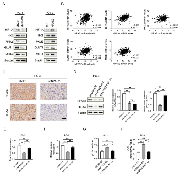
Background: Prostate cancer (PCa), one of the common malignant tumors, is the second leading cause of cancer-related deaths in men. The circadian rhythm plays a critical role in disease. Circadian disturbances are often found in patients with tumors and enable to promote tumor development and accelerate its progression. Accumulating evidence suggests that the core clock gene NPAS2 (neuronal PAS domain-containing protein 2) has been implicated in tumors initiation and progression. However, there are few studies on the association between NPAS2 and prostate cancer. The purpose of this paper is to investigate the impact of NPAS2 on cell growth and glucose metabolism in prostate cancer.

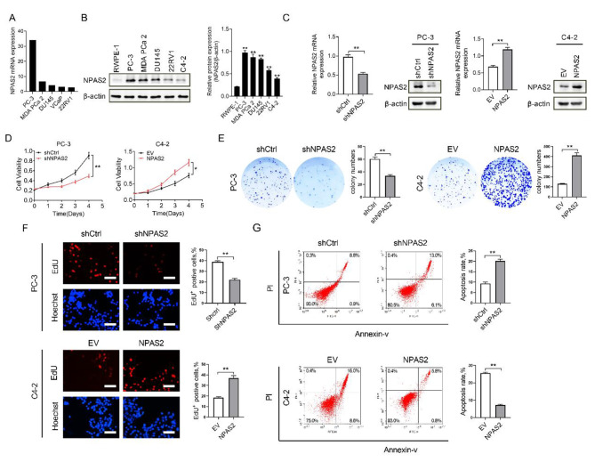
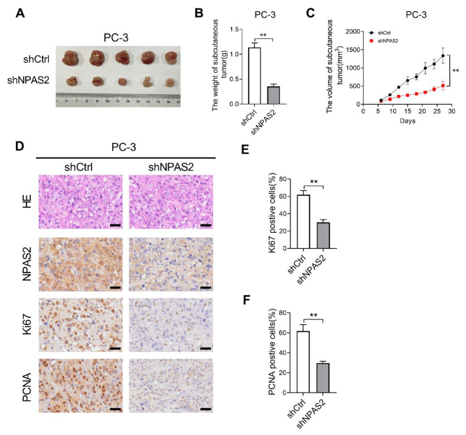
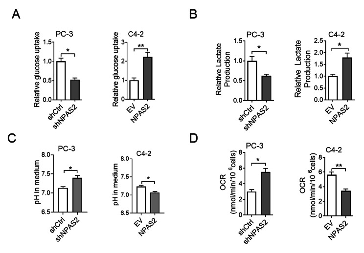
Methods: Quantitative real-time PCR (qRT-PCR), immunohistochemical (IHC) staining, western blot, GEO (Gene Expression Omnibus) and CCLE (Cancer Cell Line Encyclopedia) databases were used to analyze the expression of NPAS2 in human PCa tissues and various PCa cell lines. Cell proliferation was assessed using MTS, clonogenic assays, apoptotic analyses, and subcutaneous tumor formation experiments in nude mice. Glucose uptake, lactate production, cellular oxygen consumption rate and medium pH were measured to examine the effect of NPAS2 on glucose metabolism. The relation of NPAS2 and glycolytic genes was analyzed based on TCGA (The Cancer Genome Atlas) database.

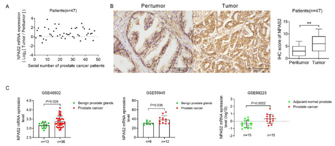
Results: Our data showed that NPAS2 expression in prostate cancer patient tissue was elevated compared with that in normal prostate tissue. NPAS2 knockdown inhibited cell proliferation and promoted cell apoptosis in vitro and suppressed tumor growth in a nude mouse model in vivo. NPAS2 knockdown led to glucose uptake and lactate production diminished, oxygen consumption rate and pH elevated. NPAS2 increased HIF-1A (hypoxia-inducible factor-1A) expression, leading to enhanced glycolytic metabolism. There was a positive correlation with the expression of NPAS2 and glycolytic genes, these genes were upregulated with overexpression of NPAS2 while knockdown of NPAS2 led to a lower level.

Conclusion: NPAS2 is upregulated in prostate cancer and promotes cell survival by promoting glycolysis and inhibiting oxidative phosphorylation in PCa cells.

Keywords: Glycolysis; HIF-1A; NPAS2; Oxidative phosphorylation; Prostate cancer (PCa).


Conflict of interest statement

The authors declare that they have no competing interests.

Figures

Fig. 1
Fig. 1
NPAS2 is significantly upregulated in prostate cancer (A) qRT-PCR analysis the expression of NPAS2 mRNA in 47 PCa tissues (B) IHC analysis the expression of NPAS2 in 47 PCa tissues. Scale bar, 100 μm (C) Bioinformatics analysis of NPAS2 mRNA expression level in prostate cancer GSE46602, GSE55945 and GSE69223 databases.*P<0.05; **P<0.01
Fig. 2
Fig. 2
NPAS2 promotes PCa cell survival in vitro (A) Expression of NPAS2 in PCa cell lines in the CCLE database (B) Western blot analysis of NPAS2 expression in PCa cell lines and a normal prostate epithelial cell (C) NPAS2 expression levels in the transfected PCa cell lines were detected through western blot and qRT-PCR analysis (D) MTS growth experiments in cell transfection models (E) Colony formation experiments in cell transfection models (F) EdU proliferation assay in cell transfection models. Scale bar: 50 μm (G) Apoptosis detection in cell transfection models. *P<0.05; **P<0.01. Data were shown as the mean ± S.E.M. from three independent experiments
Fig. 3
Fig. 3
NPAS2 knockdown inhibits PCa tumor growth in vivo (A) Subcutaneous xenograft model for PC-3 cells treated as indicated and dissected tumors from sacrificed mice were shown (B) Weight of tumors in nude mice (C) Tumor volume changes curve of nude mice (D) HE staining, immunohistochemical staining of NPAS2, Ki67 and PCNA in nude mice tumors. Scale bar: 50 μm (E) Comparison of Ki-67-positive cells in tumor tissues of nude mice xenograft model with different treatment as indicated (F) Comparison of PCNA-positive cells in tumor tissues of nude mice xenograft model with different treatment as indicated. *P<0.05; **P<0.01
Fig. 4
Fig. 4
NPAS2 promotes glycolysis and inhibits oxidative phosphorylation in PCa cells (A) Glucose uptake in cell transfection models (B) Lactate production in cell transfection models (C) Medium pH of cell transfection models (D) Oxygen consumption level of cell transfection models. *P<0.05; **P<0.01. Data were shown as the mean ± S.E.M. from three independent experiments
Fig. 5
Fig. 5
NPAS2 enhances glycolysis by increasing the expression of key molecules in the glycolytic pathway (A) Western blot analysis of the expression levels of NPAS2 and key glycolysis genes in PCa cells (B) Correlation between NPAS2 expression and key molecules of glucose metabolism in prostate cancer tissues (TCGA database) (C) Immunohistochemical staining of NPAS2 and HIF-1A in nude mice tumors (D) The transfection efficiency was examined with western blot (ANOVA, P<0.0001) (E) Glucose uptake in cell transfection models (ANOVA, P = 0.007) (F) Lactate production in cell transfection models (ANOVA, P = 0.012) (G) Medium pH of cell transfection models (ANOVA, P = 0.036) (H) Oxygen consumption level of cell transfection models (ANOVA, P<0.0001). *P<0.05; **P<0.01. Data were shown as the mean ± S.E.M. from three independent experiments

References

    1. Liao Y, Liu Y, Xia X, Shao Z, Huang C, He J, Jiang L, Tang D, Liu J, Huang H. Targeting GRP78-dependent AR-V7 protein degradation overcomes castration-resistance in prostate cancer therapy. Theranostics. 2020;10(8):3366–81. doi: 10.7150/thno.41849. - DOI - PMC - PubMed
    1. Bhatia V, Yadav A, Tiwari R, Nigam S, Goel S, Carskadon S, Gupta N, Goel A, Palanisamy N, Ateeq B. Epigenetic silencing of miRNA-338-5p and miRNA-421 drives SPINK1-Positive prostate Cancer. Clin cancer research: official J Am Association Cancer Res. 2019;25(9):2755–68. doi: 10.1158/1078-0432.CCR-18-3230. - DOI - PMC - PubMed
    1. Shah S, Rachmat R, Enyioma S, Ghose A, Revythis A, Boussios S. BRCA Mutations in Prostate Cancer: Assessment, Implications and Treatment Considerations.International journal of molecular sciences2021, 22(23). - PMC - PubMed
    1. Castelo-Branco P, Passer BJ, Buhrman JS, Antoszczyk S, Marinelli M, Zaupa C, Rabkin SD, Martuza RL. Oncolytic herpes simplex virus armed with xenogeneic homologue of prostatic acid phosphatase enhances antitumor efficacy in prostate cancer. Gene Ther. 2010;17(6):805–10. doi: 10.1038/gt.2010.20. - DOI - PMC - PubMed
    1. Boussios S, Rassy E, Shah S, Ioannidou E, Sheriff M, Pavlidis N. Aberrations of DNA repair pathways in prostate cancer: a cornerstone of precision oncology. Expert Opin Ther Targets. 2021;25(5):329–33. doi: 10.1080/14728222.2021.1951226. - DOI - PubMed

LinkOut - more resources