. 2023 Mar 28;21(1):225.
doi: 10.1186/s12967-023-04061-2.

M1 polarization enhances the antitumor activity of chimeric antigen receptor macrophages in solid tumors

Affiliations

M1 polarization enhances the antitumor activity of chimeric antigen receptor macrophages in solid tumors

Yi Huo et al. J Transl Med. .

Abstract

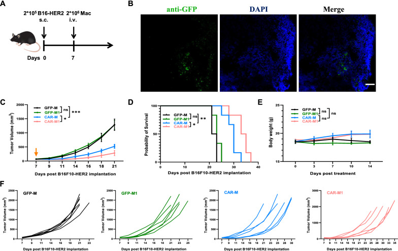
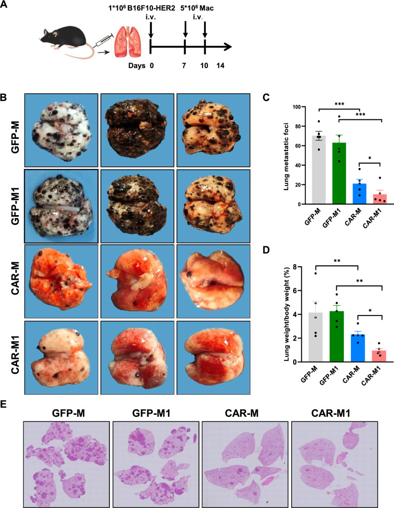
Background: Chimeric antigen receptor macrophage (CAR-M) therapy is a novel cancer immunotherapy approach that integrates CAR structure and macrophage functions. CAR-M therapy has shown unique and impressive antitumor effects in immunotherapy for solid tumors. However, the polarization state of macrophages can affect the antitumor effect of CAR-M. We hypothesized that the antitumor activity of CAR-Ms may be further improved after inducing M1-type polarization.

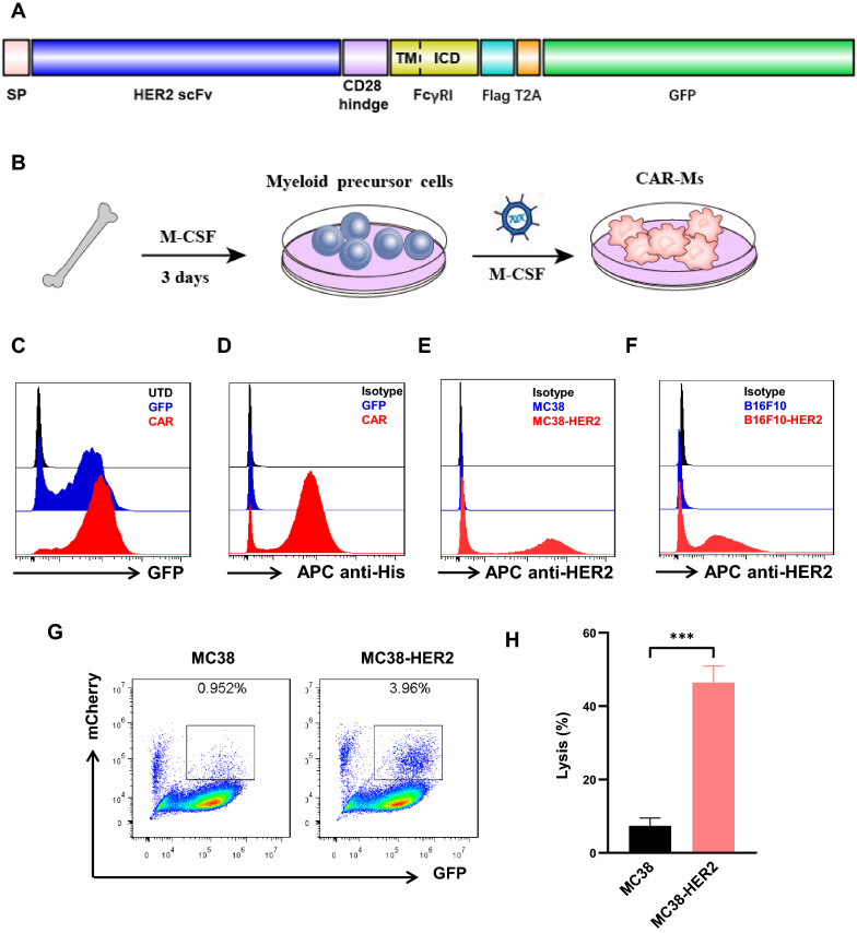
Methods: In this report, we constructed a novel HER2-targeting CAR-M, which was composed of humanized anti-HER2 scFv, CD28 hinge region and FcγRI transmembrane domain and intracellular domain. Phagocytosis, tumor-killing capacities, and cytokine release of CAR-Ms were detected with or without M1-polarization pretreatment. Several syngeneic tumor models were used to monitor the in vivo antitumor activity of M1-polarized CAR-Ms.

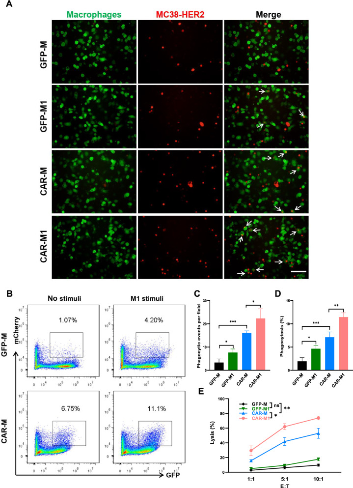
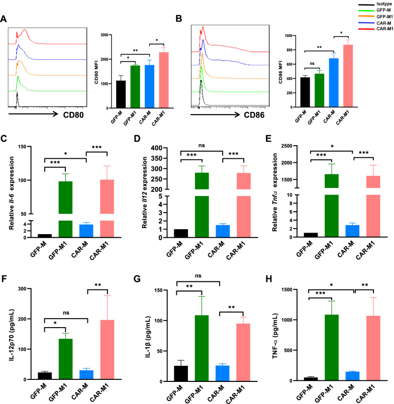
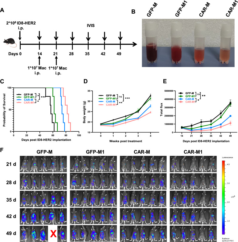
Results: After polarization with LPS combined with interferon-γ in vitro, we found that the phagocytic and tumor-killing capacities of CAR-Ms against target cells were significantly enhanced. The expression of costimulatory molecules and proinflammatory cytokines was also significantly increased after polarization. By establishing several syngeneic tumor models in vivo, we also demonstrated that infusing polarized M1-type CAR-Ms could effectively suppress tumor progression and prolong the survival of tumor-bearing mice with enhanced cytotoxicity.

Conclusions: We demonstrated that our novel CAR-M can effectively eliminate HER2-positive tumor cells both in vitro and in vivo, and M1 polarization significantly enhanced the antitumor ability of CAR-M, resulting in a stronger therapeutic effect in solid cancer immunotherapy.

Keywords: CAR-M; Cancer immunotherapy; Chimeric antigen receptor; HER2; M1 polarization; Macrophages.


Conflict of interest statement

The authors declare that they have no competing interests.

Figures

Fig. 1
Fig. 1
Anti-HER2 CAR-Ms specifically target HER2-positive cancer cells. A A diagram of the anti-HER2-CAR construct. B Overview of primary macrophage differentiation from bone marrow and the lentivirus transduction protocol. C Representative flow cytometry analysis of the CAR transduction frequency in lentivirus-transduced primary macrophages after 72 h. D Representative flow cytometry analysis of CAR expression on macrophages after lentivirus transduction, with recombinant HER2 protein binding and staining with an APC-conjugated anti-His tag antibody. E–F Representative flow cytometry analysis of HER2 expression in sorted cells of the murine tumor cell lines MC38 (E) and B16F10 (F) after stable transduction with the truncated HER2 lentivirus. G Flow cytometry-based phagocytosis of mCherry+ MC38-wild type or mCherry+ MC38-HER2 target cells by anti-HER2 CAR-Ms at an E:T = 5:1 ratio. The double-positive cell population represents the target cells engulfed by macrophages. H Luciferase-based killing assay of Luc+ MC38-wild type or Luc+ MC38-HER2 target cells by anti-HER2 CAR-Ms at an E:T = 5:1 ratio in vitro. ***, p < 0.001
Fig. 2
Fig. 2
CAR-M-mediated phagocytosis and cytotoxicity were enhanced after M1-type polarization. A Representative fluorescence microscopy images showing GFP-positive macrophage-mediated phagocytosis after 4 h of coincubation with mCherry+ MC38-HER2 cells, scale bar: 50 µm. B Representative flow cytometry analysis of the phagocytosis of mCherry+ MC38-HER2 target cells by control or M1-polarized macrophages at an E:T = 5:1 ratio. The double-positive cell population represents the target cells engulfed by macrophages. C Quantitative analysis of the data from (A). Three random fields of view were assessed per replicate, and data are represented as the mean ± SEM. of three independent experiments. D Statistical analysis of the percentage of phagocytosis by flow cytometry. Three independent experiments were performed. Data are shown as the mean ± SEM. E Cytotoxicity was evaluated by a luciferase-based killing assay with Luc+ MC38-HER2 target cells and macrophages after 24 h of coculture at different E:T ratios in vitro. Data are represented as the mean ± SEM. of three independent experiments. *, p < 0.05; **, p < 0.01; ***, p < 0.001
Fig. 3
Fig. 3
M1-polarized CAR-Ms exhibited stronger proinflammatory phenotypes than CAR-Ms. A-B Flow cytometry analysis and quantitative analysis of the mean fluorescence intensity (MFI) of the activation markers CD80 (A) and CD86 (B) on macrophages after coculture with MC38-HER2 target cells. C-E Normalized gene expression analysis of the proinflammatory cytokines Il-6 (C), Il-12 (D), and Tnf-α (E) by qRT‒PCR. FH ELISA to detect secreted proinflammatory cytokines, including IL-12p70 (F), IL-1β (G) and TNF-α (H), in the supernatants of cocultures of macrophages with target cells after 24 h. *, p < 0.05; **, p < 0.01; ***, p < 0.001
Fig. 4
Fig. 4
Local administration of polarized CAR-Ms suppressed the progression of ovarian cancer in vivo. A Schematic illustration of the experimental design for the ovarian cancer model. B The appearance of ascites/peritoneal lavage obtained from tumor-bearing mice after intraperitoneal administration. C Kaplan‒Meier survival curve of tumor-bearing mice treated with GFP-M, GFP-M1, CAR-M, and CAR-M1 after ID8-HER2 tumor cell inoculation. D Tumor burden assessed by body weight after the mice received GFP-M, GFP-M1, CAR-M, and CAR-M1 treatment. E Statistical analysis of bioluminescence imaging. Data are presented as the mean ± S.D. F Tumor burden was monitored by bioluminescence imaging on the indicated days after treatment. *, p < 0.05; **, p < 0.01; ***, p < 0.001
Fig. 5
Fig. 5
Systemic administration of polarized CAR-Ms induced efficient antitumor effects in a B16F10 melanoma model. A An illustration of the experimental design. B Representative fluorescent images of melanoma tissue stained with anti-GFP to detect the infiltration of GFP-positive anti-HER2 CAR-Ms, scale bar: 100 μm. C Tumor growth of C57 mice bearing subcutaneous B16F10-HER2 melanoma tumors in the GFP-M (black), GFP-M1 (green), CAR-M (blue), and CAR-M1 (red) treatment groups. The average tumor volume per treatment group is presented as the mean ± S.D. D Kaplan‒Meier survival curve for (C) mice bearing B16F10-HER2 cells after receiving GFP-Ms, GFP-M1s, CAR-Ms, and CAR-M1s (n = 6 mice/group). E Body weight changes in tumor-bearing mice posttreatment. The data are shown as the mean ± S.D. F Tumor growth of individual mice from (C). Each line represents an individual mouse. *, p < 0.05; **, p < 0.01; ***, p < 0.001
Fig. 6
Fig. 6
Systemic administration of M1-polarized CAR-Ms resulted in a potent antitumor response in a melanoma lung metastasis model. A An illustration of the experimental design. B Representative gross images of lungs excised from the indicated treatment groups at the experimental endpoint. C Quantitative analysis of metastatic foci from (B). Data are shown as the mean ± SEM. D Antitumoural efficacy was assessed by the lung weight-to-body weight ratio of mice bearing intravenous B16F10-HER2 cells posttreatment. E Lung metastatic burden assessed by HE staining. *, p < 0.05; **, p < 0.01; ***, p < 0.001

Similar articles

Cited by

References

    1. June CH, O'Connor RS, Kawalekar OU, Ghassemi S, Milone MC. CAR T cell immunotherapy for human cancer. Science. 2018;359:1361–1365. doi: 10.1126/science.aar6711. - DOI - PubMed
    1. Finck AV, Blanchard T, Roselle CP, Golinelli G, June CH. Engineered cellular immunotherapies in cancer and beyond. Nat Med. 2022;28:678–689. doi: 10.1038/s41591-022-01765-8. - DOI - PMC - PubMed
    1. Hou AJ, Chen LC, Chen YY. Navigating CAR-T cells through the solid-tumour microenvironment. Nat Rev Drug Discov. 2021;20:531–550. doi: 10.1038/s41573-021-00189-2. - DOI - PubMed
    1. Irvine DJ, Maus MV, Mooney DJ, Wong WW. The future of engineered immune cell therapies. Science. 2022;378:853–858. doi: 10.1126/science.abq6990. - DOI - PMC - PubMed
    1. Lin C, Zhang J. Chimeric antigen receptor engineered innate immune cells in cancer immunotherapy. Sci China Life Sci. 2019;62:633–639. doi: 10.1007/s11427-018-9451-0. - DOI - PubMed

Publication types