. 2023 Mar 17;13(6):1086.
doi: 10.3390/ani13061086.

Epigenetics in Canine Mammary Tumors: Upregulation of miR-18a and miR-18b Oncogenes Is Associated with Decreased ERS1 Target mRNA Expression and ERα Immunoexpression in Highly Proliferating Carcinomas

Affiliations

Epigenetics in Canine Mammary Tumors: Upregulation of miR-18a and miR-18b Oncogenes Is Associated with Decreased ERS1 Target mRNA Expression and ERα Immunoexpression in Highly Proliferating Carcinomas

Jessica Maria Abbate et al. Animals (Basel). .

Abstract

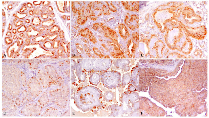
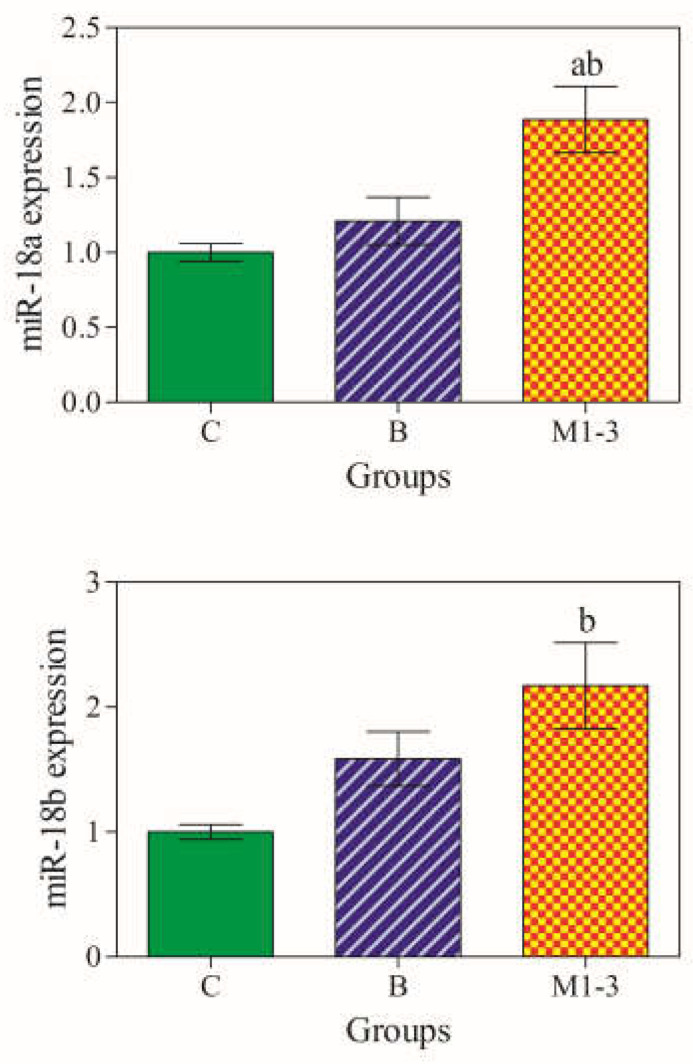
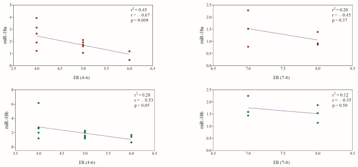
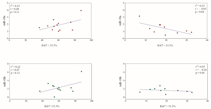
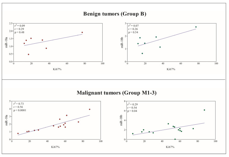
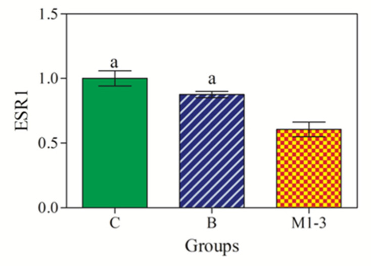
The expression of miRNAs is one of the main epigenetic mechanisms responsible for the regulation of gene expression in mammals, and in cancer, miRNAs participate by regulating the expression of protein-coding cancer-associated genes. In canine mammary tumors (CMTs), the ESR1 gene encodes for ERα, and represents a major target gene for miR-18a and miR-18b, previously found to be overexpressed in mammary carcinomas. A loss in ERα expression in CMTs is commonly associated with poor prognosis, and it is noteworthy that the downregulation of the ESR1 would appear to be more epigenetic than genetic in nature. In this study, the expression of ESR1 mRNA in formalin-fixed, paraffin-embedded (FFPE) canine mammary tumors (CMTs) was evaluated and compared with the expression levels of miR18a and miR18b, both assessed via RT-qPCR. Furthermore, the possible correlation between the miRNA expression data and the immunohistochemical prognostic factors (ERα immunoexpression; Ki67 proliferative index) was explored. A total of twenty-six FFPE mammary samples were used, including 22 CMTs (7 benign; 15 malignant) and four control samples (three normal mammary glands and one case of lobular hyperplasia). The obtained results demonstrate that miR-18a and miR-18b are upregulated in malignant CMTs, negatively correlating with the expression of target ESR1 mRNA. Of note, the upregulation of miRNAs strictly reflects the progressive loss of ERα immunoexpression and increased tumor cell proliferation as measured using the Ki67 index. The results suggest a central role of miR-18a and miR-18b in the pathophysiology of canine mammary tumors as potential epigenetic mechanisms involved in ERα downregulation. Moreover, as miRNA expression reflects ERα protein status and a high proliferative index, miR-18a and miR-18b may represent promising biomarkers with prognostic value. More detailed investigations on a larger number of cases are needed to better understand the influence of these miRNAs in canine mammary tumors.

Keywords: ERα; ESR1; Ki67 index; canine mammary tumors; epigenetics; miR-18a; miR-18b; miRNAs; oncomiR.


Conflict of interest statement

The authors declare no conflict of interest.

Figures

Figure 1
Figure 1
ERα immunoexpression in canine mammary samples. (A) Normal mammary gland, TS = 8; (B,C) tubulopapillary adenoma, TS = 8; (D) solid carcinoma, grade III, TS = 3; (E) intraductal papillary adenoma, TS = 4; (F) solid carcinoma, grade II, TS = 4.
Figure 2
Figure 2
Expression levels of miRNAs in mammary samples. miR-18a was overexpressed in malignant tumors (M1–3; orange color) compared with the control group (C; green color) and benign tumors (B; blue color), whereas miR-18b was overexpressed in malignant tumors (M1–3) compared with the benign tumors (B). Significances (p < 0.05) a vs. Group C and b vs. Group B.
Figure 3
Figure 3
Correlation between miRNA expression and ERα score. A significant negative correlation was observed for both miR-18a (red circles) and miR-18b (green circles) in CMTs with lower ERα immunoexpression. The colored circles (red; green) refer to miRNA expression levels in CMTs.
Figure 4
Figure 4
Correlation between miRNA expression and Ki67 index. A significant negative correlation was observed only between miR-18a (red circles) expression levels and Ki67 index < 33.3% (p < 0.05). The miR-18b (green circles) gene expression did not show a significant correlation with Ki67 index. The colored circles (red; green) refer to miRNA expression levels in CMTs.
Figure 5
Figure 5
Linear regression obtained between the Ki67 index and the expression of miR-18a (red circles) and miR-18b (green circles) in benign tumors (Group B) and malignant tumors (Group M1–3). A significant positive correlation was found between the Ki67 index and the miR-18a and miR-18b expression levels in malignant CMTs (p < 0.05).
Figure 6
Figure 6
RT-qPCR expression levels of ESR1 mRNA. ESR1 expression was downregulated in malignant tumors (M1–3; orange color) compared with control (C; green color) and benign tumors (B; blue color). Significances (p < 0.001): a vs. Group M1–3.

References

    1. Sharma S., Kelly T.K., Jones P.A. Epigenetics in cancer. Carcinogenesis. 2010;31:27–36. doi: 10.1093/carcin/bgp220. - DOI - PMC - PubMed
    1. Yoo C.B., Jones P.A. Epigenetic therapy of cancer: Past, present and future. Nat. Rev. Drug Discov. 2006;5:37–50. doi: 10.1038/nrd1930. - DOI - PubMed
    1. Guo H., Ingolia N.T., Weissman J.S., Bartel D.P. Mammalian microRNAs predominantly act to decrease target mRNA levels. Nature. 2010;466:835–840. doi: 10.1038/nature09267. - DOI - PMC - PubMed
    1. Bartel D.P. MicroRNAs: Target recognition and regulatory functions. Cell. 2009;136:215–233. doi: 10.1016/j.cell.2009.01.002. - DOI - PMC - PubMed
    1. Fabian M.R., Sonenberg N. The mechanisms of miRNA-mediated gene silencing: A look under the hood of miRISC. Nat. Struct. Mol. Biol. 2012;19:586–593. doi: 10.1038/nsmb.2296. - DOI - PubMed

LinkOut - more resources