. 2023 Mar 12;12(3):702.
doi: 10.3390/antiox12030702.

Different Effects and Mechanisms of Selenium Compounds in Improving Pathology in Alzheimer's Disease

Affiliations

Different Effects and Mechanisms of Selenium Compounds in Improving Pathology in Alzheimer's Disease

Zhong-Hao Zhang et al. Antioxidants (Basel). .

Abstract

Owing to the strong antioxidant capacity of selenium (Se) in vivo, a variety of Se compounds have been shown to have great potential for improving the main pathologies and cognitive impairment in Alzheimer's disease (AD) models. However, the differences in the anti-AD effects and mechanisms of different Se compounds are still unclear. Theoretically, the absorption and metabolism of different forms of Se in the body vary, which directly determines the diversification of downstream regulatory pathways. In this study, low doses of Se-methylselenocysteine (SMC), selenomethionine (SeM), or sodium selenate (SeNa) were administered to triple transgenic AD (3× Tg-AD) mice for short time periods. AD pathology, activities of selenoenzymes, and metabolic profiles in the brain were studied to explore the similarities and differences in the anti-AD effects and mechanisms of the three Se compounds. We found that all of these Se compounds significantly increased Se levels and antioxidant capacity, regulated amino acid metabolism, and ameliorated synaptic deficits, thus improving the cognitive capacity of AD mice. Importantly, SMC preferentially increased the expression and activity of thioredoxin reductase and reduced tau phosphorylation by inhibiting glycogen synthase kinase-3 beta (GSK-3β) activity. Glutathione peroxidase 1 (GPx1), the selenoenzyme most affected by SeM, decreased amyloid beta production and improved mitochondrial function. SeNa improved methionine sulfoxide reductase B1 (MsrB1) expression, reflected in AD pathology as promoting the expression of synaptic proteins and restoring synaptic deficits. Herein, we reveal the differences and mechanisms by which different Se compounds improve multiple pathologies of AD and provide novel insights into the targeted administration of Se-containing drugs in the treatment of AD.

Keywords: Alzheimer’s disease; Se-methylselenocysteine; metabolism; selenomethionine; sodium selenate; synaptic deficits.


Conflict of interest statement

There are no conflict of interest to declare.

Figures

Figure 1
Figure 1
Behavioral tests in the WT, 3× Tg-AD, SMC, SeM, and SeNa mouse groups. (ad) The learning and memory abilities of mice were tested using the Morris water maze. (a,b) The escape latency (a) and swimming trajectory (b) were recorded during the 4-day training. (c,d) The probe trial was performed 24 and 72 h after the last trial of a hidden platform task. Time in the opposite quadrant after 24 h (c) and 72 h (d) was recorded. (e) The spatial memory of mice was determined by analysis of the alternation rate in the Y-maze. (f,g) Total distance (f) and number of rearings (g) in the open-field test were used to detect the autonomous behavior ability of mice. (h,i) Mouse anxiety was assessed by the elevated plus maze. Time in the open arm (h) and number of entries in the open arm (i) were recorded. All data are presented as the means ± SEMs (n = 7–10). * p < 0.05, ** p < 0.01, *** p < 0.001 vs. AD mice; # p < 0.05, ## p < 0.01, ### p < 0.001 vs. WT mice, as determined by one-way or two-way ANOVA followed by Dunnett’s multiple comparison test.
Figure 2
Figure 2
Administration of SMC, SeM, and SeNa improved Aβ, tau, and synaptic pathology in the cortexes of 3× Tg-AD mouse brains. (a) Levels of APP, Aβ oligomer (16 kDa), and BACE1 were analyzed by Western blotting. (bd) Quantification of protein levels in (a). (e) Levels of hyperphosphorylated tau (Ser202 and Ser404) were analyzed by Western blotting. (f,g) Quantification of protein levels in (e). (h) Levels of PSD95 and synaptophysin were analyzed by Western blotting. (i,j) Quantification of protein levels in (h). α-Tubulin, β-actin, or glyceraldehyde 3-phosphate dehydrogenase (GAPDH) were used as loading controls. All data are presented as the means ± SEMs (n = 5–7). * p < 0.05, ** p < 0.01 vs. AD mice, as determined by one-way ANOVA followed by Dunnett’s multiple comparison test.
Figure 3
Figure 3
Increases in Se levels, selenoenzyme activity, and selenoprotein expression in the cortexes of 3× Tg-AD mouse brains upon SMC, SeM, and SeNa administration. (a) Levels of Se were measured by ICP-MS (n = 3–4). (b,c) Activities of GPx (b) and TrxR (c) were detected using specific assay kits (n = 5–7). (d) Levels of GPx1, TrxR1, SELENOR, SELENOP, and GPx4 proteins were analyzed by Western blotting (n = 5–7). (ei) Quantification of protein levels in (d). α-Tubulin, β-actin, or GAPDH were used as loading controls. All data are presented as the means ± SEMs. * p < 0.05, ** p < 0.01, *** p < 0.001, **** p < 0.0001 vs. AD mice, as determined by one-way ANOVA followed by Dunnett’s multiple comparison test.
Figure 4
Figure 4
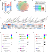
LC-MS untargeted metabolomic analysis revealed altered metabolites and metabolic pathways in the comparisons of AD VS WT, SMC VS AD, SeM VS AD, and SeNa VS AD (n = 6 mice). (a) Number of upregulated and downregulated metabolites in the four comparisons. (b) Venn diagram with the number of differential metabolites in the four comparisons. (c) Heatmap of reversed metabolites in the four comparisons. (d) Superclass of reversed metabolites with administration of the three Se compounds. (eg) KEGG enrichment analysis of differential metabolites in the three comparisons of SMC VS AD, SeM VS AD, and SeNa VS AD.
Figure 5
Figure 5
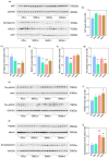
Levels of key proteins, kinases, and receptors in the signaling pathways related to AD pathologies. (a) Levels of GSK-3β and phosphorylated GSK-3β (Ser9) in the cortexes of 3× Tg-AD mouse brains were analyzed by Western blotting. (b) Quantification of protein levels in (a). (c) Levels of COX IV, Drp1, and OPA1 were analyzed by Western blotting. (df) Quantification of protein levels in (c). (g) Levels of FoxO6, FoxO3a, and phosphorylated FoxO3a (Ser253) were analyzed by Western blotting. (h,i) Quantification of protein levels in (g). (j) Levels of NMDAR1, NMDAR2A, and NMDAR2B were analyzed by Western blotting. (km) Quantification of protein levels in (j). α-Tubulin, β-actin, or GAPDH were used as loading controls. All data are presented as the means ± SEMs (n = 3). * p < 0.05 vs. AD mice, as determined by one-way ANOVA followed by Dunnett’s multiple comparison test.

References

    1. Guo T., Zhang D., Zeng Y., Huang T.Y., Xu H., Zhao Y. Molecular and cellular mechanisms underlying the pathogenesis of Alzheimer’s disease. Mol. Neurodegener. 2020;15:40. doi: 10.1186/s13024-020-00391-7. - DOI - PMC - PubMed
    1. Solovyev N.D. Importance of selenium and selenoprotein for brain function: From antioxidant protection to neuronal signalling. J. Inorg. Biochem. 2015;153:1–12. doi: 10.1016/j.jinorgbio.2015.09.003. - DOI - PubMed
    1. Rayman M.P. The importance of selenium to human health. Lancet. 2000;356:233–241. doi: 10.1016/S0140-6736(00)02490-9. - DOI - PubMed
    1. Reeves M.A., Hoffmann P.R. The human selenoproteome: Recent insights into functions and regulation. Cell Mol. Life Sci. 2009;66:2457–2478. doi: 10.1007/s00018-009-0032-4. - DOI - PMC - PubMed
    1. Li S., Sun W., Zhang D. Association of Zinc, Iron, Copper, and Selenium Intakes with Low Cognitive Performance in Older Adults: A Cross-Sectional Study from National Health and Nutrition Examination Survey (NHANES) J. Alzheimers Dis. 2019;72:1145–1157. doi: 10.3233/JAD-190263. - DOI - PubMed

LinkOut - more resources