. 2023 Mar 21;12(3):765.
doi: 10.3390/antiox12030765.

Exploring the Long-Term Tissue Accumulation and Excretion of 3 nm Cerium Oxide Nanoparticles after Single Dose Administration

Affiliations

Exploring the Long-Term Tissue Accumulation and Excretion of 3 nm Cerium Oxide Nanoparticles after Single Dose Administration

Lena M Ernst et al. Antioxidants (Basel). .

Abstract

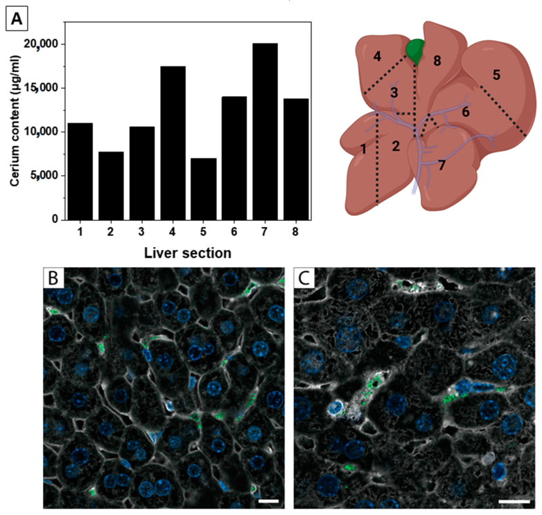
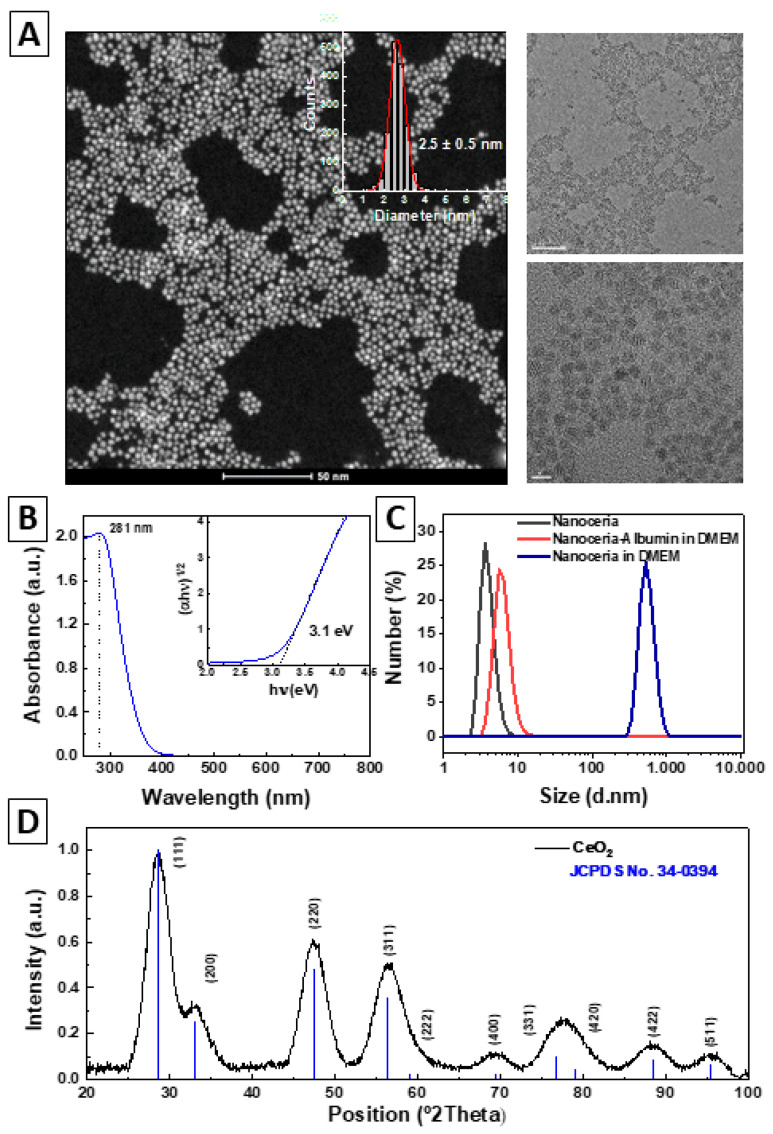
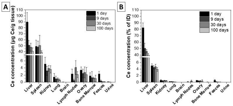
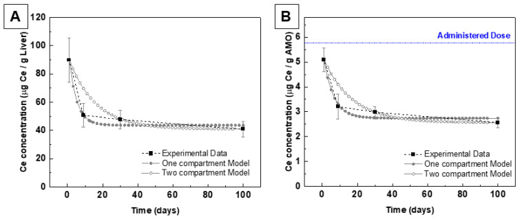
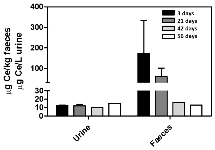
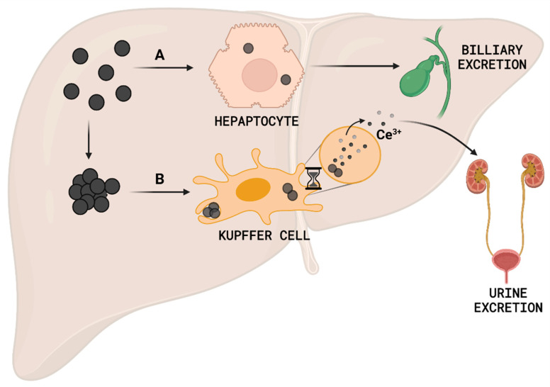
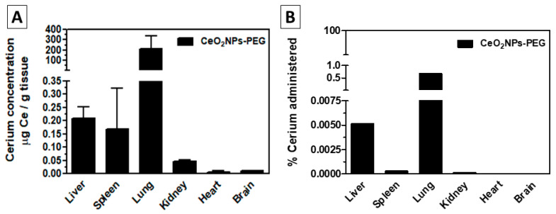
Nanoparticle (NP) pharmacokinetics significantly differ from traditional small molecule principles. From this emerges the need to create new tools and concepts to harness their full potential and avoid unnecessary risks. Nanoparticle pharmacokinetics strongly depend on size, shape, surface functionalisation, and aggregation state, influencing their biodistribution, accumulation, transformations, and excretion profile, and hence their efficacy and safety. Today, while NP biodistribution and nanoceria biodistribution have been studied often at short times, their long-term accumulation and excretion have rarely been studied. In this work, 3 nm nanoceria at 5.7 mg/kg of body weight was intravenously administrated in a single dose to healthy mice. Biodistribution was measured in the liver, spleen, kidney, lung, brain, lymph nodes, ovary, bone marrow, urine, and faeces at different time points (1, 9, 30, and 100 days). Biodistribution and urinary and faecal excretion were also studied in rats placed in metabolic cages at shorter times. The similarity of results of different NPs in different models is shown as the heterogeneous nanoceria distribution in organs. After the expectable accumulation in the liver and spleen, the concentration of cerium decays exponentially, accounting for about a 50% excretion of cerium from the body in 100 days. Cerium ions, coming from NP dissolution, are most likely excreted via the urinary tract, and ceria nanoparticles accumulated in the liver are most likely excreted via the hepatobiliary route. In addition, nanoceria looks safe and does not damage the target organs. No weight loss or apathy was observed during the course of the experiments.

Keywords: NP dissolution; NP excretion; NP long-term accumulation; nanoceria; nanoparticle biodistribution; nanopharmacokinetics; nanosafety.


Conflict of interest statement

The authors declare no conflict of interest.

Figures

Figure 1
Figure 1
Hererogenicity in organ distribution. (A) Liver biodistibution of nanoceria 24 h after IV administration, and liver schema of the digested tissue sections. Liver was divided into 8 parts (schema), and Cerium content of each section is represented in the graph. (B,C) Confocal images of AuNPs in reflectance mode of a liver thin cut after IV administration of NPs to healthy mice at (B) 40× magnification and (C) 60× magnification. Actin (white), Hoechst (blue), and NPs (green). Scale bar = 10 µm.
Figure 2
Figure 2
Synthesis and characterization of nanoceria. (A) HAADF-STEM images (left) and size distribution (insert), and HRES-TEM images (right). Scale bars are 50, 50, and 5 nm, respectively. (B) UV-visible spectrum of the nanoceria and optical bandgap energy determined by Tauc equation (insert). (C) The hydrodynamic diameter profile measured by dynamic light scattering of nanoceria (black), nanoceria-albumin in DMEM (red), and nanoceria in DMEM (blue). (D) X-ray diffraction pattern of the as-synthesized nanoceria and JCPDS No. 34-0394 standards.
Figure 3
Figure 3
Organ cerium concentration measured by ICP-MS 1, 9, 30, and 100 days after single nanoceria IV administration (5.7 mg/Kg body weight). (A) Cerium concentration (µg Ce/g tissue). (B) Cerium as a percentage of the injected dose. n = 5.
Figure 4
Figure 4
Pharmacokinetics of the CeO2 NPs after IV post-injection at 1, 9, 30, and 100 days in the liver (A) and in all measured organs (AMO) (B). Black squares are experimental data, solid grey circles are the one-compartment model fitting, and the hollow grey circles are the two-compartment model fitting. Blue lines are the administered dose.
Figure 5
Figure 5
Nanoceria excretion in rats. Cerium concentration was measured by ICP-MS in urine and faeces from CCl4-treated rats 3, 21, 42, and 56 days after IV nanoceria administration (0.1 mg/Kg bw) twice weekly for two weeks. n = 3.
Figure 6
Figure 6
Hypothetical in vivo management of 3 nm nanoceria. After liver accumulation, (A) hepatocytes rapidly process non-immunogenic nanoceria to the biliary excretion, (B) while nanoceria detected by Kupffer cells will remain inside the cell until it completely dissolves and the produced Ce3+ ions are then excreted via the urine.
Figure 7
Figure 7
Organ biodistribution after oral administration of CeO2-PEG coated nanoparticles (CeO2NPs-PEG) in healthy rats. ICP-MS measured cerium concentration. CeO2NPs-PEG were delivered daily by intragastric administration (10 mg CeO2/Kg bw) for 14 days. Major organs and serum were collected 72 h after the last administration. (A) Cerium concentration in µg Ce/g tissue. (B) Cerium as a percentage of the administered dose. n = 3.

Similar articles

Cited by

References

    1. O’Neill L.A.J., Kishton R.J., Rathmell J. A Guide to Immunometabolism for Immunologists. Nat. Rev. Immunol. 2016;16:553. doi: 10.1038/nri.2016.70. - DOI - PMC - PubMed
    1. Ernst L.M., Puntes V. How Does Immunomodulatory Nanoceria Work? ROS and Immunometabolism. Front. Immunol. 2022;13:974. doi: 10.3389/fimmu.2022.750175. - DOI - PMC - PubMed
    1. Auten R.L., Davis J.M. Oxygen Toxicity and Reactive Oxygen Species: The Devil Is in the Details. Pediatr. Res. 2009;66:121–127. doi: 10.1203/PDR.0b013e3181a9eafb. - DOI - PubMed
    1. Morris G., Gevezova M., Sarafian V., Maes M. Redox Regulation of the Immune Response. Cell. Mol. Immunol. 2022;19:1079–1101. doi: 10.1038/s41423-022-00902-0. - DOI - PMC - PubMed
    1. Lin J., Huang Z., Liu J., Huang Z., Liu Y., Liu Q., Yang Z., Li R., Wu X., Shi Z., et al. Neuroprotective Effect of Ketone Metabolism on Inhibiting Inflammatory Response by Regulating Macrophage Polarization after Acute Cervical Spinal Cord Injury in Rats. Front. Neurosci. 2020;14:583611. doi: 10.3389/fnins.2020.583611. - DOI - PMC - PubMed

LinkOut - more resources