. 2023 Mar 21;12(3):766.
doi: 10.3390/antiox12030766.

Photobiomodulation Controls Keratinocytes Inflammatory Response through Nrf2 and Reduces Langerhans Cells Activation

Affiliations

Photobiomodulation Controls Keratinocytes Inflammatory Response through Nrf2 and Reduces Langerhans Cells Activation

Sara Salman et al. Antioxidants (Basel). .

Abstract

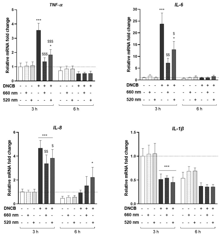
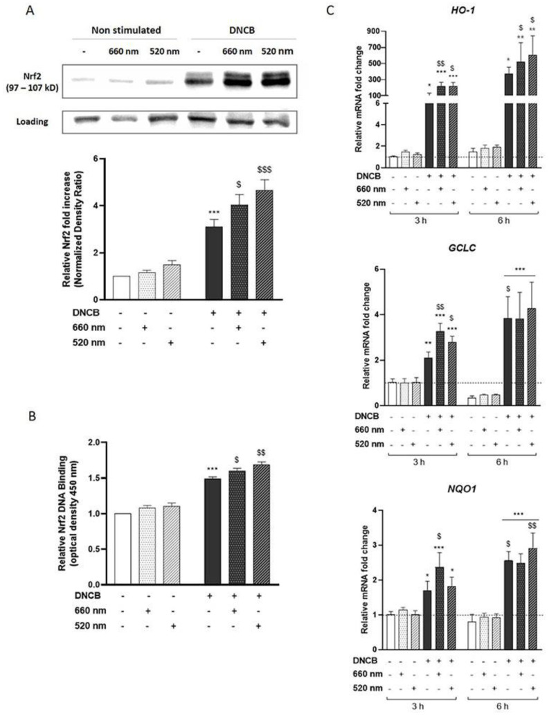
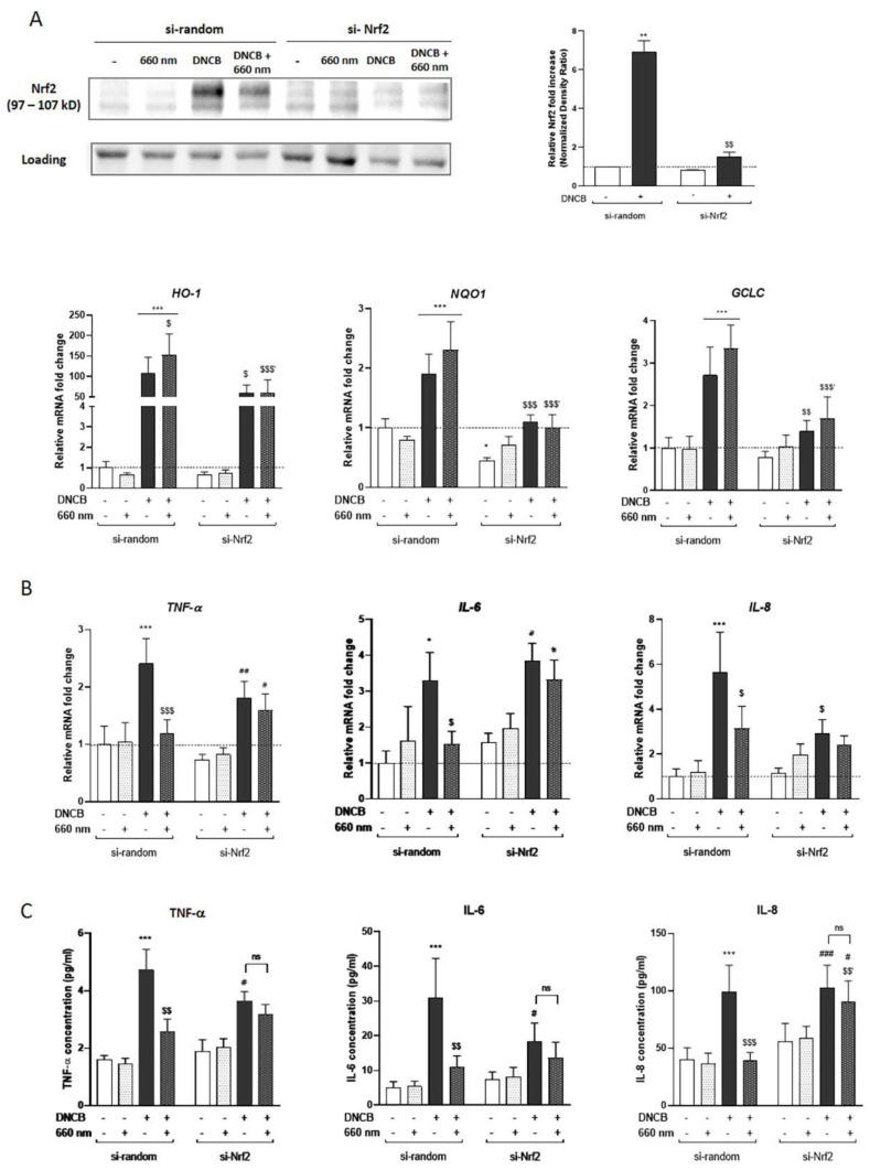
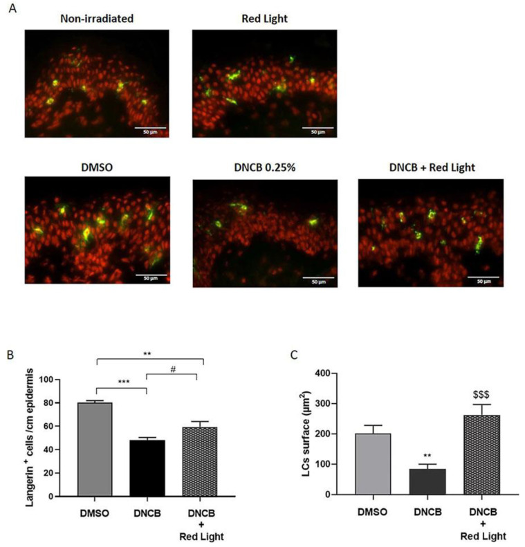
Photobiomodulation (PBM) is rapidly gaining traction as a valuable tool in dermatology for treating many inflammatory skin conditions using low levels of visible light or near-infrared radiation. However, the physiological regulatory pathways responsible for the anti-inflammatory effect of PBM have not been well defined. Since previous studies showed that nuclear factor-erythroid 2 like 2 (Nrf2) is a master regulator of the skin inflammatory response, we have addressed its role in controlling inflammation by PBM. Primary human keratinocytes (KCs) stimulated with 2,4-dinitrochlorobenzene (DNCB) to mimic pro-inflammatory stress were illuminated with two wavelengths: 660 nm or 520 nm. Both lights significantly reduced the mRNA expression of the DNCB-triggered TNF-α, IL-6, and IL-8 cytokines in KCs, while they enhanced Nrf2 pathway activation. PBM-induced Nrf2 is a key regulator of the inflammatory response in KCs since its absence abolished the regulatory effect of light on cytokines production. Further investigations of the mechanisms contributing to the immunoregulatory effect of PBM in inflamed human skin explants showed that 660 nm light prevented Langerhans cells migration into the dermis, preserving their dendricity, and decreased pro-inflammatory cytokine production compared to the DNCB-treated group. This study is the first to report that the PBM-mediated anti-inflammatory response in KCs is Nrf2-dependent and further support the role of PBM in skin immunomodulation. Therefore, PBM should be considered a promising alternative or complementary therapeutic approach for treating skin-related inflammatory diseases.

Keywords: Langerhans cells; Nrf2; immunomodulation; inflammation; keratinocyte; light; photobiomodulation; skin.


Conflict of interest statement

S.S. was funded by Lightinderm. C.G., A.R., and L.D. are employees at Lightinderm and participated in these works as scientific experts. The funders had no role in the design of the study; in the collection, analyses, or interpretation of data; in the writing of the manuscript; or in the decision to publish the results.

Figures

Figure 1
Figure 1
PBM regulates pro-inflammatory DNCB-induced cytokines in KCs. KCs were treated with DNCB or vehicle, with or without red (660 nm) or green (520 nm) light illumination (3 J/cm2, 250 s). Pro-inflammatory cytokines gene expression: TNF-α, IL-6, IL-8, and IL-1β, were measured by RT-qPCR 3 and 6 h after DNCB and light treatment. Results are expressed as fold change. Data represent mean ± SEM of 5 independent experiments. Two-Way ANOVA followed by Tukey post hoc test, * p < 0.05; *** p < 0.001 vs. the relative control at 3 h or 6 h; $ p < 0.05; $$ p < 0.01; $$$ p < 0.001 vs. DNCB at 3 h.
Figure 2
Figure 2
PBM enhances Nrf2-pathway activation in DNCB-stimulated KCs. KCs were treated with vehicle (DMSO 0.1%) or DNCB (25 µM) and illuminated with either wavelength: 660 nm (red light) or 520 nm (green light). (A) Nrf2 accumulation was assessed by Western blot 3 h after illumination. Histograms correspond to densitometric analysis relative to controls and are adjusted to the stain-free blot. (B) ELISA-based measurement of DNA-binding activity for activated Nrf2 in the nuclear extract, 4 h after KCs treatment. (C) Nrf2 target antioxidant gene expression: HO-1, GCLC, and NQO1, were assessed by RT-qPCR, 3 h or 6 h after light treatment. Data represent mean ± SEM of at least five independent experiments. Two-Way ANOVA followed by Tukey post hoc test, * p < 0.05; ** p < 0.01; *** p < 0.001 vs. the relative untreated control at 3 h or 6 h; $ p < 0.05; $$ p < 0.01; $$$ p < 0.001 vs. DNCB at 3 h.
Figure 3
Figure 3
The anti-inflammatory effect of red light in KCs is Nrf2-dependent. KCs were transfected for 48 h by si-Nrf2 or si-random, and then treated with DNCB and exposed to red light. (A) Nrf2 accumulation in the knockdown model was assessed by Western blot 3 h after treatment. The blot is representative of 3 independent experiments. Histograms correspond to densitometric analysis relative to untreated si-random and are adjusted to the stain-free blot. Quantifying the expression of Nrf2 target genes encoding HO-1, NQO1, and GCLC in transfected KCs were assessed using RT-qPCR 3 h after treatment. Induction values were the ratio between gene expressions in treated cells versus gene expression in untreated si-random control. (B) Pro-inflammatory cytokines gene expression: TNF-α, IL-6 and IL-8, were assessed by RT-qPCR, 3 h after light treatment. Results are expressed as fold change compared to the untreated si-random control. (C) The inflammatory cytokines level was determined by Meso Scaled Discovery technology in the supernatant of KCs after 6 h of exposure to DNCB with or without red light illumination. Reported data are mean ± SEM of 6 independent experiments. Two-Way ANOVA followed by Tukey post hoc test, * p < 0.05; ** p < 0.01; *** p < 0.001 vs. untreated si-random; $ p < 0.05; $$ p < 0.01; $$$ p < 0.001 vs. DNCB-treated si-random, $$′ p < 0.01; $$$′ p < 0.001 vs. DNCB + 660 nm-treated si-random; # p < 0.05, ## p < 0.01, ### p < 0.001 vs. untreated si-Nrf2. ns means non-significant different.
Figure 4
Figure 4
Red light decreases DNCB-induced LCs activation in the epidermis. Human skin explants were topically treated with the excipient (DMSO 20%) or DNCB (0.25% w/v) with or without red light exposure (660 nm, 3 J/cm2, 250 s). (A) Langerin immunostaining was performed on frozen skin sections 24 h after DNCB treatment and revealed by AlexaFluor 488 (green). The nuclei were counterstained using propidium iodide (red). Scale bars represent 50 µm. (B) The absolute number of langerin-positive cells in the epidermis was counted at 2 different zones for the 3 explants of each batch. Histogram represents the mean ± SEM of 6 measures of langerin-positive cells/cm epidermis. (C) Langerin-positive cells total surface was measured by ImageJ software (version 1.8). Histogram represents the mean ± SEM of cell surface of 10 different measures. ANOVA followed by Tukey post hoc test; ** p < 0.01; *** p < 0.001 vs. DMSO-treated control group; # p = 0.06, $$$ p < 0.001 vs. DNCB-treated group.

References

    1. Gilaberte Y., Prieto-Torres L., Pastushenko I., Juarranz Á. Chapter 1-Anatomy and Function of the Skin. In: Hamblin M.R., Avci P., Prow T.W., editors. Nanoscience in Dermatology. Academic Press; Boston, MA, USA: 2016. pp. 1–14. - DOI
    1. Jiang Y., Tsoi L.C., Billi A.C., Ward N.L., Harms P.W., Zeng C., Maverakis E., Kahlenberg J.M., Gudjonsson J.E. Cytokinocytes: The Diverse Contribution of Keratinocytes to Immune Responses in Skin. JCI Insight. 2020;5:e1420672020. doi: 10.1172/jci.insight.142067. - DOI - PMC - PubMed
    1. Eyerich S., Eyerich K., Traidl-Hoffmann C., Biedermann T. Cutaneous Barriers and Skin Immunity: Differentiating A Connected Network. Trends Immunol. 2018;39:315–327. doi: 10.1016/j.it.2018.02.004. - DOI - PubMed
    1. Nestle F.O., Di Meglio P., Qin J.-Z., Nickoloff B.J. Skin Immune Sentinels in Health and Disease. Nat. Rev. Immunol. 2009;9:679–691. doi: 10.1038/nri2622. - DOI - PMC - PubMed
    1. Pasparakis M., Haase I., Nestle F.O. Mechanisms Regulating Skin Immunity and Inflammation. Nat. Rev. Immunol. 2014;14:289–301. doi: 10.1038/nri3646. - DOI - PubMed

Grants and funding

LinkOut - more resources