. 2023 Mar 1;12(3):393.
doi: 10.3390/biology12030393.

Genetic Variants in Protein Tyrosine Phosphatase Non-Receptor Type 23 Are Responsible for Mesiodens Formation

Affiliations

Genetic Variants in Protein Tyrosine Phosphatase Non-Receptor Type 23 Are Responsible for Mesiodens Formation

Ploy Adisornkanj et al. Biology (Basel). .

Abstract

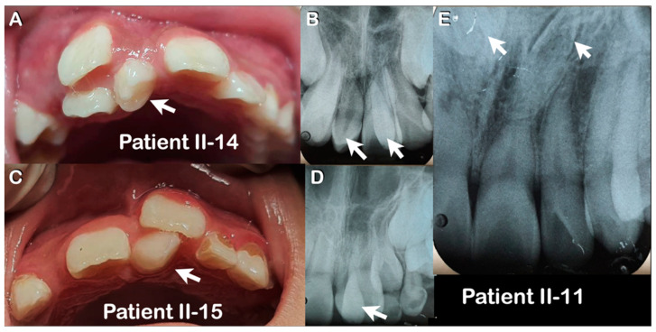
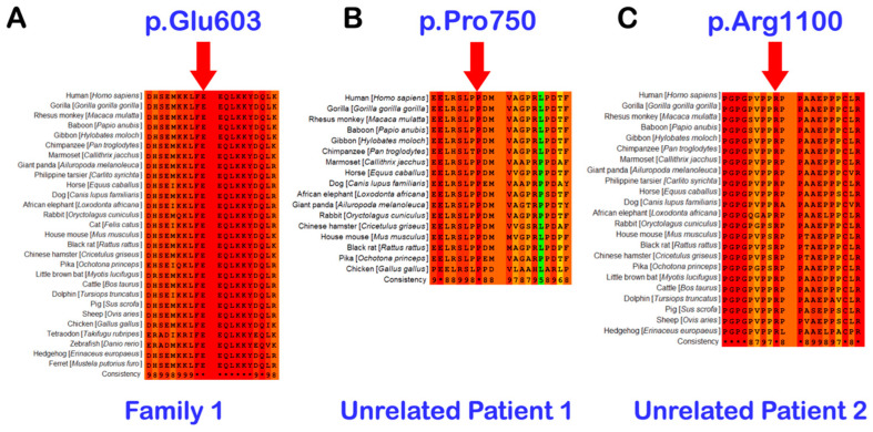
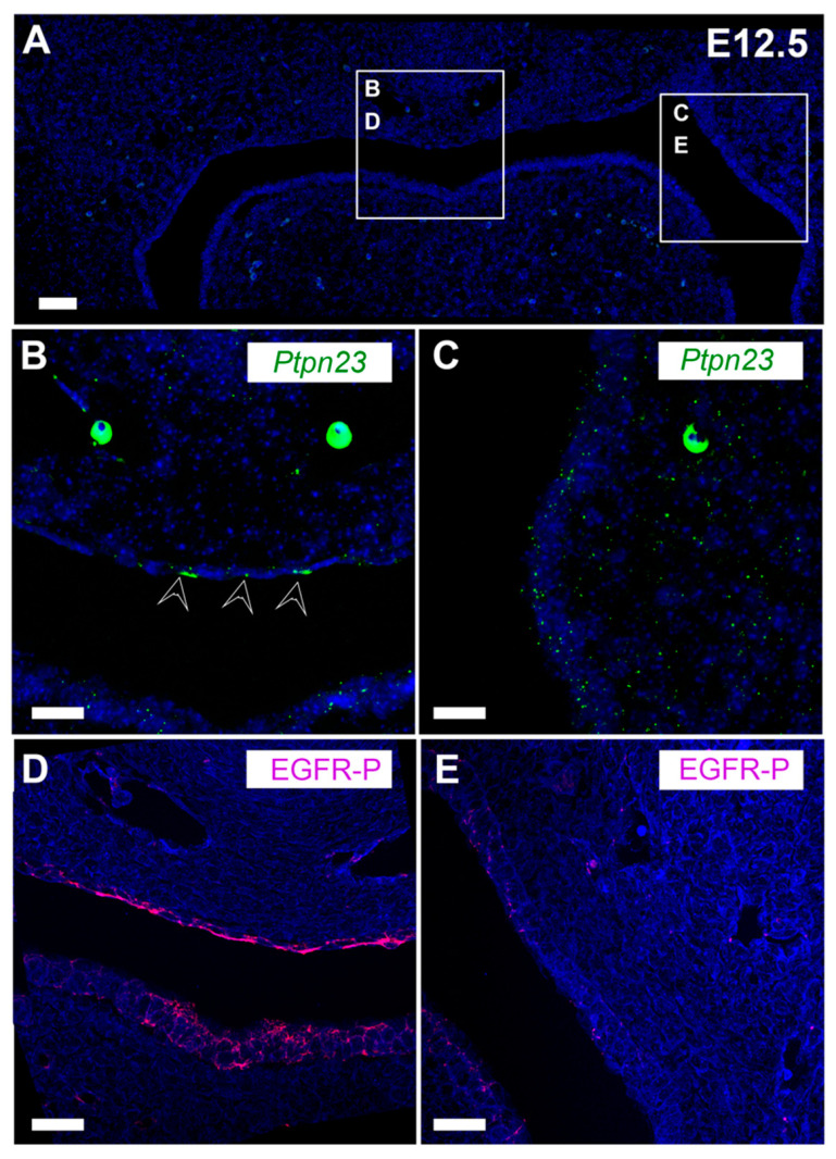
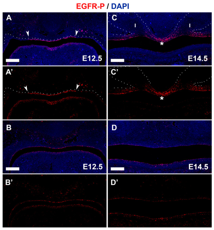
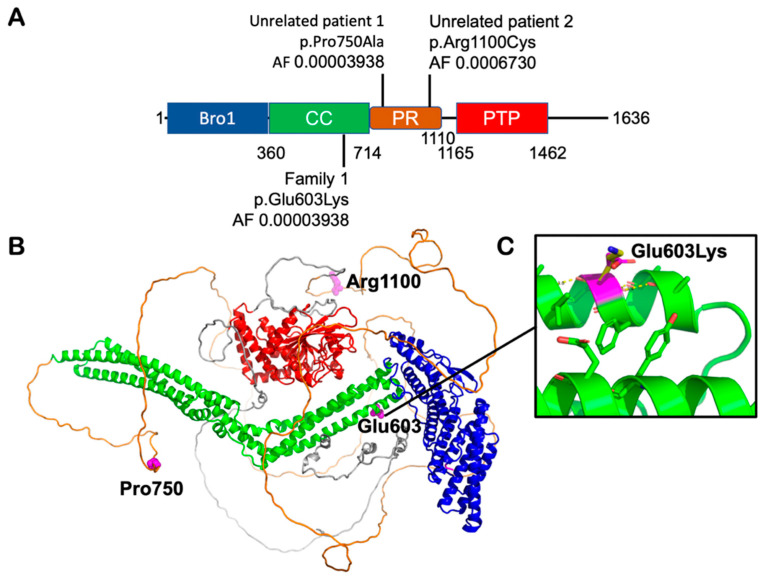
A mesiodens is a supernumerary tooth located in the midline of the premaxilla. To investigate the genetic cause of mesiodens, clinical and radiographic examination were performed on 23 family members of a two-generation Hmong family. Whole exome sequencing (WES) or Sanger sequencing were performed in 22 family members and two unrelated Thai patients with mesiodens. WES in the Hmong family revealed a missense mutation (c.1807G>A;p.Glu603Lys) in PTPN23 in seven affected members and six unaffected members. The mode of inheritance was autosomal dominance with incomplete penetrance (53.84%). Two additional mutations in PTPN23, c.2248C>G;p.Pro750Ala and c.3298C>T;p.Arg1100Cys were identified in two unrelated patients with mesiodens. PTPN23 is a regulator of endosomal trafficking functioning to move activated membrane receptors, such as EGFR, from the endosomal sorting complex towards the ESCRT-III complex for multivesicular body biogenesis, lysosomal degradation, and subsequent downregulation of receptor signaling. Immunohistochemical study and RNAscope on developing mouse embryos showed broad expression of PTPN23 in oral tissues, while immunofluorescence showed that EGFR was specifically concentrated in the midline epithelium. Importantly, PTPN23 mutant protein was shown to have reduced phosphatase activity. In conclusion, mesiodens were associated with genetic variants in PTPN23, suggesting that mesiodens may form due to defects in endosomal trafficking, leading to disrupted midline signaling.

Keywords: PTPN23; RNA expression; extra tooth; mesiodentes; mutation; phosphatase; protein expression; supernumerary tooth.


Conflict of interest statement

The authors declare no conflict of interest.

Figures

Figure 1
Figure 1
Pedigree of the two-generation Hmong family (Family 1). This family consists of 26 members, of which eight are affected with mesiodens (block red) and 18 are unaffected (white). Red dots represent non-penetrant individuals who have the variant p.Glu603Lys but not mesiodens. A number of the affected are children of the non-penetrant. Family members I-2, I-4, I-6, and II-1 were not available for genetic study. The phenotypes of the parents of I-1, I-4, I-5, I-8, I-9, and I-11 are unknown.
Figure 2
Figure 2
Clinical and radiographic pictures of mesiodens (arrows) in (A) Patient II-14. (B) Periapical radiograph in (A) shows double mesiodentes (arrows). (C) Patient II-15. (D) Periapical radiograph in (C) shows mesiodens. (E) Periapical radiograph of patient II-11. Note double inverted mesiodentes (arrows).
Figure 3
Figure 3
Chromatograms of the heterozygous missense (A) c.1807G>A; p.Glu603Lys, (B) c.2248C>G; p.Pro750Ala, and (C) c.3298C>T; p.Arg1100Cys mutations in PTPN23 gene in family 1, unrelated patient 1 and unrelated patient 2, respectively.
Figure 4
Figure 4
Conservation of amino acid residues (A) p.Glu603, (B) p.Pro750, and (C) p.Arg1100 across vertebrate species. All amino acids are highly conserved.
Figure 5
Figure 5
Periapical radiograph of mesiodens of the unrelated patient 1 (arrow).
Figure 6
Figure 6
Unrelated patient 2. Extracted double mesiodentes in (A) Frontal view. (B) Top view. (C) Periapical radiograph prior to extraction (arrows). Note screwdriver-shaped crown morphology of both mesiodentes.
Figure 7
Figure 7
Expression of PTPN23 and EGFR in the oral cavity. Murine E12.5 frontal sections at the midline at the upper incisors. Serial sections used for PTPN23 and EGFR. (A) DAPI (blue) stained section highlighting regions shown in (BE). (B,C) PTPN23 RNAscope. Signal shows up as green dots. Large circular patches are autofluorescence from blood cells. (D,E) Immunofluorescence EGFR protein in pink. (B) PTPN23 is expressed in the midline epithelium (arrows) and (C) robustly in the more lateral epithelium and mesenchyme. (D) EGFR is expressed in the midline in the forming incisor region, similar to PTPN23 expression. (E) EGFR is weakly expressed more laterally. Scale bar in (A): 100 μm. Scale bar in (BE): 50 μm.
Figure 8
Figure 8
EGFR is expressed at high levels in the forming midline. Murine frontal sections. (AB’) E12.5. (CD’) E14.5. (AD) Immunofluorescence EGFR (pink/red) plus DAPI (blue). (A’,B’,C’,D’) EGFR only. (A,A’,C,C’) Anterior region. (B,B’,D,D’) More posterior region. Arrows in (A,A’) highlight border of expression of EGFR in the midline at E12.5. Asterix in (C,C’) highlights midline between cap stage incisors (outlined with white dots). I = incisor. Scale bar in (A,C): 100 μm (same scale in all images).
Figure 9
Figure 9
PTPN23 mutant protein has reduced phosphatase activity. (A) Cell lysates from transfected and untransfected cells were probed for PTPN23 expression. Both untransfected and control transfected cells showed low levels of endogenous PTPN23 protein expression. WT and mutant PTPN23 transfected lysates showed increased expression (n = 3). (B) Phosphatase activity was measured after no transfection and transfection of control DNA, WT PTPN23, and mutant PTPN23 (Glu603Lys). Three different cell lines were used to measure phosphatase activity: HEK293, MDPC (dental pulp cells), and LS-8 (oral epithelial cells). WT PTPN23 has low phosphatase activity in the three cell lines, compared to Potato Acid Phosphatase controls with either 1 unit (u) or 2 units. However, mutant PTPN23 has decreased phosphatase activity compared to WT PTPN23 (n = 3).
Figure 10
Figure 10
EGFR–Multivesicular Biogenesis–Lysosomal Degradation pathway.
Figure 11
Figure 11
Proposed pathogenetic pathways as a result of PTPN23 mutations that lead to mesiodens formation. Refs: [59,62,70,72,73,77,78,79,80,81,82,83,84,85].
Figure 12
Figure 12
Organization of PTPN23 domains and location of the variants. (A) Residue numbers indicate domain boundaries, and arrows show locations of the mutations found in our patients. CC: coiled-coil domain; PR: proline-rich region; PTP inactive phosphatase. (B) Theoretical Alpha fold model of the PTPN23 protein structure. Colors as in (A). The mutated residues are highlighted in magenta as sphere models. (C) Close-up view of the Glu603 wild-type residues (magenta stick model) and its lysine substitution (yellow).

Similar articles

Cited by

References

    1. Bolk L. Die Uberzahligen oberen incisivi des Menschen. Dtsch. Mscf. 2hk. 1917;35:185.
    1. Thilander B., Myrberg N. The prevalence of malocclusion in Swedish schoolchildren. Eur. J. Oral Sci. 1973;81:12–21. doi: 10.1111/j.1600-0722.1973.tb01489.x. - DOI - PubMed
    1. Gündüz K., Çelenk P., Zengin Z., Sümer P. Mesiodens: A radiographic study in children. J. Oral Sci. 2008;50:287–291. doi: 10.2334/josnusd.50.287. - DOI - PubMed
    1. Kositbowornchai S., Keinprasit C., Poomat N. Prevalence and distribution of dental anomalies in pretreatment orthodontic Thai patients. Khon Kaen Dent. J. 2010;13:92–100.
    1. Mukhopadhyay S. Mesiodens: A clinical and radiographic study in children. J. Indian Soc. Pedod. Prev. Dent. 2011;29:34–38. doi: 10.4103/0970-4388.79928. - DOI - PubMed