. 2023 Mar 15;12(3):453.
doi: 10.3390/biology12030453.

Origin of Neuroblasts in the Avian Otic Placode and Their Distributions in the Acoustic and Vestibular Ganglia

Affiliations

Origin of Neuroblasts in the Avian Otic Placode and Their Distributions in the Acoustic and Vestibular Ganglia

Matías Hidalgo-Sánchez et al. Biology (Basel). .

Abstract

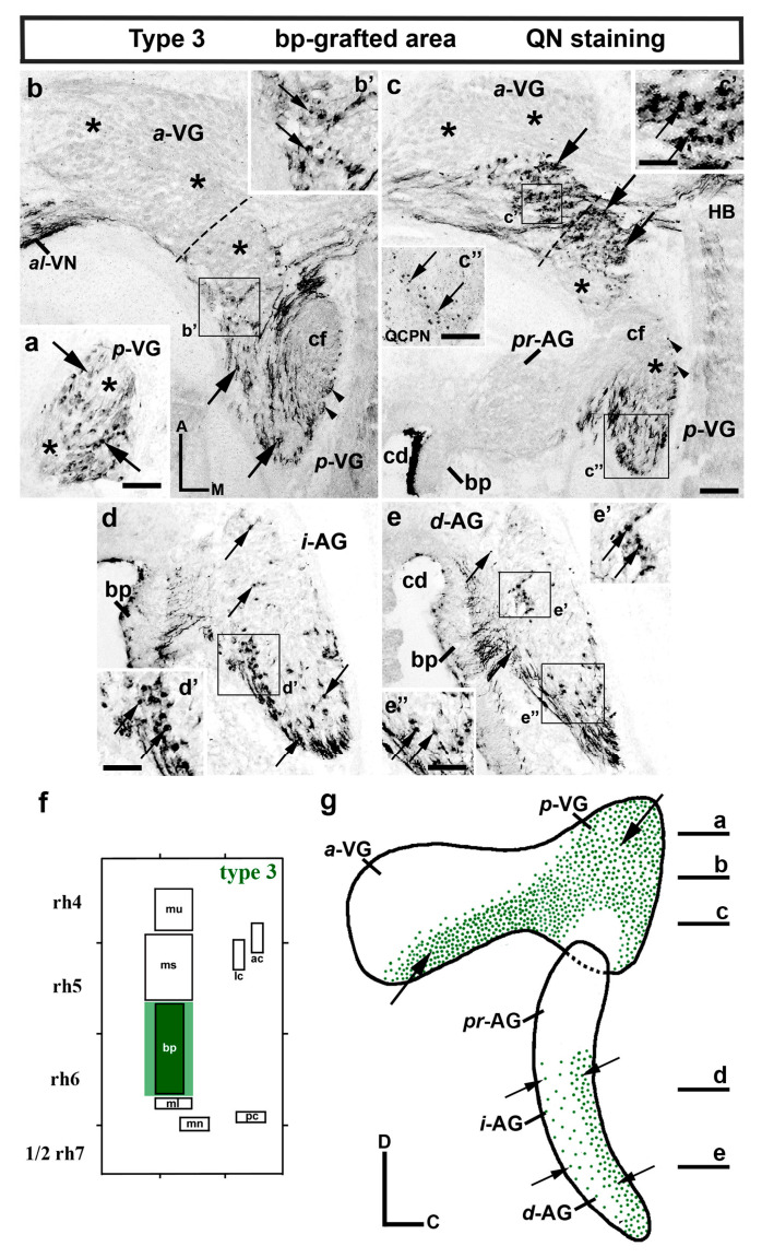
The inner ear is a complex three-dimensional sensorial structure with auditory and vestibular functions. This intricate sensory organ originates from the otic placode, which generates the sensory elements of the membranous labyrinth, as well as all the ganglionic neuronal precursors. How auditory and vestibular neurons establish their fate identities remains to be determined. Their topological origin in the incipient otic placode could provide positional information before they migrate, to later segregate in specific portions of the acoustic and vestibular ganglia. To address this question, transplants of small portions of the avian otic placode were performed according to our previous fate map study, using the quail/chick chimeric graft model. All grafts taking small areas of the neurogenic placodal domain contributed neuroblasts to both acoustic and vestibular ganglia. A differential distribution of otic neurons in the anterior and posterior lobes of the vestibular ganglion, as well as in the proximal, intermediate, and distal portions of the acoustic ganglion, was found. Our results clearly show that, in birds, there does not seem to be a strict segregation of acoustic and vestibular neurons in the incipient otic placode.

Keywords: acoustic ganglion; chick/quail chimaeric embryos; neuroblasts; sensory patch; vestibular ganglion.


Conflict of interest statement

The authors declare no conflict of interest.

Figures

Figure 1
Figure 1
Graft-derived quail ganglionic neurons from the extended area of the macula utriculi (type-1 grafts; n = 9). (ah) Serial horizontal sections stained with QN (ae) and QCPN (fh) antibodies through the vestibular (VG in (a,b,f)) and acoustic (AG in (ce,g,h)) ganglia. (a,b,f) The dotted lines define the separation between the anterior and posterior lobes of the VG (a-VG and p-VG). The four nerves arising from the VG were also indicated in (a,b,f) (a-VN, al-VN, pl-VN, and p-VN). QN- and QCPN-stained grafted quail neurons were undoubtedly detected in the VG (a-VG and p-VG; QN, arrows in (a,b); QCPN, arrows in (f)). (a,b) The black asterisks indicate the areas of the VG devoid of mu-grated neurons. A higher density of grafted quail neurons was observed in the entire AG (pr-AG, i-AG, and d-AG; QN, arrows in (ce); QCPN, arrows in (g,h)). (ae) The white asterisks mark the QN-stained axonal fascicle from the basilar papilla (bp) and macula lagena, associated to the cochlear nerve fascicle (cf in (b,b′)). (a,b)The arrowheads point to grafted-neuron axons towards the hindbrain (HB in (a,b)). (i) Schematic representation of type-1 grafts performed at the 10-somite stage, taking a small portion of the chick otic containing the expanded sensory area of the macula utriculi (mu; dark and light blue areas). (j) Diagram showing a lateral view of the VG (dorsal) and AG (ventral), summarizing all these results in a lateral view of the VG and AG, in which the horizontal sections are indicated. The arrows point to the distribution of mu-grafted quail neurons. The afferent fibers of the basilar papilla were also observed (bp in (ce); [48]). Orientation: A, anterior; C, caudal; D, dorsal; M, Medial. Additional abbreviations: cd, cochlear duct. Scale bar = 19 µm in (e) (ce); 23 µm in (a,b,f); 17 µm in (g,h).
Figure 2
Figure 2
Graft-derived quail ganglionic neurons from the extended area of the macula sacculi (type-2 grafts; n = 9). (af) serial horizontal sections stained with QN antibodies through the vestibular (VG in (ac)) and acoustic (AG in (df)) ganglia. (ac) The dotted lines define the separation between the anterior and posterior lobes of the VG (a-VG and p-VG). QN-stained grafted quail neurons were detected in areas of the a-VG and p-VG (arrows in (ac)), with other areas devoid of ms-grafted neurons (asterisks in (ac)). In the AG, the i-AG and d-AG displayed a reduced number of QN-positive neurons (arrows in (e,f)), with the pr-AG without any QN-positive quail neurons (pr-AG in (d)). (ac) The arrowheads point to axons in the cochlear nerve fascicle (cf). (g) Schematic representation of type-2 grafts performed at the 10-somite stage, considering the expanded sensory area of the macula sacculi (ms; dark and light red areas). (h) Diagram summarizing the results, in which horizontal sections are indicated. The arrows point to the most significant accumulations of ms-grafted quail neurons. Orientation: A, anterior; C, caudal; D, dorsal; M, Medial. Additional abbreviations: al-VN, anterolateral vestibular nerve; ap-VN, anteroposterior vestibular nerve; bp, basilar papilla; cd, cochlear duct; HB, hindbrain; ms, macula sacculi; p-VN, posterior vestibular nerve; pl-VN, posterolateral vestibular nerve. Scale bar = 11 µm in (a′,b′); 20 µm in (a,b,c); 14 µm in (c′); 12 µm in (d,e,f); 8,5 µm in (f′).
Figure 3
Figure 3
Graft-derived quail ganglionic neurons from the extended area of the basilar papilla (type-3 grafts; n = 8). (ae) Horizontal sections stained with QN antibodies through the vestibular (VG in (ac″)) and acoustic (AG in (ce′)) ganglia. The dotted lines in b and c define the separation between the anterior and posterior lobes of the VG (a-VG and p-VG). QN-stained grafted quail neurons were detected in the vestibular ganglion (a-VG and p-VG; arrows in (ac″)) and in the i-AG and d-AG (arrows in (de′)). The insert (c″) confirms the presence of QCPN-stained neurons in the p-VG. (ac) The asterisks indicate the areas of the VG devoid of bp-grafted neurons. (b) The arrowheads point to axons in the cochlear fascicle (cf). (f) Schematic representation of type-3 grafts performed at the 10-somite stage, considering the expanded sensory area of the basilar papilla (bp; dark and light green areas). (g) Diagram summarizing these results, in which horizontal sections are indicated. The arrows point to the most significant accumulations of grafted quail neurons. Orientation: A, anterior; C, caudal; D, dorsal; M, Medial. Additional abbreviations: al-VN, anterolateral vestibular nerve; bp, basilar papilla; cd, cochlear duct; HB, hindbrain. Scale bar = 14 µm in (a); 18 µm in (b,c); 6.5 µm in (b′,c′); 12 µm in (d); 7 µm in (d′); 15 µm in (e); 10 µm in (e′,e″).
Figure 4
Figure 4
Graft-derived quail ganglionic neurons from the extended area of the macula lagena and macula neglecta (type-4 grafts; n = 8). (af) Horizontal sections through the vestibular (ac) and acoustic (df) ganglia. QN-stained neurons were detected in the vestibular ganglion (a-VG and p-VG; arrows in (ac″)). (ac) The asterisks indicate the portions of the VG devoid of quail grafted neurons. In the AG, quail-grafted neurons were found in the periphery of the intermediate and distal subdivisions (i-AG and d-AG; arrows in (e′,f′)). Inserts (c″,e′) confirm the presence of QCPN-stained neurons in the p-VG and i-AG, respectively. Note that the proximal pr-AG was completely devoid of QN-positive neurons (d). The arrowheads in b point to a few axons in the cochlear fascicle (cf). (g) Schematic representation of type-4 grafts, considering the expanded sensory area of the macula lagena and macula neglecta (ml and mn; dark and light grey areas). (h) Diagram summarizing all these results, in which horizontal sections are indicated. The arrows point to the most significant accumulations of ml/mn-grafted neurons. Orientation: A, anterior; C, caudal; D, dorsal; M, Medial. Additional abbreviations: pl-VN, posterolateral vestibular nerve; bp, basilar papilla; cd, cochlear duct; HB, hindbrain. Scale bar = 16 µm in (a); 32 µm in (b,c); 11 µm in (b′,b″,c′,c″); 19 µm in (df); 8 µm in (e′,f′); 16 µm in (f″).
Figure 5
Figure 5
Graft-derived quail ganglionic neurons from the extended area of the anterior and lateral cristae (type-5 grafts; n = 7). (af) Horizontal sections through the vestibular (VG in (ac) and acoustic (AG in (df)) ganglia. QN-stained grafted quail neurons occupied a large portion of both the anterior and posterior vestibular ganglion (a-VG and p-VG; arrows in (ac′)). The asterisks in b and c indicate the areas devoid of ac/lc-grafted neurons. In the AG (df), the pr-AG did not display any quail neurons (d). In the i-AG and d-AG, QN-positive neurons were clearly observed (arrows in (ef′)). (c″) The white asterisk indicates axons in the cochlear fascicle (cf). (g) Schematic representation of the type-5 grafts performed at the 10-somite stage, considering the expanded sensory area of the anterior and lateral cristae (ac and lc; dark and light orange areas). (h) Diagram summarizing the results, in which horizontal sections are indicated. The arrows point to the most significant accumulations of grafted quail neurons. Orientation: A, anterior; C, caudal; D, dorsal; M, Medial. Additional abbreviations: al-VN, anterolateral vestibular nerve; bp, basilar papilla; cd, cochlear duct; HB, hindbrain; pl-VN, posterolateral vestibular nerve. Scale bar = 9 µm in (a); 8 µm in (b′,c′); 10 µm in (c″); 19 µm in (b,c); 12 µm in (df); 6 µm in (f′).
Figure 6
Figure 6
Graft-derived quail ganglionic neurons from the extended area of the posterior crista (type-6 grafts; n = 6). (a,b) Horizontal sections through the VG. The dotted lines in b define the separation between the anterior and posterior lobes of the VG (a-VG and p-VG). A few QN-stained grafted quail neurons were detected exclusively in the p-VG (arrows in (a′,b′)). The a-VG (b) and the AG were devoid of pc-grafted neurons. (c) Schematic representation of the type-6 grafts performed at the 10-somite stage, considering the expanded sensory area of the posterior crista (pc; purple areas). (d) Diagram summarizing the results, the arrow points to the restricted location of grafted neurons in the VG. Orientation: A, anterior; C, caudal; D, dorsal; M, Medial. Additional abbreviations: ms, macula sacculi; s, saccule. Scale bar = 19 µm in (a,b); 9.5 µm in (a′,b′).
Figure 7
Figure 7
Avian otic placode schematic representation at stage HH10 (10 somites), showing the grafted ectoderm portions of each transplant type. (b) Schematic representation displaying the neuronal contribution of each graft type to the different acoustic–vestibular ganglion portions (a-VG, p-VG, pr-AG, i-AG, and d-AG). Each graft type is represented by the same color code used in (a). The neuronal contribution of each graft is denoted as high (full colored), medium (stripes), low (dots), or no contribution (white).

Similar articles

References

    1. Streit A. Extensive Cell Movements Accompany Formation of the Otic Placode. Dev. Biol. 2002;249:237–254. doi: 10.1006/dbio.2002.0739. - DOI - PubMed
    1. Ohyama T., Groves A.K., Martin K. The First Steps towards Hearing: Mechanisms of Otic Placode Induction. Int. J. Dev. Biol. 2007;51:463–472. doi: 10.1387/ijdb.072320to. - DOI - PubMed
    1. Ladher R.K., O′Neill P., Begbie J. From Shared Lineage to Distinct Functions: The Development of the Inner Ear and Epibranchial Placodes. Development. 2010;137:1777–1785. doi: 10.1242/dev.040055. - DOI - PubMed
    1. Chen J., Streit A. Induction of the Inner Ear: Stepwise Specification of Otic Fate from Multipotent Progenitors. Hear. Res. 2013;297:3–12. doi: 10.1016/j.heares.2012.11.018. - DOI - PubMed
    1. Sánchez-Guardado L.Ó., Puelles L., Hidalgo-Sánchez M. Fate Map of the Chicken Otic Placode. Development. 2014;141:2302–2312. doi: 10.1242/dev.101667. - DOI - PubMed

LinkOut - more resources