Review
. 2023 Mar 9;13(3):502.
doi: 10.3390/biom13030502.

Osteopontin: A Bone-Derived Protein Involved in Rheumatoid Arthritis and Osteoarthritis Immunopathology

Affiliations
Review

Osteopontin: A Bone-Derived Protein Involved in Rheumatoid Arthritis and Osteoarthritis Immunopathology

Beatriz Teresita Martín-Márquez et al. Biomolecules. .

Abstract

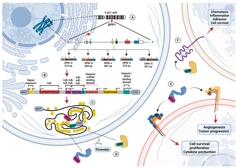
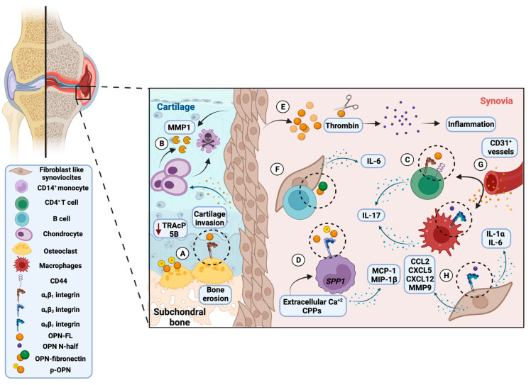
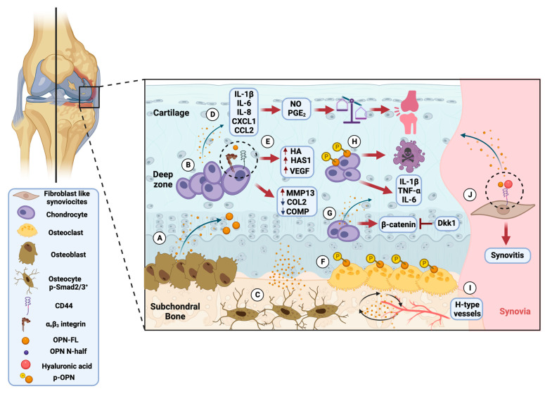
Osteopontin (OPN) is a bone-derived phosphoglycoprotein related to physiological and pathological mechanisms that nowadays has gained relevance due to its role in the immune system response to chronic degenerative diseases, including rheumatoid arthritis (RA) and osteoarthritis (OA). OPN is an extracellular matrix (ECM) glycoprotein that plays a critical role in bone remodeling. Therefore, it is an effector molecule that promotes joint and cartilage destruction observed in clinical studies, in vitro assays, and animal models of RA and OA. Since OPN undergoes multiple modifications, including posttranslational changes, proteolytic cleavage, and binding to a wide range of receptors, the mechanisms by which it produces its effects, in some cases, remain unclear. Although there is strong evidence that OPN contributes significantly to the immunopathology of RA and OA when considering it as a common denominator molecule, some experimental trial results argue for its protective role in rheumatic diseases. Elucidating in detail OPN involvement in bone and cartilage degeneration is of interest to the field of rheumatology. This review aims to provide evidence of the OPN's multifaceted role in promoting joint and cartilage destruction and propose it as a common denominator of AR and OA immunopathology.

Keywords: joint and cartilage degeneration; osteoarthritis; osteopontin; rheumatoid arthritis.


Conflict of interest statement

The authors declare no conflict of interest.

Figures

Figure 1
Figure 1
Representation of osteopontin gene, protein structure, and receptor binding. (A) Human OPN-encoding gene (SPP1) is located on the chromosome 4q21-25 and gives five messenger RNA, where OPN-a is the version with coding information from all exons. (B) OPN full-length (OPN-FL) is a 314 amino acid glycosylated phosphoprotein that contains the Arg-Gly-Asp (RGD) motif sequence, SVVYGLR domain, thrombin cleavage sequence, heparan sulfate binding domain, and heparin/CD44 binding domain. (C) OPN is heavily posttranslationally modified by serine/threonine phosphorylation, O-glycosylation, and tyrosine sulfation. (D) Thrombin cleavage changes the OPN conforming N-terminal fragment (OPN N-half), which allows access to the RGD motif. (E) OPN N-half interacts with integrins αvβ1 and αvβ3 to exert physiological and pathological functions. (F) OPN interaction with CD44 occurs through its putative binding domain located in the last 18 amino acids at the highly conserved C terminus. Created by Biorender with the CT24XI5TS7 agreement number.
Figure 2
Figure 2
Osteopontin’s role in the rheumatoid arthritis immunopathology, based on clinical, in vitro, and experimental observations. (A) Osteoclast overexpresses αvβ3 integrin that binds OPN and promotes bone erosion. Phosphorylated OPN (p-OPN) activates osteoclast due to insufficient tartrate-resistant acid phosphatase (TRAcP) 5B production. (B) Fibroblast-like synoviocytes (FSL) produce OPN in the synovial lining layer and at the sites of cartilage invasion, contribute to matrix degradation by stimulation of metalloproteinase-1 (MMP-1) secretion by chondrocytes, and drive chondrocyte apoptosis. (C) CD4+ T synovial cells express OPN that, through αvβ1 integrin and CD44 binding, induce T cell differentiation towards TH1 and TH17, and secrete IL-17. Furthermore, through α9β1 integrin binding with OPN N-half, synovial macrophages promote IL-17 production. (D) In CD14+ monocytes, OPN induces the expression of monocyte chemoattractant protein-1 (MCP-1) and macrophage inflammatory protein 1 beta (MIP-1β), while p-OPN induces its activation. Extracellular Ca+2 and calciprotein particles (CPPs) differentiate monocytes into SPP1/OPN calcium macrophages. (E) Higher thrombin levels and OPN N-terminal fragment (OPN N-half) were detected in synovial fluid. (F) OPN/fibronectin supports the adhesion of B lymphocytes to FLS and enhances IL-6 production. (G) OPN promotes angiogenesis through new CD31+ vessel formation. (H) OPN N-half binds α9β1 integrin expressed on FLS and promotes the TH17-related cytokines by conventional dendritic cells (DC) and macrophages. Created by Biorender with the MN24XI1DEE agreement number.
Figure 3
Figure 3
Osteopontin’s role in the osteoarthritis immunopathology based on clinical, in vitro, and experimental observations. (A) Osteoblast expresses OPN in the subchondral bone. (B) OPN expression is found in deep zone chondrocytes. (C) OPN expression correlates with p-Smad2/3+ osteocytes associated with OA severity. (D) OPN in cartilage produces cytokines and chemokines that lead to the production of nitric oxide (NO) and prostaglandin E2 (PGE2), which leads to an imbalance in cartilage homeostasis and is associated with joint pain. (E) OPN promotes the expression of hyaluronan synthase 1 (HAS1) and hyaluronic acid (HA) in chondrocytes through CD44 and αvβ3. In addition, OPN can upregulate metalloproteinase-13 (MMP-13), downregulate collagen type 2 (COL2) and cartilage oligomeric matrix protein (COMP), and enhance the production of vascular endothelial growth factor (VEGF). (F) Phosphorylated OPN (p-OPN) is important for interaction with the osteoclast. (G) OPN and β-catenin are increased and could be inhibited by Dickkopf-related protein 1 (Dkk1) in chondrocytes. (H) p-OPN causes apoptosis and production of inflammatory cytokines in chondrocytes. (I) OPN accelerates the subchondral bone turnover and remodeling and induces the formation of h-type vessels. (J) In fibroblast-like synoviocytes (FLS), HA can upregulate OPN and promote synovitis. Created by Biorender with the FQ24XI1ZZF agreement number.

References

    1. Bandopadhyay M., Bulbule A., Butti R., Chakraborty G., Ghorpade P., Ghosh P., Gorain M., Kale S., Kumar D., Kumar S., et al. Osteopontin as a therapeutic target for cancer. Expert Opin. Ther. Targets. 2014;18:883–895. doi: 10.1517/14728222.2014.925447. - DOI - PubMed
    1. Kahles F., Findeisen H.M., Bruemmer D. Osteopontin: A novel regulator at the cross roads of inflammation, obesity and diabetes. Mol. Metab. 2014;3:384–393. doi: 10.1016/j.molmet.2014.03.004. - DOI - PMC - PubMed
    1. Franzen A., Heinegård D. Isolation and characterization of two sialoproteins present only in bone calcified matrix. Biochem. J. 1985;232:715–724. doi: 10.1042/bj2320715. - DOI - PMC - PubMed
    1. Oldberg A., Franzén A., Heinegård D. Cloning and sequence analysis of rat bone sialoprotein (osteopontin) cDNA reveals an Arg-Gly-Asp cell-binding sequence. Proc. Natl. Acad. Sci. USA. 1986;83:8819–8823. doi: 10.1073/pnas.83.23.8819. - DOI - PMC - PubMed
    1. Reinholt F.P., Hultenby K., Oldberg A., Heinegård D. Osteopontin—A possible anchor of osteoclasts to bone. Proc. Natl. Acad. Sci. USA. 1990;87:4473–4475. doi: 10.1073/pnas.87.12.4473. - DOI - PMC - PubMed