Review
. 2023 Mar 20;13(3):405.
doi: 10.3390/bios13030405.

Recent Advancements of LSPR Fiber-Optic Biosensing: Combination Methods, Structure, and Prospects

Affiliations
Review

Recent Advancements of LSPR Fiber-Optic Biosensing: Combination Methods, Structure, and Prospects

Hongxin Zhang et al. Biosensors (Basel). .

Abstract

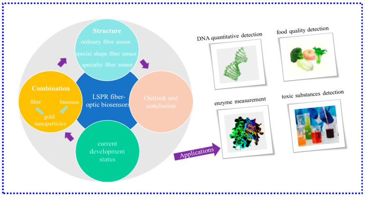
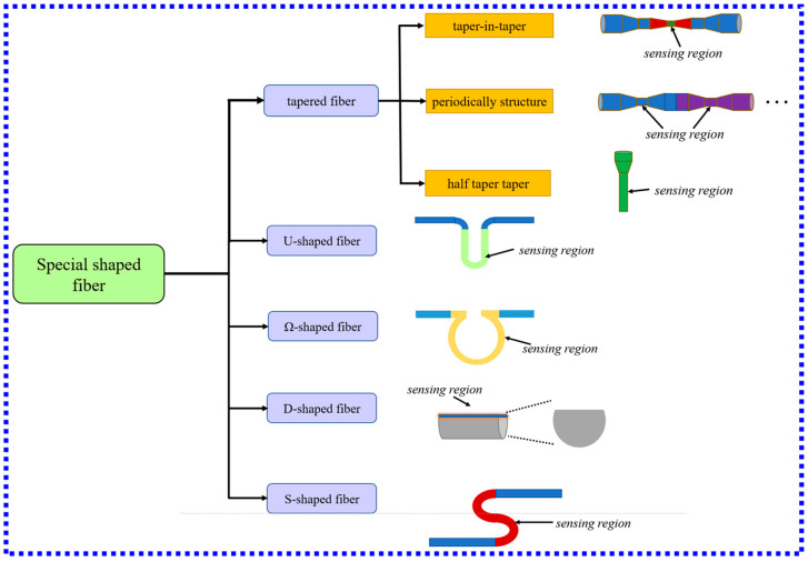
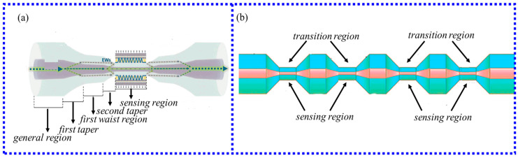
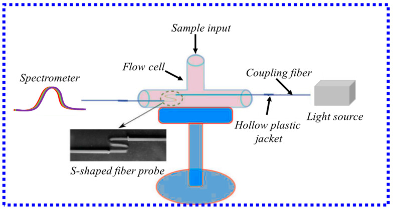
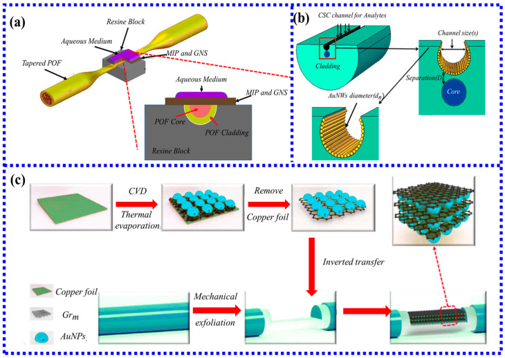
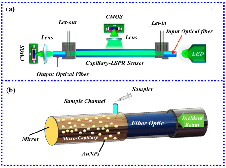
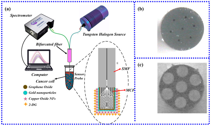
Fiber-optic biosensors based on localized surface plasmon resonance (LSPR) have the advantages of great biocompatibility, label-free, strong stability, and real-time monitoring of various analytes. LSPR fiber-optic biosensors have attracted extensive research attention in the fields of environmental science, clinical medicine, disease diagnosis, and food safety. The latest development of LSPR fiber-optic biosensors in recent years has focused on the detection of clinical disease markers and the detection of various toxic substances in the environment and the progress of new sensitization mechanisms in LSPR fiber-optic sensors. Therefore, this paper reviews the LSPR fiber-optic sensors from the aspects of working principle, structure, and application fields in biosensors. According to the structure, the sensor can be divided into three categories: traditional ordinary optical fiber, special shape optical fiber, and specialty optical fiber. The advantages and disadvantages of existing and future LSPR fiber-optic biosensors are discussed in detail. Additionally, the prospect of future development of fiber-optic biosensors based on LSPR is addressed.

Keywords: LSPR; biosensor; label-free; optical fiber sensor.


Conflict of interest statement

The authors declare no conflict of interest.

Figures

Figure 6
Figure 6
(a) Fiber-optic sensing probe for uric acid detection. (b) Gold nanodisk array sensing probe [64]. (c) Schematic diagram of ZnO nanowires and AuNPs composite structure [66]. Copyright 2023, Elsevier; (d) Schematic diagram of the PS-b-P4VP-templated citrate-AuNR monolayer [48]. Copyright 2023, Elsevier.
Figure 16
Figure 16
(a) Cross-sectional view of the MOF coated with gold nanoparticles on the inner wall. (b) Schematic diagram of the single-channel sensor structure; (c) Schematic diagram of the dual-channel sensor structure [105]. Copyright 2023, Elsevier.
Figure 1
Figure 1
Schematic diagram of the main content of this review.
Figure 2
Figure 2
(a) Transmissive structure; (b) Reflective structure; (c) Hollow core fiber LSPR sensor; (d) Fiber end face LSPR sensor; (e) Fiber core mismatch LSPR sensor.
Figure 3
Figure 3
(a) aminosilanization process; (b) mercaptosilane modified method’s process.
Figure 4
Figure 4
(a) The method of increasing the volume of gold nanoparticles by the secondary immersion in citrate; (b) Comparison of the BCP method with traditional electrostatic self-assembly and silane method [48]. Copyright 2023, Elsevier.
Figure 5
Figure 5
(a) Periodic arrays of FIB fabricated gold nanopillars on the cleaved end-faces of four-mode optical fibers; (b) Arrays of square nanopillars on the tip of a multimode optical fiber. (c)The fabrication process of the microtip-array-based LSPR sensor [53]. Copyright 2023, Elsevier.
Figure 7
Figure 7
Schematic diagram of the fabrication process of fiber-optic LSPR sensors based on monomer (without benzenethiol treatment) and dimer (with benzenethiol treatment) [67]. Copyright 2023, Elsevier.
Figure 8
Figure 8
Schematic diagram of the classification and typical structure of LSPR biosensors with special shapes.
Figure 9
Figure 9
(a) Schematic diagram of taper-in-taper fiber structure [74]; (b) Schematic diagram of four periodic tapered fibers [75]. Copyright 2023, Elsevier.
Figure 10
Figure 10
Chitosan-capped gold nanoparticles synthesized and immobilized on bovine serum albumin functionalized optical fiber [77]. Copyright 2023, Elsevier.
Figure 11
Figure 11
(a) Schematic diagram of Ω-type sensing probe; (b) The relationship between the outer diameter of U-type fiber and Ω-type fiber and the RI sensitivity [80]. Copyright 2023, Elsevier. (c) Schematic illustration of the Ω-FOLSPR for Salmonella typhimurium detection. AuNPs tag is the conjugation of the SH-apt and AuNPs [82]. Copyright 2023, Elsevier. (d) Hybridization and plasmonic photothermal treatment [83]. Copyright 2023, Elsevier.
Figure 12
Figure 12
Schematic diagram of S-type optical fiber sensing device [85]. Copyright 2023, Elsevier.
Figure 13
Figure 13
Schematic diagram of the optical sensor system based on LSPR in D-type fiber: (a) tapered platform with sensing layer of five-branched gold nanostars and molecularly imprinted polymers [90]. Copyright 2023, Elsevier; (b) fiber-optic sensor covered with multiple Au nanowires [91]. (c) Schematic of the preparation procedure of n*(Grm/AuNPs)/D-POF [92].
Figure 14
Figure 14
(a) Schematic diagram of the plasmonic sensing device based on capillary LSPR sensor [95]; (b) Schematic diagram of reflective structure based on HCF.
Figure 15
Figure 15
(a) Schematic diagram of the MCF-based LSPR cell sensing experimental device [99]; (b) MCF cross-section of multicore fiber before HF corrosion [99]; (c) MCF cross-section view after HF corrosion [99]. Copyright 2023, Elsevier.
Figure 17
Figure 17
(a) The process of the probe capturing Pb2+ [107]. (b) Linear fit for intensity spectrum [107]. (c) Functionalization of E. coli captured by T4 phage; (d) Assay scheme of oligonucleotides captured by sandwich DNA hybridization.
Figure 18
Figure 18
Schematic diagram of the experimental setup for measuring ascorbic acid.
Figure 19
Figure 19
The outlook of LSPR fiber-optic biosensors.

References

    1. Chang T.-C., Sun A.Y., Huang Y.-C., Wang C.-H., Wang S.-C., Chau L.-K. Integration of Power-Free and Self-Contained Microfluidic Chip with Fiber Optic Particle Plasmon Resonance Aptasensor for Rapid Detection of SARS-CoV-2 Nucleocapsid Protein. Biosensors. 2022;12:785. doi: 10.3390/bios12100785. - DOI - PMC - PubMed
    1. Li X., Chen N., Zhou X., Zhang Y., Zhao Y., Nguyen L.V., Ebendorff-Heidepriem H., Warren-Smith S.C.J.S., Chemical A.B. In-situ DNA detection with an interferometric-type optical sensor based on tapered exposed core microstructured optical fiber. Sens. Actuators B Chem. 2022;351:130942. doi: 10.1016/j.snb.2021.130942. - DOI
    1. Sypabekova M., Aitkulov A., Blanc W., Tosi D. Reflector-less nanoparticles doped optical fiber biosensor for the detection of Case thrombin. Biosens. Bioelectron. 2020;165:112365. doi: 10.1016/j.bios.2020.112365. - DOI - PubMed
    1. Arjmand M., Aray A., Saghafifar H., Alijanianzadeh M. Quantitative Analysis of Methyl-Parathion Pesticide in Presence of Enzyme Substrate Using Tapered Fiber Optic Biosensor. IEEE Sens. J. 2020;20:5243–5250. doi: 10.1109/JSEN.2020.2968895. - DOI
    1. Mahani M., Alimohamadi F., Torkzadeh-Mahani M., Hassani Z., Khakbaz F., Divsar F., Yoosefian M. LSPR biosensing for the early-stage prostate cancer detection using hydrogen bonds between PSA and antibody: Molecular dynamic and experimental study. J. Mol. Liq. 2021;324:114736. doi: 10.1016/j.molliq.2020.114736. - DOI

LinkOut - more resources